Enimvaadatud - Posterior Uveitis - Chorioretinitis
|

Acute Annular Outer Retinopathy for 3 Months - 25 YO Female Fluorescein Angiogramvaatamisi: 52125-year-old female who, about three months ago, put on her sunglasses and noticed a smudge in the vision and at that point she noticed the right eye was hazy. She had a cold a week or two before the vision loss, but she didn’t have any fever. She has no focal neurological problems in her past history. VA 20/20 OU
|
|
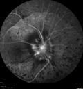
Vasculitis - Retinitis - Uveitis - Vision NLP right eye , 20/50 left eyevaatamisi: 521
|
|
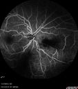
Toxoplasmosis Retinitis - Vitritis and Anerior Uveitisvaatamisi: 521
|
|

West Nile Virus Multifocal Choroiditis and diabetic retinopathyvaatamisi: 521Multifocal and clustered focal lesions are from west nile virus. The retinopathy is probably diabetic but could also be related to west nile virus
|
|

Multifocal Choroidits Vitiligo ( Voght- Kayanagi-Harada Syndrome )vaatamisi: 52036-year old patient with decreased vision in both eyes with fatigue for over a year. VA 20/25-OD, 20/60 -OS. Patient was treated with Avastin and oral prednisone.
|
|
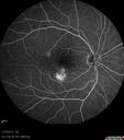
Choroidal Neovascular Membrane - Multifocal Scars - PIC vs MFC vs OHS vaatamisi: 520
|
|

Macular Star, Toxoplasmosisvaatamisi: 520Young male with visual disturbance in the right eye. Fundus photo shows swelling around the optic nerve with macular star visible.
|
|

West Nile Virus Multifocal Choroiditis and diabetic retinopathyvaatamisi: 520Multifocal and clustered focal lesions are from west nile virus. The retinopathy is probably diabetic but could also be related to west nile virus
|
|

Syphillitic Optic Neuropathy and Maculopathyvaatamisi: 52046 year old woman with 2 weeks of vision loss OD. VA 20/200 OD; 20/20 OS. there are posterior vitreous cells on OCT in both eyes. ICG, FAF, and FA show an abnormal macula OD. VA improved to 20/30 within a month with treatment with PCN.
|
|

Multifocal Choroiditis - Linear Scars - Schlaegel linesvaatamisi: 52066 year old woman with macular pucker right eye. Images show typical linear peripheral scars for MFC or POHS - this is unilateral.
|
|

Pigmented Paravenous Retinochoroidal Atrophy - Left Eyevaatamisi: 52070 year old woman lost vision OS 20 years ago.
|
|

Autoimmune Retinopathyvaatamisi: 52091 year old female with severe vision loss over the past 3 years. She has shimmery scotomas. The main findings are outer retinal atrophy evident on OCT and FAF. VA 20/80 OD and 20/125 OS
|
|

DUSN - Diffuse Unilateral Subacute Neuroretinitis - Nematodevaatamisi: 519
|
|

Cytomeglovirusvaatamisi: 51925 year old male experiencing blurry vision OS for two days. Onset very rapid. VA 20/40 OS 20/30 OD. After HIV titer test, determined the pt. had HIV and Cytomeglovirus.
|
|

Choroidal Neovascular Membrane - Multifocal Scars - PIC vs MFC vs OHS vaatamisi: 519
|
|

Birdshot Chorioretinitis - Fresh Presentationvaatamisi: 519Ultawidefield ICG shows best the multifocal lesions. 61 year old woman with floaters for less than 6 months. HLA-A29 Positive
|
|

Syphilitic Chorioretinitisvaatamisi: 519Severe outer retinal atrophy and RPE non-geographic atrophy from syphilis. Vision improved from 20/63 to 20/32 with therapy. Images look a lot like DUSN. Patient had several surgeries (vitrectomy for floaters, vitrectomy for macular pucker, Avastin injections and macular laser for edema) prior by other retina specialists prior to being correctly diagnosed and treated.
|
|

Recurrent MEWDS Left Eye - Teenage Girlvaatamisi: 519Patient presented in 2014 (first images) 2 months following vision loss left eye. Vision went from 20/60 - 20/20. OCT showed outer retinal irregularities which normalized. Then 3 years later in 2017 she had full blown MEWDS. She presented acutely and all the images are typical. Both episodes where in the same time of year, about 3 months after her flu shot for sports.
|
|

Toxoplasmosis - Recurrent - Moderate Vitritisvaatamisi: 51932 year old woman with 20/80 vision and moderately painful red eye in the left eye only. The left eye had some redness about 10 years ago. She is a native of Sierra Leone
|
|

Toxoplasmosis - Recurrent - Moderate Vitritisvaatamisi: 51932 year old woman with 20/80 vision and moderately painful red eye in the left eye only. The left eye had some redness about 10 years ago. She is a native of Sierra Leone
|
|

Sarcoidosis Multifocal Choroiditisvaatamisi: 51966 year old man with dense cataracts and recurrent uveitis. Images show multifocal choroidal granulomas from sarcoidosis more in the right eye than the left eye.
|
|

Autoimmune Retinopathyvaatamisi: 51991 year old female with severe vision loss over the past 3 years. She has shimmery scotomas. The main findings are outer retinal atrophy evident on OCT and FAF. VA 20/80 OD and 20/125 OS
|
|

Uveitis - Retinitis - Vasculitis - possibly Syphilitisvaatamisi: 518Visit 1: 12-06-12
74-year-old man 12-3-12 visit. 2 months ago noticed vision loss, first in the right eye than the left. He is getting tired easily. He can’t walk for a long distance. He has lost five pounds within the last month. He also had a severe itchy episode, but not quite a rash a few days ago, which went away after he took a shower. FTA-ABS is positive but a spinal tap, which was negative. OD 1/200, OS 20/63.
|
|

Choroidal Neovascular Membrane - Multifocal Scars - PIC vs MFC vs OHS vaatamisi: 518
|
|

Unilateral Acute Idiopathic Maculopathy (UAIM)vaatamisi: 51841 year old with serous retinal detachment OS and sudden vision loss. He had a viral prodrome. Coxscackie IgG was high but no acute infection was documented. Vision improved in about 2 months without therapy. - Right eye has spot nasal to the optic nerve
|
|

Multiple Evanescent White Dot Syndrome - Atypical Yellow Foveal Spot Right Eyevaatamisi: 51712-year-old had decreased vision in the left eye from reported amblyopia since around the year 2000. His right eye was doing fine until two days ago, when he had sudden severe vision loss. He sees a blurred spot in the middle of the vision in the right eye wherever he looks.
OD 20/60 with eccentric movement, OS 20/200. Pinhole 20/70. IOP: OD 14, OS 10.
|
|

Birdshot Chorioretinitis - Retinal Vasculitis - HLA A29 Positive vaatamisi: 51764-year-old woman three or four year’s fluctuations in her vision. She mostly notices them in bright light her vision glares out and she has difficulty seeing. PMHx: malaria in 1948 that was treated. She was told at one point she could possibly have had Familial Mediterranean fever.
She does intermittently get rashes around her arms. She has arthritis in her knees and ankles.
VISUAL ACUITY: OD 20/40, OS 20/50 - HLA - A29 Positive
|
|

DUSN - Diffuse Unilateral Subacute Neuroretinitis - Nematodevaatamisi: 517
|
|
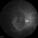
Syphillitic Optic Neuropathy and Maculopathyvaatamisi: 51746 year old woman with 2 weeks of vision loss OD. VA 20/200 OD; 20/20 OS. there are posterior vitreous cells on OCT in both eyes. ICG, FAF, and FA show an abnormal macula OD. VA improved to 20/30 within a month with treatment with PCN.
|
|

Recurrent MEWDS Left Eye - Teenage Girlvaatamisi: 517Patient presented in 2014 (first images) 2 months following vision loss left eye. Vision went from 20/60 - 20/20. OCT showed outer retinal irregularities which normalized. Then 3 years later in 2017 she had full blown MEWDS. She presented acutely and all the images are typical. Both episodes where in the same time of year, about 3 months after her flu shot for sports.
|
|

Recurrent MEWDS Left Eye - Teenage Girlvaatamisi: 517Patient presented in 2014 (first images) 2 months following vision loss left eye. Vision went from 20/60 - 20/20. OCT showed outer retinal irregularities which normalized. Then 3 years later in 2017 she had full blown MEWDS. She presented acutely and all the images are typical. Both episodes where in the same time of year, about 3 months after her flu shot for sports.
|
|

Recurrent MEWDS Left Eye - Teenage Girlvaatamisi: 517Patient presented in 2014 (first images) 2 months following vision loss left eye. Vision went from 20/60 - 20/20. OCT showed outer retinal irregularities which normalized. Then 3 years later in 2017 she had full blown MEWDS. She presented acutely and all the images are typical. Both episodes where in the same time of year, about 3 months after her flu shot for sports.
|
|

Toxoplasmosis Retinitis - Vitritis and Anerior Uveitisvaatamisi: 516Anterior Uveitis
|
|

Unilateral Acute Idiopathic Maculopathy (UAIM) - VIDEO TAKES TIME TO LOAD - raster oct scanvaatamisi: 51641 year old with serous retinal detachment OS and sudden vision loss. He had a viral prodrome. Coxscackie IgG was high but no acute infection was documented. Vision improved in about 2 months without therapy.
|
|

Retinal Atrophy 1 year following Streptococcus sanguinis endophthalmitis - Vision 5/200vaatamisi: 51679 year old woman with sleep apnea who sleeps with CPAP machine
|
|

Syphillitic Optic Neuropathy and Maculopathyvaatamisi: 51646 year old woman with 2 weeks of vision loss OD. VA 20/200 OD; 20/20 OS. there are posterior vitreous cells on OCT in both eyes. ICG, FAF, and FA show an abnormal macula OD. VA improved to 20/30 within a month with treatment with PCN.
|
|

Multifocal Choroiditis Panuveitis - New Choroidal Neovascular Membrane Right Eyevaatamisi: 51674 year old female had vitrectomy elsewhere for possible ocular lymphoma - 5 years of shimmery vision changes then 3 weeks ago noticed vision loss right eye.
|
|

Acute Annular Outer Retinopathy for 3 Months - 25 YO Female Early ICTvaatamisi: 51525-year-old female who, about three months ago, put on her sunglasses and noticed a smudge in the vision and at that point she noticed the right eye was hazy. She had a cold a week or two before the vision loss, but she didn’t have any fever. She has no focal neurological problems in her past history. VA 20/20 OU
|
|

Choroidal Neovascular Membrane - Multifocal Scars - PIC vs MFC vs OHS vaatamisi: 515
|
|

Toxoplasmosis - Recurrent - Moderate Vitritisvaatamisi: 51532 year old woman with 20/80 vision and moderately painful red eye in the left eye only. The left eye had some redness about 10 years ago. She is a native of Sierra Leone
|
|

Sarcoidosis Multifocal Choroiditisvaatamisi: 51566 year old man with dense cataracts and recurrent uveitis. Images show multifocal choroidal granulomas from sarcoidosis more in the right eye than the left eye.
|
|

Multifocal Choroiditis Panuveitis with Subretinal Fibrosis Acute Phasevaatamisi: 51536 year old latin american female 3 weeks of vision loss and new headaches. MRI was normal. VA initially 20/32 OD; 20/20 OS Final vision was 20/63 OD; 20/20 OS
|
|

Autoimmune Retinopathyvaatamisi: 51591 year old female with severe vision loss over the past 3 years. She has shimmery scotomas. The main findings are outer retinal atrophy evident on OCT and FAF. VA 20/80 OD and 20/125 OS
|
|

Acute Annular Outer Retinopathy for 3 Months - 25 YO Female Fundus Photographvaatamisi: 51425-year-old female who, about three months ago, put on her sunglasses and noticed a smudge in the vision and at that point she noticed the right eye was hazy. She had a cold a week or two before the vision loss, but she didn’t have any fever. She has no focal neurological problems in her past history. VA 20/20 OU
|
|

Uveitis - Retinitis - Vasculitis - possibly Syphilitisvaatamisi: 514Visit 1: 12-06-12
74-year-old man 12-3-12 visit. 2 months ago noticed vision loss, first in the right eye than the left. He is getting tired easily. He can’t walk for a long distance. He has lost five pounds within the last month. He also had a severe itchy episode, but not quite a rash a few days ago, which went away after he took a shower. FTA-ABS is positive but a spinal tap, which was negative. OD 1/200, OS 20/63.
|
|

Vasculitis - Retinitis - Uveitis - Vision NLP right eye , 20/50 left eyevaatamisi: 514
|
|

Multifocal Choroiditis Panuveitis with Subretinal Fibrosis Acute Phasevaatamisi: 51436 year old latin american female 3 weeks of vision loss and new headaches. MRI was normal. VA initially 20/32 OD; 20/20 OS Final vision was 20/63 OD; 20/20 OS
|
|

Birdshot Chorioretintis and PAMMvaatamisi: 514Patient presented with paracentral acute middle maculopathy as the presenting symptom of birdshot. This case was published. RETINAL CASES & BRIEF REPORTS 10:151–153, 2016.
|
|

West Nile Virus Multifocal Choroiditis and diabetic retinopathyvaatamisi: 513Multifocal and clustered focal lesions are from west nile virus. The retinopathy is probably diabetic but could also be related to west nile virus
|
|

Syphillitic Optic Neuropathy and Maculopathyvaatamisi: 51346 year old woman with 2 weeks of vision loss OD. VA 20/200 OD; 20/20 OS. there are posterior vitreous cells on OCT in both eyes. ICG, FAF, and FA show an abnormal macula OD. VA improved to 20/30 within a month with treatment with PCN.
|
|

Multifocal Choroiditis Panuveitis with Subretinal Fibrosis Acute Phasevaatamisi: 51336 year old latin american female 3 weeks of vision loss and new headaches. MRI was normal. VA initially 20/32 OD; 20/20 OS Final vision was 20/63 OD; 20/20 OS
|
|

Birdshot Chorioretinitis - Retinal Vasculitis - HLA A29 Positive vaatamisi: 51264-year-old woman three or four year’s fluctuations in her vision. She mostly notices them in bright light her vision glares out and she has difficulty seeing. PMHx: malaria in 1948 that was treated. She was told at one point she could possibly have had Familial Mediterranean fever.
She does intermittently get rashes around her arms. She has arthritis in her knees and ankles.
VISUAL ACUITY: OD 20/40, OS 20/50 - HLA - A29 Positive
|
|

Acute Annular Outer Retinopathy for 3 Months - 25 YO Female Late FAvaatamisi: 51225-year-old female who, about three months ago, put on her sunglasses and noticed a smudge in the vision and at that point she noticed the right eye was hazy. She had a cold a week or two before the vision loss, but she didn’t have any fever. She has no focal neurological problems in her past history. VA 20/20 OU
|
|

Vasculitis - Retinitis - Uveitis - Vision NLP right eye , 20/50 left eyevaatamisi: 512
|
|

Syphillitic Optic Neuropathy and Maculopathyvaatamisi: 51146 year old woman with 2 weeks of vision loss OD. VA 20/200 OD; 20/20 OS. there are posterior vitreous cells on OCT in both eyes. ICG, FAF, and FA show an abnormal macula OD. VA improved to 20/30 within a month with treatment with PCN.
|
|

Syphilitic Chorioretinitisvaatamisi: 511Severe outer retinal atrophy and RPE non-geographic atrophy from syphilis. Vision improved from 20/63 to 20/32 with therapy. Images look a lot like DUSN. Patient had several surgeries (vitrectomy for floaters, vitrectomy for macular pucker, Avastin injections and macular laser for edema) prior by other retina specialists prior to being correctly diagnosed and treated.
|
|
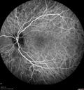
Recurrent MEWDS Left Eye - Teenage Girlvaatamisi: 511Patient presented in 2014 (first images) 2 months following vision loss left eye. Vision went from 20/60 - 20/20. OCT showed outer retinal irregularities which normalized. Then 3 years later in 2017 she had full blown MEWDS. She presented acutely and all the images are typical. Both episodes where in the same time of year, about 3 months after her flu shot for sports.
|
|

AZOOR - unilateral pseudo-retinitis pigmentosavaatamisi: 51174 year old woman with unilateral vision loss 40 years ago.
|
|

Multifocal Choroiditis - Pneumocystis - aspergillis - cryptococcusvaatamisi: 51131 year old male with anaplastic astrocytoma on chemotherapy with mild vision loss in the right eye. LP did not reveal organism. He was placed on a trial of anti-fungal medications and lost to follow-up
|
|
Autoimmune Retinopathyvaatamisi: 51191 year old female with severe vision loss over the past 3 years. She has shimmery scotomas. The main findings are outer retinal atrophy evident on OCT and FAF. VA 20/80 OD and 20/125 OS
|
|

DUSN - Diffuse Unilateral Subacute Neuroretinitis - Nematodevaatamisi: 510
|
|

Uveitis - Retinitis - Vasculitis - possibly Syphilitisvaatamisi: 510Visit 1: 12-06-12
74-year-old man 12-3-12 visit. 2 months ago noticed vision loss, first in the right eye than the left. He is getting tired easily. He can’t walk for a long distance. He has lost five pounds within the last month. He also had a severe itchy episode, but not quite a rash a few days ago, which went away after he took a shower. FTA-ABS is positive but a spinal tap, which was negative. OD 1/200, OS 20/63.
|
|
Toxoplasmosis Retinitis - Vitritis and Anerior Uveitisvaatamisi: 510Anterior Uveitis
|
|

Recurrent MEWDS Left Eye - Teenage Girlvaatamisi: 510Patient presented in 2014 (first images) 2 months following vision loss left eye. Vision went from 20/60 - 20/20. OCT showed outer retinal irregularities which normalized. Then 3 years later in 2017 she had full blown MEWDS. She presented acutely and all the images are typical. Both episodes where in the same time of year, about 3 months after her flu shot for sports.
|
|
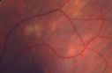
Sarcoidosis Multifocal Choroiditisvaatamisi: 51066 year old man with dense cataracts and recurrent uveitis. Images show multifocal choroidal granulomas from sarcoidosis more in the right eye than the left eye.
|
|

Pigmented Paravenous Retinochoroidal Atrophy - Left Eyevaatamisi: 51070 year old woman lost vision OS 20 years ago.
|
|
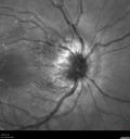
Cat Scratch - Neuroretinitis OD AND retinitis OS - Bartonella henselae positivevaatamisi: 51027 year old female with exposure to cats and prior fever. Reports with recent vision loss right eye. She was treated with doxycycline and within 2 weeks the right eye looks better. Then she skipped all follow-up visits. The right eye presented 20/200 and the left 20/16
|
|

Acute Annular Outer Retinopathy for 3 Months - 25 YO Female Spectral Domain Optical Coherence Tomgraphyvaatamisi: 50925-year-old female who, about three months ago, put on her sunglasses and noticed a smudge in the vision and at that point she noticed the right eye was hazy. She had a cold a week or two before the vision loss, but she didn’t have any fever. She has no focal neurological problems in her past history. VA 20/20 OU
|
|

Uveitis - Retinitis - Vasculitis - possibly Syphilitisvaatamisi: 509Visit 1: 12-06-12
74-year-old man 12-3-12 visit. 2 months ago noticed vision loss, first in the right eye than the left. He is getting tired easily. He can’t walk for a long distance. He has lost five pounds within the last month. He also had a severe itchy episode, but not quite a rash a few days ago, which went away after he took a shower. FTA-ABS is positive but a spinal tap, which was negative. OD 1/200, OS 20/63.
|
|

Syphillitic Optic Neuropathy and Maculopathyvaatamisi: 50946 year old woman with 2 weeks of vision loss OD. VA 20/200 OD; 20/20 OS. there are posterior vitreous cells on OCT in both eyes. ICG, FAF, and FA show an abnormal macula OD. VA improved to 20/30 within a month with treatment with PCN.
|
|

Syphillitic Optic Neuropathy and Maculopathyvaatamisi: 50946 year old woman with 2 weeks of vision loss OD. VA 20/200 OD; 20/20 OS. there are posterior vitreous cells on OCT in both eyes. ICG, FAF, and FA show an abnormal macula OD. VA improved to 20/30 within a month with treatment with PCN.
|
|

Recurrent MEWDS Left Eye - Teenage Girlvaatamisi: 509Patient presented in 2014 (first images) 2 months following vision loss left eye. Vision went from 20/60 - 20/20. OCT showed outer retinal irregularities which normalized. Then 3 years later in 2017 she had full blown MEWDS. She presented acutely and all the images are typical. Both episodes where in the same time of year, about 3 months after her flu shot for sports.
|
|

Recurrent MEWDS Left Eye - Teenage Girlvaatamisi: 509Patient presented in 2014 (first images) 2 months following vision loss left eye. Vision went from 20/60 - 20/20. OCT showed outer retinal irregularities which normalized. Then 3 years later in 2017 she had full blown MEWDS. She presented acutely and all the images are typical. Both episodes where in the same time of year, about 3 months after her flu shot for sports.
|
|

Autoimmune Retinopathyvaatamisi: 50991 year old female with severe vision loss over the past 3 years. She has shimmery scotomas. The main findings are outer retinal atrophy evident on OCT and FAF. VA 20/80 OD and 20/125 OS
|
|

Choroidal Neovascular Membrane - Multifocal Scars - PIC vs MFC vs OHS vaatamisi: 508
|
|
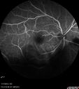
Toxoplasmosis Retinitis - Vitritis and Anerior Uveitisvaatamisi: 508
|
|
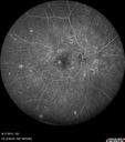
West Nile Virus Multifocal Choroiditis and diabetic retinopathyvaatamisi: 508Multifocal and clustered focal lesions are from west nile virus. The retinopathy is probably diabetic but could also be related to west nile virus
|
|

Birdshot Chorioretinitis - Fresh Presentationvaatamisi: 508Ultawidefield ICG shows best the multifocal lesions. 61 year old woman with floaters for less than 6 months. HLA-A29 Positive
|
|

Multifocal Choroiditis - Linear Scars - Schlaegel linesvaatamisi: 50866 year old woman with macular pucker right eye. Images show typical linear peripheral scars for MFC or POHS - this is unilateral.
|
|
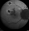
Multifocal Choroiditis Panuveitis - New Choroidal Neovascular Membrane Right Eyevaatamisi: 50874 year old female had vitrectomy elsewhere for possible ocular lymphoma - 5 years of shimmery vision changes then 3 weeks ago noticed vision loss right eye.
|
|

Multifocal Choroiditis Panuveitis with Subretinal Fibrosis Acute Phasevaatamisi: 50836 year old latin american female 3 weeks of vision loss and new headaches. MRI was normal. VA initially 20/32 OD; 20/20 OS Final vision was 20/63 OD; 20/20 OS
|
|
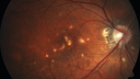
Punctate Inner Choroidopathy - New CNVM right eyevaatamisi: 50842 year old female with recent vision loss in the right eye - Right eye is 20/80 - Vision improved to 20/16 with Ranabizumab injections over 3 months
|
|

Cat Scratch - Neuroretinitis OD AND retinitis OS - Bartonella henselae positivevaatamisi: 50827 year old female with exposure to cats and prior fever. Reports with recent vision loss right eye. She was treated with doxycycline and within 2 weeks the right eye looks better. Then she skipped all follow-up visits. The right eye presented 20/200 and the left 20/16
|
|

Unilateral Acute Idiopathic Maculopathy (UAIM)vaatamisi: 50741 year old with serous retinal detachment OS and sudden vision loss. He had a viral prodrome. Coxscackie IgG was high but no acute infection was documented. Vision improved in about 2 months without therapy. - Right eye has spot nasal to the optic nerve
|
|
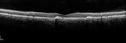
Acute Posterior Multifocal Placoid Pigment Epitheliopathy - 4 years after onsetvaatamisi: 5074 years after presentation - vision 20/80 OD 20/16 OS and there is subretinal fibrosis
|
|

Birdshot Chorioretinitis - Fresh Presentationvaatamisi: 507Ultawidefield ICG shows best the multifocal lesions. 61 year old woman with floaters for less than 6 months. HLA-A29 Positive
|
|

Multifocal Choroiditis Panuveitis with Subretinal Fibrosis Acute Phasevaatamisi: 50736 year old latin american female 3 weeks of vision loss and new headaches. MRI was normal. VA initially 20/32 OD; 20/20 OS Final vision was 20/63 OD; 20/20 OS
|
|

Multiple Evanescent White Dot Syndrome - Atypical Yellow Foveal Spot Right Eyevaatamisi: 50612-year-old had decreased vision in the left eye from reported amblyopia since around the year 2000. His right eye was doing fine until two days ago, when he had sudden severe vision loss. He sees a blurred spot in the middle of the vision in the right eye wherever he looks.
OD 20/60 with eccentric movement, OS 20/200. Pinhole 20/70. IOP: OD 14, OS 10.
|
|

Acute Annular Outer Retinopathy for 3 Months - 25 YO Female Spectral Domain Optical Coherence Tomgraphyvaatamisi: 50625-year-old female who, about three months ago, put on her sunglasses and noticed a smudge in the vision and at that point she noticed the right eye was hazy. She had a cold a week or two before the vision loss, but she didn’t have any fever. She has no focal neurological problems in her past history. VA 20/20 OU
|
|

Retinal Atrophy 1 year following Streptococcus sanguinis endophthalmitis - Vision 5/200vaatamisi: 50679 year old woman with sleep apnea who sleeps with CPAP machine
|
|

Retinal Atrophy 1 year following Streptococcus sanguinis endophthalmitis - Vision 5/200vaatamisi: 50679 year old woman with sleep apnea who sleeps with CPAP machine
|
|

Retinal Atrophy 1 year following Streptococcus sanguinis endophthalmitis - Vision 5/200vaatamisi: 50679 year old woman with sleep apnea who sleeps with CPAP machine
|
|
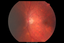
Sarcoidosis Multifocal Choroiditisvaatamisi: 50666 year old man with dense cataracts and recurrent uveitis. Images show multifocal choroidal granulomas from sarcoidosis more in the right eye than the left eye.
|
|

Neuroretinitis - Negative cat scratch serology twice - Possible Behcetsvaatamisi: 50665 year old man with no direct cat exposure and vision loss from neuroretinitis. His work up was positive for HLA B51. He had negative cat scratch titers twice. Vision dropped from 20/60 - 20/200 and then improved to 20/40 over 2 months
|
|
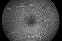
Multiple Evanescent White Dot Syndrome - Atypical Yellow Foveal Spot Right Eyevaatamisi: 50512-year-old had decreased vision in the left eye from reported amblyopia since around the year 2000. His right eye was doing fine until two days ago, when he had sudden severe vision loss. He sees a blurred spot in the middle of the vision in the right eye wherever he looks.
OD 20/60 with eccentric movement, OS 20/200. Pinhole 20/70. IOP: OD 14, OS 10.
|
|

Acute Annular Outer Retinopathy for 3 Months - 25 YO Female InfraRed Imagevaatamisi: 50525-year-old female who, about three months ago, put on her sunglasses and noticed a smudge in the vision and at that point she noticed the right eye was hazy. She had a cold a week or two before the vision loss, but she didn’t have any fever. She has no focal neurological problems in her past history. VA 20/20 OU
|
|

Recurrent MEWDS Left Eye - Teenage Girlvaatamisi: 505Patient presented in 2014 (first images) 2 months following vision loss left eye. Vision went from 20/60 - 20/20. OCT showed outer retinal irregularities which normalized. Then 3 years later in 2017 she had full blown MEWDS. She presented acutely and all the images are typical. Both episodes where in the same time of year, about 3 months after her flu shot for sports.
|
|

Multifocal Choroiditis - Linear Scars - Schlaegel linesvaatamisi: 50566 year old woman with macular pucker right eye. Images show typical linear peripheral scars for MFC or POHS - this is unilateral.
|
|

Neuroretinitis and Multifocal Retinitisvaatamisi: 50562 year old female with vision loss in the left eye to 20/160. Positive Bartonellas IgG. Vision recovered in 2 months. She was treated with Oral Erythromycin BID for 2 weeks.
|
|

Neuroretinitis Positive IgM Bartonella henselaevaatamisi: 50533 year old with vision loss. Her vision improved and she was treated with ciprofloxacen
|
|
|
|


