Most viewed - Posterior Uveitis - Chorioretinitis
|

Multifocal Choroiditis Panuveitis with Subretinal Fibrosis Acute Phase496 views36 year old latin american female 3 weeks of vision loss and new headaches. MRI was normal. VA initially 20/32 OD; 20/20 OS Final vision was 20/63 OD; 20/20 OS
|
|

Punctate Inner Choroidopathy - New CNVM right eye496 views42 year old female with recent vision loss in the right eye - Right eye is 20/80 - Vision improved to 20/16 with Ranabizumab injections over 3 months
|
|

Cat Scratch - Neuroretinitis OD AND retinitis OS - Bartonella henselae positive496 views27 year old female with exposure to cats and prior fever. Reports with recent vision loss right eye. She was treated with doxycycline and within 2 weeks the right eye looks better. Then she skipped all follow-up visits. The right eye presented 20/200 and the left 20/16
|
|

Multifocal Choroiditis - Pneumocystis - aspergillis - cryptococcus496 views31 year old male with anaplastic astrocytoma on chemotherapy with mild vision loss in the right eye. LP did not reveal organism. He was placed on a trial of anti-fungal medications and lost to follow-up
|
|

Multifocal Choroiditis and Subretinal Fibrosis with new Subretinal Neovascular Membrane496 views26 year old African American Girl Saw me at age 10 (16 years ago) with acute multifocal choroiditis and panuveitis which evolved into subretinal fibrosis. She was initially treated with 3 years of methotrexate. Vision ended up 20/20 in both eyes. Her eyes have been inactive since and she was lost to follow-up.
A week ago she noticed sudden decreased vision in the right eye. Within the past few weeks there are more floaters in the right eye.
VA OD: sc6’/200 PHNI
VA OS: sc20/16
|
|

Punctate Inner Choroidopathy (PIC) Mild496 views24 year old female with high myopia. Noticing some distortion on grid. Vision 20/20 OU. Was followed frequently for several months with no progression and no CNVM.
|
|
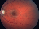
Punctate Inner Choroidopathy (PIC) Mild496 views24 year old female with high myopia. Noticing some distortion on grid. Vision 20/20 OU. Was followed frequently for several months with no progression and no CNVM.
|
|

Acute Annular Outer Retinopathy for 3 Months - 25 YO Female Spectral Domain Optical Coherence Tomgraphy495 views25-year-old female who, about three months ago, put on her sunglasses and noticed a smudge in the vision and at that point she noticed the right eye was hazy. She had a cold a week or two before the vision loss, but she didn’t have any fever. She has no focal neurological problems in her past history. VA 20/20 OU
|
|

Vasculitis - Retinitis - Uveitis - Vision NLP right eye , 20/50 left eye495 views
|
|
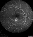
Choroidal Neovascular Membrane - Multifocal Scars - PIC vs MFC vs OHS 495 views
|
|

Syphillitic Optic Neuropathy and Maculopathy495 views46 year old woman with 2 weeks of vision loss OD. VA 20/200 OD; 20/20 OS. there are posterior vitreous cells on OCT in both eyes. ICG, FAF, and FA show an abnormal macula OD. VA improved to 20/30 within a month with treatment with PCN.
|
|

Birdshot Chorioretinitis - Fresh Presentation495 viewsUltawidefield ICG shows best the multifocal lesions. 61 year old woman with floaters for less than 6 months. HLA-A29 Positive
|
|

Birdshot Chorioretinitis - Fresh Presentation495 viewsUltawidefield ICG shows best the multifocal lesions. 61 year old woman with floaters for less than 6 months. HLA-A29 Positive
|
|

Syphilitic Chorioretinitis495 viewsSevere outer retinal atrophy and RPE non-geographic atrophy from syphilis. Vision improved from 20/63 to 20/32 with therapy. Images look a lot like DUSN. Patient had several surgeries (vitrectomy for floaters, vitrectomy for macular pucker, Avastin injections and macular laser for edema) prior by other retina specialists prior to being correctly diagnosed and treated.
|
|

Unilteral Acute Idiopathic Maculopathy - Initial Presentation495 views
|
|

Multifocal Choroiditis Panuveitis - New Choroidal Neovascular Membrane Right Eye495 views74 year old female had vitrectomy elsewhere for possible ocular lymphoma - 5 years of shimmery vision changes then 3 weeks ago noticed vision loss right eye.
|
|
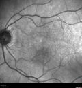
AZOOR - unilateral pseudo-retinitis pigmentosa495 views74 year old woman with unilateral vision loss 40 years ago.
|
|

AZOOR - unilateral pseudo-retinitis pigmentosa495 views74 year old woman with unilateral vision loss 40 years ago.
|
|

Exogenous Fungal Endophthalmitis495 views69 year old female with endophthalmitis 2 months after ECCE. Treated with vitrectomy and 3 injections of amphotericin B. Vision improved from 5/200 - 20/50
|
|

Recurrent MEWDS Left Eye - Teenage Girl494 viewsPatient presented in 2014 (first images) 2 months following vision loss left eye. Vision went from 20/60 - 20/20. OCT showed outer retinal irregularities which normalized. Then 3 years later in 2017 she had full blown MEWDS. She presented acutely and all the images are typical. Both episodes where in the same time of year, about 3 months after her flu shot for sports.
|
|

Multifocal Choroiditis - Linear Scars - Schlaegel lines494 views66 year old woman with macular pucker right eye. Images show typical linear peripheral scars for MFC or POHS - this is unilateral.
|
|

Vasculitis - Retinitis - Uveitis - Vision NLP right eye , 20/50 left eye493 views
|
|

Choroidal Neovascular Membrane - Multifocal Scars - PIC vs MFC vs OHS 493 views
|
|

Choroidal Neovascular Membrane - Multifocal Scars - PIC vs MFC vs OHS 493 views
|
|

Birdshot Chorioretinitis - Fresh Presentation493 viewsUltawidefield ICG shows best the multifocal lesions. 61 year old woman with floaters for less than 6 months. HLA-A29 Positive
|
|

Recurrent MEWDS Left Eye - Teenage Girl493 viewsPatient presented in 2014 (first images) 2 months following vision loss left eye. Vision went from 20/60 - 20/20. OCT showed outer retinal irregularities which normalized. Then 3 years later in 2017 she had full blown MEWDS. She presented acutely and all the images are typical. Both episodes where in the same time of year, about 3 months after her flu shot for sports.
|
|
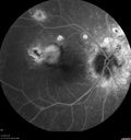
Multifocal Choroiditis Panuveitis - New Choroidal Neovascular Membrane Right Eye493 views74 year old female had vitrectomy elsewhere for possible ocular lymphoma - 5 years of shimmery vision changes then 3 weeks ago noticed vision loss right eye.
|
|

Autoimmune Retinopathy493 views91 year old female with severe vision loss over the past 3 years. She has shimmery scotomas. The main findings are outer retinal atrophy evident on OCT and FAF. VA 20/80 OD and 20/125 OS
|
|

Autoimmune Retinopathy493 views91 year old female with severe vision loss over the past 3 years. She has shimmery scotomas. The main findings are outer retinal atrophy evident on OCT and FAF. VA 20/80 OD and 20/125 OS
|
|

Unilateral Acute Idiopathic Maculopathy (UAIM)492 views41 year old with serous retinal detachment OS and sudden vision loss. He had a viral prodrome. Coxscackie IgG was high but no acute infection was documented. Vision improved in about 2 months without therapy. - Right eye has spot nasal to the optic nerve
|
|

Multifocal Choroiditis Panuveitis with Subretinal Fibrosis Acute Phase492 views36 year old latin american female 3 weeks of vision loss and new headaches. MRI was normal. VA initially 20/32 OD; 20/20 OS Final vision was 20/63 OD; 20/20 OS
|
|

Vasculitis - Retinitis - Uveitis - Vision NLP right eye , 20/50 left eye491 views
|
|

Birdshot Chorioretinitis - Fresh Presentation491 viewsUltawidefield ICG shows best the multifocal lesions. 61 year old woman with floaters for less than 6 months. HLA-A29 Positive
|
|
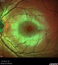
Recurrent MEWDS Left Eye - Teenage Girl491 viewsPatient presented in 2014 (first images) 2 months following vision loss left eye. Vision went from 20/60 - 20/20. OCT showed outer retinal irregularities which normalized. Then 3 years later in 2017 she had full blown MEWDS. She presented acutely and all the images are typical. Both episodes where in the same time of year, about 3 months after her flu shot for sports.
|
|
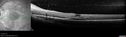
Neuroretinitis - NOT cat scratch - 5 year old491 viewsPresented with nerve swelling, serous RD and uveitis. Ultimately responded to steroids and methotrexate. ANA is positive. All other extensive testing negative including MRI worried about MS
|
|

Sarcoidosis Multifocal Choroiditis491 views66 year old man with dense cataracts and recurrent uveitis. Images show multifocal choroidal granulomas from sarcoidosis more in the right eye than the left eye.
|
|
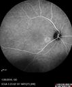
Multifocal Choroiditis Panuveitis with Subretinal Fibrosis Acute Phase491 views36 year old latin american female 3 weeks of vision loss and new headaches. MRI was normal. VA initially 20/32 OD; 20/20 OS Final vision was 20/63 OD; 20/20 OS
|
|

Cat Scratch - Neuroretinitis OD AND retinitis OS - Bartonella henselae positive491 views27 year old female with exposure to cats and prior fever. Reports with recent vision loss right eye. She was treated with doxycycline and within 2 weeks the right eye looks better. Then she skipped all follow-up visits. The right eye presented 20/200 and the left 20/16
|
|

Cat Scratch - Neuroretinitis OD AND retinitis OS - Bartonella henselae positive491 views27 year old female with exposure to cats and prior fever. Reports with recent vision loss right eye. She was treated with doxycycline and within 2 weeks the right eye looks better. Then she skipped all follow-up visits. The right eye presented 20/200 and the left 20/16
|
|

Birdshot Chorioretintis and PAMM491 viewsPatient presented with paracentral acute middle maculopathy as the presenting symptom of birdshot. This case was published. RETINAL CASES & BRIEF REPORTS 10:151–153, 2016.
|
|

Uveitis - Retinitis - Vasculitis - possibly Syphilitis490 viewsVisit 1: 12-06-12
74-year-old man 12-3-12 visit. 2 months ago noticed vision loss, first in the right eye than the left. He is getting tired easily. He can’t walk for a long distance. He has lost five pounds within the last month. He also had a severe itchy episode, but not quite a rash a few days ago, which went away after he took a shower. FTA-ABS is positive but a spinal tap, which was negative. OD 1/200, OS 20/63.
|
|

Vasculitis - Retinitis - Uveitis - Vision NLP right eye , 20/50 left eye490 views
|
|

Unilateral Hemorrhagic Retinopathy490 viewsUnilateral Hemorrhagic Retinopathy
|
|
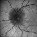
Syphillitic Optic Neuropathy and Maculopathy490 views46 year old woman with 2 weeks of vision loss OD. VA 20/200 OD; 20/20 OS. there are posterior vitreous cells on OCT in both eyes. ICG, FAF, and FA show an abnormal macula OD. VA improved to 20/30 within a month with treatment with PCN.
|
|

Syphillitic Optic Neuropathy and Maculopathy490 views46 year old woman with 2 weeks of vision loss OD. VA 20/200 OD; 20/20 OS. there are posterior vitreous cells on OCT in both eyes. ICG, FAF, and FA show an abnormal macula OD. VA improved to 20/30 within a month with treatment with PCN.
|
|

Birdshot Chorioretinitis - Fresh Presentation490 viewsUltawidefield ICG shows best the multifocal lesions. 61 year old woman with floaters for less than 6 months. HLA-A29 Positive
|
|

Pigmented Paravenous Retinochoroidal Atrophy - Left Eye490 views70 year old woman lost vision OS 20 years ago.
|
|
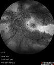
Pigmented Paravenous Retinochoroidal Atrophy - Left Eye490 views70 year old woman lost vision OS 20 years ago.
|
|

Neuroretinitis and Multifocal Retinitis490 views62 year old female with vision loss in the left eye to 20/160. Positive Bartonellas IgG. Vision recovered in 2 months. She was treated with Oral Erythromycin BID for 2 weeks.
|
|

Multifocal Choroiditis Panuveitis with Subretinal Fibrosis Acute Phase490 views36 year old latin american female 3 weeks of vision loss and new headaches. MRI was normal. VA initially 20/32 OD; 20/20 OS Final vision was 20/63 OD; 20/20 OS
|
|

Cat Scratch - Neuroretinitis OD AND retinitis OS - Bartonella henselae positive490 views27 year old female with exposure to cats and prior fever. Reports with recent vision loss right eye. She was treated with doxycycline and within 2 weeks the right eye looks better. Then she skipped all follow-up visits. The right eye presented 20/200 and the left 20/16
|
|

Autoimmune Retinopathy490 views91 year old female with severe vision loss over the past 3 years. She has shimmery scotomas. The main findings are outer retinal atrophy evident on OCT and FAF. VA 20/80 OD and 20/125 OS
|
|

Autoimmune Retinopathy490 views91 year old female with severe vision loss over the past 3 years. She has shimmery scotomas. The main findings are outer retinal atrophy evident on OCT and FAF. VA 20/80 OD and 20/125 OS
|
|

Cytomeglovirus489 views25 year old male experiencing blurry vision OS for two days. Onset very rapid. VA 20/40 OS 20/30 OD. After HIV titer test, determined the pt. had HIV and Cytomeglovirus.
|
|

Vasculitis - Retinitis - Uveitis - Vision NLP right eye , 20/50 left eye489 views
|
|

Vasculitis - Retinitis - Uveitis - Vision NLP right eye , 20/50 left eye489 views
|
|

489 views
|
|

Unilateral Acute Idiopathic Maculopathy (UAIM)489 views41 year old with serous retinal detachment OS and sudden vision loss. He had a viral prodrome. Coxscackie IgG was high but no acute infection was documented. Vision improved in about 2 months without therapy. - Right eye has spot nasal to the optic nerve
|
|

West Nile Virus Multifocal Choroiditis and diabetic retinopathy489 viewsMultifocal and clustered focal lesions are from west nile virus. The retinopathy is probably diabetic but could also be related to west nile virus
|
|

West Nile Virus Multifocal Choroiditis and diabetic retinopathy489 viewsMultifocal and clustered focal lesions are from west nile virus. The retinopathy is probably diabetic but could also be related to west nile virus
|
|

Birdshot Chorioretinitis - Fresh Presentation489 viewsUltawidefield ICG shows best the multifocal lesions. 61 year old woman with floaters for less than 6 months. HLA-A29 Positive
|
|

Multifocal Choroiditis Panuveitis - New Choroidal Neovascular Membrane Right Eye489 views74 year old female had vitrectomy elsewhere for possible ocular lymphoma - 5 years of shimmery vision changes then 3 weeks ago noticed vision loss right eye.
|
|

Neuroretinitis and Multifocal Retinitis489 views62 year old female with vision loss in the left eye to 20/160. Positive Bartonellas IgG. Vision recovered in 2 months. She was treated with Oral Erythromycin BID for 2 weeks.
|
|

AZOOR - unilateral pseudo-retinitis pigmentosa489 views74 year old woman with unilateral vision loss 40 years ago.
|
|

Birdshot Chorioretintis and PAMM489 viewsPatient presented with paracentral acute middle maculopathy as the presenting symptom of birdshot. This case was published. RETINAL CASES & BRIEF REPORTS 10:151–153, 2016.
|
|

Multifocal Choroiditis - Pneumocystis - aspergillis - cryptococcus489 views31 year old male with anaplastic astrocytoma on chemotherapy with mild vision loss in the right eye. LP did not reveal organism. He was placed on a trial of anti-fungal medications and lost to follow-up
|
|

488 views
|
|

Birdshot Chorioretinitis - Fresh Presentation488 viewsUltawidefield ICG shows best the multifocal lesions. 61 year old woman with floaters for less than 6 months. HLA-A29 Positive
|
|

Cat Scratch - Neuroretinitis OD AND retinitis OS - Bartonella henselae positive488 views27 year old female with exposure to cats and prior fever. Reports with recent vision loss right eye. She was treated with doxycycline and within 2 weeks the right eye looks better. Then she skipped all follow-up visits. The right eye presented 20/200 and the left 20/16
|
|

Autoimmune Retinopathy488 views91 year old female with severe vision loss over the past 3 years. She has shimmery scotomas. The main findings are outer retinal atrophy evident on OCT and FAF. VA 20/80 OD and 20/125 OS
|
|

Acute Annular Outer Retinopathy for 3 Months - 25 YO Female InfraRed Image487 views25-year-old female who, about three months ago, put on her sunglasses and noticed a smudge in the vision and at that point she noticed the right eye was hazy. She had a cold a week or two before the vision loss, but she didn’t have any fever. She has no focal neurological problems in her past history. VA 20/20 OU
|
|

Uveitis - Retinitis - Vasculitis - possibly Syphilitis487 viewsVisit 1: 12-06-12
74-year-old man 12-3-12 visit. 2 months ago noticed vision loss, first in the right eye than the left. He is getting tired easily. He can’t walk for a long distance. He has lost five pounds within the last month. He also had a severe itchy episode, but not quite a rash a few days ago, which went away after he took a shower. FTA-ABS is positive but a spinal tap, which was negative. OD 1/200, OS 20/63.
|
|

Uveitis - Retinitis - Vasculitis - possibly Syphilitis487 viewsVisit 1: 12-06-12
74-year-old man 12-3-12 visit. 2 months ago noticed vision loss, first in the right eye than the left. He is getting tired easily. He can’t walk for a long distance. He has lost five pounds within the last month. He also had a severe itchy episode, but not quite a rash a few days ago, which went away after he took a shower. FTA-ABS is positive but a spinal tap, which was negative. OD 1/200, OS 20/63.
|
|

Birdshot Chorioretinitis - Fresh Presentation487 viewsUltawidefield ICG shows best the multifocal lesions. 61 year old woman with floaters for less than 6 months. HLA-A29 Positive
|
|

Birdshot Chorioretinitis - Fresh Presentation487 viewsUltawidefield ICG shows best the multifocal lesions. 61 year old woman with floaters for less than 6 months. HLA-A29 Positive
|
|

Neuroretinitis - NOT cat scratch - 5 year old487 viewsPresented with nerve swelling, serous RD and uveitis. Ultimately responded to steroids and methotrexate. ANA is positive. All other extensive testing negative including MRI worried about MS
|
|

Punctate Inner Choroidopathy (PIC) Mild487 views24 year old female with high myopia. Noticing some distortion on grid. Vision 20/20 OU. Was followed frequently for several months with no progression and no CNVM.
|
|

Unilateral Acute Idiopathic Maculopathy (UAIM)486 views41 year old with serous retinal detachment OS and sudden vision loss. He had a viral prodrome. Coxscackie IgG was high but no acute infection was documented. Vision improved in about 2 months without therapy. - Right eye has spot nasal to the optic nerve
|
|

West Nile Virus Multifocal Choroiditis and diabetic retinopathy486 viewsMultifocal and clustered focal lesions are from west nile virus. The retinopathy is probably diabetic but could also be related to west nile virus
|
|
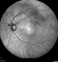
Birdshot Chorioretinitis - Fresh Presentation486 viewsUltawidefield ICG shows best the multifocal lesions. 61 year old woman with floaters for less than 6 months. HLA-A29 Positive
|
|

Recurrent MEWDS Left Eye - Teenage Girl486 viewsPatient presented in 2014 (first images) 2 months following vision loss left eye. Vision went from 20/60 - 20/20. OCT showed outer retinal irregularities which normalized. Then 3 years later in 2017 she had full blown MEWDS. She presented acutely and all the images are typical. Both episodes where in the same time of year, about 3 months after her flu shot for sports.
|
|

Sarcoidosis Multifocal Choroiditis486 views66 year old man with dense cataracts and recurrent uveitis. Images show multifocal choroidal granulomas from sarcoidosis more in the right eye than the left eye.
|
|
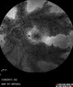
Pigmented Paravenous Retinochoroidal Atrophy - Left Eye486 views70 year old woman lost vision OS 20 years ago.
|
|

AZOOR - unilateral pseudo-retinitis pigmentosa486 views74 year old woman with unilateral vision loss 40 years ago.
|
|

Cat Scratch - Neuroretinitis OD AND retinitis OS - Bartonella henselae positive486 views27 year old female with exposure to cats and prior fever. Reports with recent vision loss right eye. She was treated with doxycycline and within 2 weeks the right eye looks better. Then she skipped all follow-up visits. The right eye presented 20/200 and the left 20/16
|
|

Birdshot Chorioretintis and PAMM486 viewsPatient presented with paracentral acute middle maculopathy as the presenting symptom of birdshot. This case was published. RETINAL CASES & BRIEF REPORTS 10:151–153, 2016.
|
|

Multifocal Choroiditis - Pneumocystis - aspergillis - cryptococcus486 views31 year old male with anaplastic astrocytoma on chemotherapy with mild vision loss in the right eye. LP did not reveal organism. He was placed on a trial of anti-fungal medications and lost to follow-up
|
|

Uveitis - Retinitis - Vasculitis - possibly Syphilitis485 viewsVisit 1: 12-06-12
74-year-old man 12-3-12 visit. 2 months ago noticed vision loss, first in the right eye than the left. He is getting tired easily. He can’t walk for a long distance. He has lost five pounds within the last month. He also had a severe itchy episode, but not quite a rash a few days ago, which went away after he took a shower. FTA-ABS is positive but a spinal tap, which was negative. OD 1/200, OS 20/63.
|
|

Choroidal Neovascular Membrane - Multifocal Scars - PIC vs MFC vs OHS 485 views
|
|

Choroidal Neovascular Membrane - Multifocal Scars - PIC vs MFC vs OHS 485 views
|
|

Toxoplasmosis Retinitis - Vitritis and Anerior Uveitis485 views
|
|

Toxoplasmosis Retinitis - Vitritis and Anerior Uveitis485 views
|
|
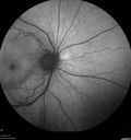
Birdshot Chorioretinitis - Fresh Presentation485 viewsUltawidefield ICG shows best the multifocal lesions. 61 year old woman with floaters for less than 6 months. HLA-A29 Positive
|
|

Birdshot Chorioretinitis - Fresh Presentation485 viewsUltawidefield ICG shows best the multifocal lesions. 61 year old woman with floaters for less than 6 months. HLA-A29 Positive
|
|

Recurrent MEWDS Left Eye - Teenage Girl485 viewsPatient presented in 2014 (first images) 2 months following vision loss left eye. Vision went from 20/60 - 20/20. OCT showed outer retinal irregularities which normalized. Then 3 years later in 2017 she had full blown MEWDS. She presented acutely and all the images are typical. Both episodes where in the same time of year, about 3 months after her flu shot for sports.
|
|
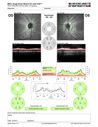
Recurrent MEWDS Left Eye - Teenage Girl485 viewsPatient presented in 2014 (first images) 2 months following vision loss left eye. Vision went from 20/60 - 20/20. OCT showed outer retinal irregularities which normalized. Then 3 years later in 2017 she had full blown MEWDS. She presented acutely and all the images are typical. Both episodes where in the same time of year, about 3 months after her flu shot for sports.
|
|

485 views
|
|

Multifocal Choroiditis Panuveitis with Subretinal Fibrosis Acute Phase485 views36 year old latin american female 3 weeks of vision loss and new headaches. MRI was normal. VA initially 20/32 OD; 20/20 OS Final vision was 20/63 OD; 20/20 OS
|
|
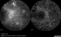
Multifocal Choroiditis Panuveitis with Subretinal Fibrosis Acute Phase485 views36 year old latin american female 3 weeks of vision loss and new headaches. MRI was normal. VA initially 20/32 OD; 20/20 OS Final vision was 20/63 OD; 20/20 OS
|
|

Neuroretinitis Positive IgM Bartonella henselae485 views33 year old with vision loss. Her vision improved and she was treated with ciprofloxacen
|
|
| 2409 files on 25 page(s) |
 |
|
|
|
|
12 | |
|
|
|
|


