Most viewed - Retina Vascular Occlusions
|

Branch Retinal Vein Occlusion with Edema - Failed avastin (bevacizumab) and did well with Eylea (Aflibercept)386 views67 year old female The vision in the left eye was good until about 4 months ago. Prior to that it was OK. The vision in the right eye is fine. She is not sure if she has had strokes from studies taken in December. She was, at that time, sent to the ER for high blood pressure. Â
VA OD: Dcc20/40 OS: 20/100
Four treatments with Avastin she was still 20/100 with edema. With two Eylea treatments she improved to 20/32 and has remained dry and with good vision
|
|

Branch Retinal Vein Occlusion - Both Eyes - Edema OD and NVE OS - Severe Non-perfusion Both Eyes386 views83 year old man with vision loss for 3 months not sure which eye. VA 20/100 OD and 20/50 OS. Tried avastin once without much improvement in the right eye and then lost to follow-up. FA in the left eye shows leaking NVE
|
|

Branch Retinal Vein Occlusion with NVE and vitreous hemorrhage and non-perfusion386 views50 year old man The right eye was fine and then there was a sudden change in the vision. When he is outside looking at something bright there are thousands of dots with squiggly lines through them.Â
PMHx: Type II DM (1 year), HTN, AnemiaÂ
VA OD: sc20/40 PH20/32 NscJ7
VA OS: sc20/160 PH20/25 NscJ2
Myelinate RNFL in the left eye
|
|

Branch Retinal Vein Occlusion with NVE and vitreous hemorrhage and non-perfusion386 views50 year old man The right eye was fine and then there was a sudden change in the vision. When he is outside looking at something bright there are thousands of dots with squiggly lines through them.Â
PMHx: Type II DM (1 year), HTN, AnemiaÂ
VA OD: sc20/40 PH20/32 NscJ7
VA OS: sc20/160 PH20/25 NscJ2
Myelinate RNFL in the left eye
|
|

Branch Retinal Vein Occlusion with NVE and vitreous hemorrhage and non-perfusion385 views50 year old man The right eye was fine and then there was a sudden change in the vision. When he is outside looking at something bright there are thousands of dots with squiggly lines through them.Â
PMHx: Type II DM (1 year), HTN, AnemiaÂ
VA OD: sc20/40 PH20/32 NscJ7
VA OS: sc20/160 PH20/25 NscJ2
Myelinate RNFL in the left eye
|
|

Central Retinal Vein Occlusion following Cataract Surgery385 views79 year old man with decreased vision The patient had cataract surgery in the right eye 9 days ago and in the left eye 21 days ago and presents with vision loss in the right eye.
Meds: HCTZ, Garlic, Tumeric
VA 20/60 OD, 20/25 OS
|
|

Central Retinal Vein Occlusion following Cataract Surgery385 views79 year old man with decreased vision The patient had cataract surgery in the right eye 9 days ago and in the left eye 21 days ago and presents with vision loss in the right eye.
Meds: HCTZ, Garlic, Tumeric
VA 20/60 OD, 20/25 OS
|
|

Central Retinal Vein Occlusion with Macular Edema and Retinal Hemorrhages 384 viewsFemale patient of 86-years of age presents a central retinal vein occlusion with macular edema in the right eye. Patient's VA was 20/70 in the right eye. Proceeded with laser treatment to the right eye.
|
|

Combined Artery and Vein Occlusion383 viewsPatient comes in with decreased vision in both eyes. VA 20/200-OD, 20/60-OS. Fundus exam shows great amount of macular edema due to artery and vein occlusions. There is some neovascularization on the optic nerve in the right eye. Patient was treated with Eylea Injection in the left eye today and will return for Eylea Injection in the right eye.
|
|
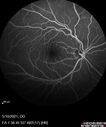
Branch Retinal Vein Occlusion with Neovascularization Elsewhere and Vitreous Hemorrhage383 views57 year old man New floaters in the left eye about a month ago. Now they are getting better. PMHx A-Fib on Pradaxa
VA 20/32 OD, 20/40 OS
1+ NS OU
BP 137/77, 200LBs, 5’8â€
PRP laser was done
|
|

Central Retinal Vein Occlusion following Cataract Surgery383 views79 year old man with decreased vision The patient had cataract surgery in the right eye 9 days ago and in the left eye 21 days ago and presents with vision loss in the right eye.
Meds: HCTZ, Garlic, Tumeric
VA 20/60 OD, 20/25 OS
|
|

382 views
|
|

Branch Retinal Vein Occlusion - Both Eyes - Edema OD and NVE OS - Severe Non-perfusion Both Eyes382 views83 year old man with vision loss for 3 months not sure which eye. VA 20/100 OD and 20/50 OS. Tried avastin once without much improvement in the right eye and then lost to follow-up. FA in the left eye shows leaking NVE
|
|

Central Retinal Vein Occlusion following Cataract Surgery382 views79 year old man with decreased vision The patient had cataract surgery in the right eye 9 days ago and in the left eye 21 days ago and presents with vision loss in the right eye.
Meds: HCTZ, Garlic, Tumeric
VA 20/60 OD, 20/25 OS
|
|

Branch Retinal Vein Occlusion with Neovascularization Elsewhere and Vitreous Hemorrhage381 views57 year old man New floaters in the left eye about a month ago. Now they are getting better. PMHx A-Fib on Pradaxa
VA 20/32 OD, 20/40 OS
1+ NS OU
BP 137/77, 200LBs, 5’8â€
PRP laser was done
|
|
379 views
|
|

Central Retinal Artery Occlusion379 views80 Year old man Sudden severe vision loss about 2 weeks ago. Today he can see light for the first time. The right eye is OK. His neuroimaging showed a brain stroke along with the eye stroke. He was stepping in the shower and the vision was suddenly worse. He went, later, to the ER and had a full evaluation. VA 20/25 right eye, LP left eye
|
|

Branch Retinal Vein Occlusion with Neovascularization Elsewhere and Vitreous Hemorrhage379 views57 year old man New floaters in the left eye about a month ago. Now they are getting better. PMHx A-Fib on Pradaxa
VA 20/32 OD, 20/40 OS
1+ NS OU
BP 137/77, 200LBs, 5’8â€
PRP laser was done
|
|

Elevated Episcleral Venous Pressure and Retinal Hemorrhages in both eyes379 views78 year old female with chronic red eye. The eyes are red all the time now. She is on Xeralto because of a history of blood clots in her lungs (2005 about). She has not had problems since. She has had floaters for many years. Ever since her eyes got funny she sees more floaters in both eyes. She had a brain scan over 6 months ago which was OK. She also had double vision when this first started which went away. She could not drive for a few months.
VA OD: Dcc20/32-1
VA OS: Dcc20/125
IOP: TP: OD:17 OS:16
Neuroimaging did not show a CC fistula
|
|
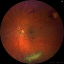
Branch Retinal Vein Occlusion with Neovascularization Elsewhere and Vitreous Hemorrhage - 1 month post laser379 views57 year old man New floaters in the left eye about a month ago. Now they are getting better. PMHx A-Fib on Pradaxa
VA 20/32 OD, 20/40 OS
1+ NS OU
BP 137/77, 200LBs, 5’8â€
PRP laser was done
|
|
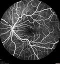
Elevated Episcleral Venous Pressure and Retinal Hemorrhages in both eyes378 views78 year old female with chronic red eye. The eyes are red all the time now. She is on Xeralto because of a history of blood clots in her lungs (2005 about). She has not had problems since. She has had floaters for many years. Ever since her eyes got funny she sees more floaters in both eyes. She had a brain scan over 6 months ago which was OK. She also had double vision when this first started which went away. She could not drive for a few months.
VA OD: Dcc20/32-1
VA OS: Dcc20/125
IOP: TP: OD:17 OS:16
Neuroimaging did not show a CC fistula
|
|

BRVO with good vision378 views87 year old man with good vision (diabetes without retinopathy). VA 20/25 OU. Vision never worsened from BRVO OS.
|
|

Branch Retinal Vein Occlusion with Venous Macroaneurysm (collateral)377 views66 year old female Did not notice vision loss in the right eye. The right eye has been a little worse than the left but now is much worse. Picked up during annual eye examination. VA had been 20/20 in 5 years ago.
History of brain and spinal cord radiation 1986 for T-cell lymphoma. Now with breast cancer, ablation for tachycardia, HTN
VA OD 6/200, OS 20/32
|
|

Central retinal artery occlusion and paracentral acute middle maculopathy and emboli377 views86 year old female with sudden vision loss a week ago and imaging shows PAMM lesions and emboli. Her vision is 20/200 in the left eye
|
|
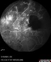
Branch Retinal Vein Occlusion with Neovascularization Elsewhere and Vitreous Hemorrhage376 views57 year old man New floaters in the left eye about a month ago. Now they are getting better. PMHx A-Fib on Pradaxa
VA 20/32 OD, 20/40 OS
1+ NS OU
BP 137/77, 200LBs, 5’8â€
PRP laser was done
|
|
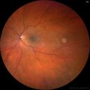
Elevated Episcleral Venous Pressure and Retinal Hemorrhages in both eyes376 views78 year old female with chronic red eye. The eyes are red all the time now. She is on Xeralto because of a history of blood clots in her lungs (2005 about). She has not had problems since. She has had floaters for many years. Ever since her eyes got funny she sees more floaters in both eyes. She had a brain scan over 6 months ago which was OK. She also had double vision when this first started which went away. She could not drive for a few months.
VA OD: Dcc20/32-1
VA OS: Dcc20/125
IOP: TP: OD:17 OS:16
Neuroimaging did not show a CC fistula
|
|

Central Retinal Vein Occlusion following Cataract Surgery376 views79 year old man with decreased vision The patient had cataract surgery in the right eye 9 days ago and in the left eye 21 days ago and presents with vision loss in the right eye.
Meds: HCTZ, Garlic, Tumeric
VA 20/60 OD, 20/25 OS
|
|
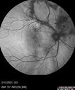
Branch Retinal Vein Occlusion with Venous Macroaneurysm (collateral)375 views66 year old female Did not notice vision loss in the right eye. The right eye has been a little worse than the left but now is much worse. Picked up during annual eye examination. VA had been 20/20 in 5 years ago.
History of brain and spinal cord radiation 1986 for T-cell lymphoma. Now with breast cancer, ablation for tachycardia, HTN
VA OD 6/200, OS 20/32
|
|

Central retinal artery occlusion and paracentral acute middle maculopathy and emboli375 views86 year old female with sudden vision loss a week ago and imaging shows PAMM lesions and emboli. Her vision is 20/200 in the left eye
|
|

Branch Retinal Vein Occlusion - Both Eyes - Edema OD and NVE OS - Severe Non-perfusion Both Eyes374 views83 year old man with vision loss for 3 months not sure which eye. VA 20/100 OD and 20/50 OS. Tried avastin once without much improvement in the right eye and then lost to follow-up. FA in the left eye shows leaking NVE
|
|
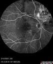
Branch Retinal Vein Occlusion with Venous Macroaneurysm (collateral)374 views66 year old female Did not notice vision loss in the right eye. The right eye has been a little worse than the left but now is much worse. Picked up during annual eye examination. VA had been 20/20 in 5 years ago.
History of brain and spinal cord radiation 1986 for T-cell lymphoma. Now with breast cancer, ablation for tachycardia, HTN
VA OD 6/200, OS 20/32
|
|

Branch Retinal Vein Occlusion with NVE and vitreous hemorrhage and non-perfusion374 views50 year old man The right eye was fine and then there was a sudden change in the vision. When he is outside looking at something bright there are thousands of dots with squiggly lines through them.Â
PMHx: Type II DM (1 year), HTN, AnemiaÂ
VA OD: sc20/40 PH20/32 NscJ7
VA OS: sc20/160 PH20/25 NscJ2
Myelinate RNFL in the left eye
|
|

Central Retinal Artery Occlusion - Fluorescein Angiogram Video showing slow fill374 views63 year old man Sudden severe vision loss in the left eye for 3 days. Six months following macula off retinal detachment repair, Pneumatic then PPV, then PPV buckle. VA was 20/63 on 4/2021 (3 months post RD repair).
PMHx – HTN. Meds: HCT, Meloxicam
VA OD 20/25, OS HM
|
|
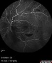
Branch Retinal Vein Occlusion with Neovascularization Elsewhere and Vitreous Hemorrhage373 views57 year old man New floaters in the left eye about a month ago. Now they are getting better. PMHx A-Fib on Pradaxa
VA 20/32 OD, 20/40 OS
1+ NS OU
BP 137/77, 200LBs, 5’8â€
PRP laser was done
|
|
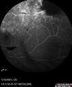
Branch Retinal Vein Occlusion with Neovascularization Elsewhere and Vitreous Hemorrhage373 views57 year old man New floaters in the left eye about a month ago. Now they are getting better. PMHx A-Fib on Pradaxa
VA 20/32 OD, 20/40 OS
1+ NS OU
BP 137/77, 200LBs, 5’8â€
PRP laser was done
|
|
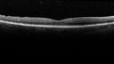
Elevated Episcleral Venous Pressure and Retinal Hemorrhages in both eyes373 views78 year old female with chronic red eye. The eyes are red all the time now. She is on Xeralto because of a history of blood clots in her lungs (2005 about). She has not had problems since. She has had floaters for many years. Ever since her eyes got funny she sees more floaters in both eyes. She had a brain scan over 6 months ago which was OK. She also had double vision when this first started which went away. She could not drive for a few months.
VA OD: Dcc20/32-1
VA OS: Dcc20/125
IOP: TP: OD:17 OS:16
Neuroimaging did not show a CC fistula
|
|

Branch Retinal Vein Occlusion with Venous Macroaneurysm (collateral)373 views66 year old female Did not notice vision loss in the right eye. The right eye has been a little worse than the left but now is much worse. Picked up during annual eye examination. VA had been 20/20 in 5 years ago.
History of brain and spinal cord radiation 1986 for T-cell lymphoma. Now with breast cancer, ablation for tachycardia, HTN
VA OD 6/200, OS 20/32
|
|

Branch Retinal Vein Occlusion with NVE and vitreous hemorrhage and non-perfusion373 views50 year old man The right eye was fine and then there was a sudden change in the vision. When he is outside looking at something bright there are thousands of dots with squiggly lines through them.Â
PMHx: Type II DM (1 year), HTN, AnemiaÂ
VA OD: sc20/40 PH20/32 NscJ7
VA OS: sc20/160 PH20/25 NscJ2
Myelinate RNFL in the left eye
|
|

Central Retinal Artery Occlusion - Fluorescein Angiogram Video373 views63 year old man Sudden severe vision loss in the left eye for 3 days. Six months following macula off retinal detachment repair, Pneumatic then PPV, then PPV buckle. VA was 20/63 on 4/2021 (3 months post RD repair).
PMHx – HTN. Meds: HCT, Meloxicam
VA OD 20/25, OS HM
|
|
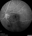
Central Retinal Vein Occlusion following Cataract Surgery373 views79 year old man with decreased vision The patient had cataract surgery in the right eye 9 days ago and in the left eye 21 days ago and presents with vision loss in the right eye.
Meds: HCTZ, Garlic, Tumeric
VA 20/60 OD, 20/25 OS
|
|
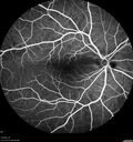
Elevated Episcleral Venous Pressure and Retinal Hemorrhages in both eyes372 views78 year old female with chronic red eye. The eyes are red all the time now. She is on Xeralto because of a history of blood clots in her lungs (2005 about). She has not had problems since. She has had floaters for many years. Ever since her eyes got funny she sees more floaters in both eyes. She had a brain scan over 6 months ago which was OK. She also had double vision when this first started which went away. She could not drive for a few months.
VA OD: Dcc20/32-1
VA OS: Dcc20/125
IOP: TP: OD:17 OS:16
Neuroimaging did not show a CC fistula
|
|

Central Retinal Vein Occlusion following Cataract Surgery372 views79 year old man with decreased vision The patient had cataract surgery in the right eye 9 days ago and in the left eye 21 days ago and presents with vision loss in the right eye.
Meds: HCTZ, Garlic, Tumeric
VA 20/60 OD, 20/25 OS
|
|

372 views
|
|

Central Retinal Vein Occlusion with Macular Edema and Retinal Hemorrhages 371 viewsFemale patient of 86-years of age presents a central retinal vein occlusion with macular edema in the right eye. Patient's VA was 20/70 in the right eye. Proceeded with laser treatment to the right eye.
|
|
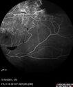
Branch Retinal Vein Occlusion with Neovascularization Elsewhere and Vitreous Hemorrhage371 views57 year old man New floaters in the left eye about a month ago. Now they are getting better. PMHx A-Fib on Pradaxa
VA 20/32 OD, 20/40 OS
1+ NS OU
BP 137/77, 200LBs, 5’8â€
PRP laser was done
|
|

BRVO with good vision371 views87 year old man with good vision (diabetes without retinopathy). VA 20/25 OU. Vision never worsened from BRVO OS.
|
|
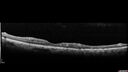
Branch Retinal Vein Occlusion - Both Eyes - Edema OD and NVE OS - Severe Non-perfusion Both Eyes370 views83 year old man with vision loss for 3 months not sure which eye. VA 20/100 OD and 20/50 OS. Tried avastin once without much improvement in the right eye and then lost to follow-up. FA in the left eye shows leaking NVE
|
|
369 views
|
|

Branch Retinal Vein Occlusion with Neovascularization Elsewhere and Vitreous Hemorrhage369 views57 year old man New floaters in the left eye about a month ago. Now they are getting better. PMHx A-Fib on Pradaxa
VA 20/32 OD, 20/40 OS
1+ NS OU
BP 137/77, 200LBs, 5’8â€
PRP laser was done
|
|

Branch Retinal Vein Occlusion with NVE and vitreous hemorrhage and non-perfusion369 views50 year old man The right eye was fine and then there was a sudden change in the vision. When he is outside looking at something bright there are thousands of dots with squiggly lines through them.Â
PMHx: Type II DM (1 year), HTN, AnemiaÂ
VA OD: sc20/40 PH20/32 NscJ7
VA OS: sc20/160 PH20/25 NscJ2
Myelinate RNFL in the left eye
|
|

Branch Retinal Vein Occlusion with NVE and vitreous hemorrhage and non-perfusion369 views50 year old man The right eye was fine and then there was a sudden change in the vision. When he is outside looking at something bright there are thousands of dots with squiggly lines through them.Â
PMHx: Type II DM (1 year), HTN, AnemiaÂ
VA OD: sc20/40 PH20/32 NscJ7
VA OS: sc20/160 PH20/25 NscJ2
Myelinate RNFL in the left eye
|
|

Cilioretinal Artery Occlusion - non central369 views81 year old female She noticed sudden vision loss in the right eye just before Christmas while watching television. This was about one week ago. She noticed when she closed her left eye and looked out of her right eye there was a darkness in the vision. The left eye is OK. She has no history of stroke and no neurological problems. She had a few headaches recently but they went away. Â
PMHx: HTN, Breast Cancer
Meds: HCTZ, Atorvastatin, Atenolol, Anastrozole
VA 20/80 OD, 20/32 OS
VA improved to 20/40 in one month in the involved eye
|
|

Central retinal artery occlusion and paracentral acute middle maculopathy and emboli369 views86 year old female with sudden vision loss a week ago and imaging shows PAMM lesions and emboli. Her vision is 20/200 in the left eye
|
|
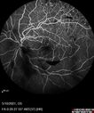
Branch Retinal Vein Occlusion with Neovascularization Elsewhere and Vitreous Hemorrhage368 views57 year old man New floaters in the left eye about a month ago. Now they are getting better. PMHx A-Fib on Pradaxa
VA 20/32 OD, 20/40 OS
1+ NS OU
BP 137/77, 200LBs, 5’8â€
PRP laser was done
|
|
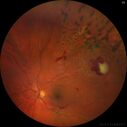
Branch Retinal Vein Occlusion with Neovascularization Elsewhere and Vitreous Hemorrhage - 1 month post laser368 views57 year old man New floaters in the left eye about a month ago. Now they are getting better. PMHx A-Fib on Pradaxa
VA 20/32 OD, 20/40 OS
1+ NS OU
BP 137/77, 200LBs, 5’8â€
PRP laser was done
|
|

Central Retinal Artery Occlusion - Fluorescein Angiogram Video368 views63 year old man Sudden severe vision loss in the left eye for 3 days. Six months following macula off retinal detachment repair, Pneumatic then PPV, then PPV buckle. VA was 20/63 on 4/2021 (3 months post RD repair).
PMHx – HTN. Meds: HCT, Meloxicam
VA OD 20/25, OS HM
|
|
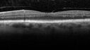
Branch Retinal Vein Occlusion with Neovascularization Elsewhere and Vitreous Hemorrhage367 views57 year old man New floaters in the left eye about a month ago. Now they are getting better. PMHx A-Fib on Pradaxa
VA 20/32 OD, 20/40 OS
1+ NS OU
BP 137/77, 200LBs, 5’8â€
PRP laser was done
|
|
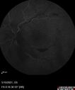
Branch Retinal Vein Occlusion with Neovascularization Elsewhere and Vitreous Hemorrhage367 views57 year old man New floaters in the left eye about a month ago. Now they are getting better. PMHx A-Fib on Pradaxa
VA 20/32 OD, 20/40 OS
1+ NS OU
BP 137/77, 200LBs, 5’8â€
PRP laser was done
|
|
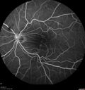
Elevated Episcleral Venous Pressure and Retinal Hemorrhages in both eyes367 views78 year old female with chronic red eye. The eyes are red all the time now. She is on Xeralto because of a history of blood clots in her lungs (2005 about). She has not had problems since. She has had floaters for many years. Ever since her eyes got funny she sees more floaters in both eyes. She had a brain scan over 6 months ago which was OK. She also had double vision when this first started which went away. She could not drive for a few months.
VA OD: Dcc20/32-1
VA OS: Dcc20/125
IOP: TP: OD:17 OS:16
Neuroimaging did not show a CC fistula
|
|

Central Retinal Vein Occlusion following Cataract Surgery367 views79 year old man with decreased vision The patient had cataract surgery in the right eye 9 days ago and in the left eye 21 days ago and presents with vision loss in the right eye.
Meds: HCTZ, Garlic, Tumeric
VA 20/60 OD, 20/25 OS
|
|
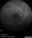
Branch Retinal Vein Occlusion with Neovascularization Elsewhere and Vitreous Hemorrhage366 views57 year old man New floaters in the left eye about a month ago. Now they are getting better. PMHx A-Fib on Pradaxa
VA 20/32 OD, 20/40 OS
1+ NS OU
BP 137/77, 200LBs, 5’8â€
PRP laser was done
|
|

Branch Retinal Vein Occlusion with Venous Macroaneurysm (collateral)366 views66 year old female Did not notice vision loss in the right eye. The right eye has been a little worse than the left but now is much worse. Picked up during annual eye examination. VA had been 20/20 in 5 years ago.
History of brain and spinal cord radiation 1986 for T-cell lymphoma. Now with breast cancer, ablation for tachycardia, HTN
VA OD 6/200, OS 20/32
|
|

Central retinal artery occlusion and paracentral acute middle maculopathy and emboli366 views86 year old female with sudden vision loss a week ago and imaging shows PAMM lesions and emboli. Her vision is 20/200 in the left eye
|
|
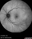
Branch Retinal Vein Occlusion with Venous Macroaneurysm (collateral)365 views66 year old female Did not notice vision loss in the right eye. The right eye has been a little worse than the left but now is much worse. Picked up during annual eye examination. VA had been 20/20 in 5 years ago.
History of brain and spinal cord radiation 1986 for T-cell lymphoma. Now with breast cancer, ablation for tachycardia, HTN
VA OD 6/200, OS 20/32
|
|

Branch Retinal Vein Occlusion with NVE and vitreous hemorrhage and non-perfusion365 views50 year old man The right eye was fine and then there was a sudden change in the vision. When he is outside looking at something bright there are thousands of dots with squiggly lines through them.Â
PMHx: Type II DM (1 year), HTN, AnemiaÂ
VA OD: sc20/40 PH20/32 NscJ7
VA OS: sc20/160 PH20/25 NscJ2
Myelinate RNFL in the left eye
|
|

Central Retinal Artery Occlusion - Fluorescein Angiogram Video365 views63 year old man Sudden severe vision loss in the left eye for 3 days. Six months following macula off retinal detachment repair, Pneumatic then PPV, then PPV buckle. VA was 20/63 on 4/2021 (3 months post RD repair).
PMHx – HTN. Meds: HCT, Meloxicam
VA OD 20/25, OS HM
|
|
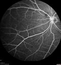
Elevated Episcleral Venous Pressure and Retinal Hemorrhages in both eyes364 views78 year old female with chronic red eye. The eyes are red all the time now. She is on Xeralto because of a history of blood clots in her lungs (2005 about). She has not had problems since. She has had floaters for many years. Ever since her eyes got funny she sees more floaters in both eyes. She had a brain scan over 6 months ago which was OK. She also had double vision when this first started which went away. She could not drive for a few months.
VA OD: Dcc20/32-1
VA OS: Dcc20/125
IOP: TP: OD:17 OS:16
Neuroimaging did not show a CC fistula
|
|

Central Retinal Vein Occlusion following Cataract Surgery364 views79 year old man with decreased vision The patient had cataract surgery in the right eye 9 days ago and in the left eye 21 days ago and presents with vision loss in the right eye.
Meds: HCTZ, Garlic, Tumeric
VA 20/60 OD, 20/25 OS
|
|

Central Retinal Vein Occlusion following Cataract Surgery364 views79 year old man with decreased vision The patient had cataract surgery in the right eye 9 days ago and in the left eye 21 days ago and presents with vision loss in the right eye.
Meds: HCTZ, Garlic, Tumeric
VA 20/60 OD, 20/25 OS
|
|
364 views
|
|
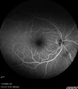
Central retinal artery occlusion and paracentral acute middle maculopathy and emboli364 views86 year old female with sudden vision loss a week ago and imaging shows PAMM lesions and emboli. Her vision is 20/200 in the left eye
|
|
363 views
|
|

Branch Retinal Vein Occlusion with Neovascularization Elsewhere and Vitreous Hemorrhage362 views57 year old man New floaters in the left eye about a month ago. Now they are getting better. PMHx A-Fib on Pradaxa
VA 20/32 OD, 20/40 OS
1+ NS OU
BP 137/77, 200LBs, 5’8â€
PRP laser was done
|
|
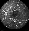
Elevated Episcleral Venous Pressure and Retinal Hemorrhages in both eyes362 views78 year old female with chronic red eye. The eyes are red all the time now. She is on Xeralto because of a history of blood clots in her lungs (2005 about). She has not had problems since. She has had floaters for many years. Ever since her eyes got funny she sees more floaters in both eyes. She had a brain scan over 6 months ago which was OK. She also had double vision when this first started which went away. She could not drive for a few months.
VA OD: Dcc20/32-1
VA OS: Dcc20/125
IOP: TP: OD:17 OS:16
Neuroimaging did not show a CC fistula
|
|
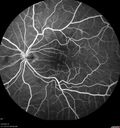
Elevated Episcleral Venous Pressure and Retinal Hemorrhages in both eyes362 views78 year old female with chronic red eye. The eyes are red all the time now. She is on Xeralto because of a history of blood clots in her lungs (2005 about). She has not had problems since. She has had floaters for many years. Ever since her eyes got funny she sees more floaters in both eyes. She had a brain scan over 6 months ago which was OK. She also had double vision when this first started which went away. She could not drive for a few months.
VA OD: Dcc20/32-1
VA OS: Dcc20/125
IOP: TP: OD:17 OS:16
Neuroimaging did not show a CC fistula
|
|

Branch Retinal Vein Occlusion with Neovascularization Elsewhere and Vitreous Hemorrhage361 views57 year old man New floaters in the left eye about a month ago. Now they are getting better. PMHx A-Fib on Pradaxa
VA 20/32 OD, 20/40 OS
1+ NS OU
BP 137/77, 200LBs, 5’8â€
PRP laser was done
|
|

Branch Retinal Vein Occlusion with Venous Macroaneurysm (collateral)361 views66 year old female Did not notice vision loss in the right eye. The right eye has been a little worse than the left but now is much worse. Picked up during annual eye examination. VA had been 20/20 in 5 years ago.
History of brain and spinal cord radiation 1986 for T-cell lymphoma. Now with breast cancer, ablation for tachycardia, HTN
VA OD 6/200, OS 20/32
|
|

Central Retinal Artery Occlusion - Fluorescein Angiogram Video361 views63 year old man Sudden severe vision loss in the left eye for 3 days. Six months following macula off retinal detachment repair, Pneumatic then PPV, then PPV buckle. VA was 20/63 on 4/2021 (3 months post RD repair).
PMHx – HTN. Meds: HCT, Meloxicam
VA OD 20/25, OS HM
|
|

Central Retinal Vein Occlusion following Cataract Surgery361 views79 year old man with decreased vision The patient had cataract surgery in the right eye 9 days ago and in the left eye 21 days ago and presents with vision loss in the right eye.
Meds: HCTZ, Garlic, Tumeric
VA 20/60 OD, 20/25 OS
|
|

Branch Retinal Vein Occlusion with Venous Macroaneurysm (collateral)360 views66 year old female Did not notice vision loss in the right eye. The right eye has been a little worse than the left but now is much worse. Picked up during annual eye examination. VA had been 20/20 in 5 years ago.
History of brain and spinal cord radiation 1986 for T-cell lymphoma. Now with breast cancer, ablation for tachycardia, HTN
VA OD 6/200, OS 20/32
|
|

Branch Retinal Vein Occlusion with Venous Macroaneurysm (collateral)360 views66 year old female Did not notice vision loss in the right eye. The right eye has been a little worse than the left but now is much worse. Picked up during annual eye examination. VA had been 20/20 in 5 years ago.
History of brain and spinal cord radiation 1986 for T-cell lymphoma. Now with breast cancer, ablation for tachycardia, HTN
VA OD 6/200, OS 20/32
|
|

Branch Retinal Vein Occlusion with NVE and vitreous hemorrhage and non-perfusion360 views50 year old man The right eye was fine and then there was a sudden change in the vision. When he is outside looking at something bright there are thousands of dots with squiggly lines through them.Â
PMHx: Type II DM (1 year), HTN, AnemiaÂ
VA OD: sc20/40 PH20/32 NscJ7
VA OS: sc20/160 PH20/25 NscJ2
Myelinate RNFL in the left eye
|
|
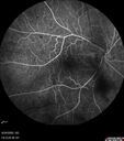
Central Retinal Vein Occlusion following Cataract Surgery360 views79 year old man with decreased vision The patient had cataract surgery in the right eye 9 days ago and in the left eye 21 days ago and presents with vision loss in the right eye.
Meds: HCTZ, Garlic, Tumeric
VA 20/60 OD, 20/25 OS
|
|

Central Retinal Vein Occlusion following Cataract Surgery360 views79 year old man with decreased vision The patient had cataract surgery in the right eye 9 days ago and in the left eye 21 days ago and presents with vision loss in the right eye.
Meds: HCTZ, Garlic, Tumeric
VA 20/60 OD, 20/25 OS
|
|
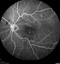
Elevated Episcleral Venous Pressure and Retinal Hemorrhages in both eyes359 views78 year old female with chronic red eye. The eyes are red all the time now. She is on Xeralto because of a history of blood clots in her lungs (2005 about). She has not had problems since. She has had floaters for many years. Ever since her eyes got funny she sees more floaters in both eyes. She had a brain scan over 6 months ago which was OK. She also had double vision when this first started which went away. She could not drive for a few months.
VA OD: Dcc20/32-1
VA OS: Dcc20/125
IOP: TP: OD:17 OS:16
Neuroimaging did not show a CC fistula
|
|

Branch Retinal Vein Occlusion with Venous Macroaneurysm (collateral)359 views66 year old female Did not notice vision loss in the right eye. The right eye has been a little worse than the left but now is much worse. Picked up during annual eye examination. VA had been 20/20 in 5 years ago.
History of brain and spinal cord radiation 1986 for T-cell lymphoma. Now with breast cancer, ablation for tachycardia, HTN
VA OD 6/200, OS 20/32
|
|

Branch Retinal Vein Occlusion with Venous Macroaneurysm (collateral)359 views66 year old female Did not notice vision loss in the right eye. The right eye has been a little worse than the left but now is much worse. Picked up during annual eye examination. VA had been 20/20 in 5 years ago.
History of brain and spinal cord radiation 1986 for T-cell lymphoma. Now with breast cancer, ablation for tachycardia, HTN
VA OD 6/200, OS 20/32
|
|

Central Retinal Vein Occlusion following Cataract Surgery359 views79 year old man with decreased vision The patient had cataract surgery in the right eye 9 days ago and in the left eye 21 days ago and presents with vision loss in the right eye.
Meds: HCTZ, Garlic, Tumeric
VA 20/60 OD, 20/25 OS
|
|

Central Retinal Vein Occlusion following Cataract Surgery359 views79 year old man with decreased vision The patient had cataract surgery in the right eye 9 days ago and in the left eye 21 days ago and presents with vision loss in the right eye.
Meds: HCTZ, Garlic, Tumeric
VA 20/60 OD, 20/25 OS
|
|
358 views
|
|
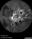
Branch Retinal Vein Occlusion with Venous Macroaneurysm (collateral)358 views66 year old female Did not notice vision loss in the right eye. The right eye has been a little worse than the left but now is much worse. Picked up during annual eye examination. VA had been 20/20 in 5 years ago.
History of brain and spinal cord radiation 1986 for T-cell lymphoma. Now with breast cancer, ablation for tachycardia, HTN
VA OD 6/200, OS 20/32
|
|

Central retinal artery occlusion and paracentral acute middle maculopathy and emboli357 views86 year old female with sudden vision loss a week ago and imaging shows PAMM lesions and emboli. Her vision is 20/200 in the left eye
|
|
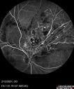
Branch Retinal Vein Occlusion with Venous Macroaneurysm (collateral)356 views66 year old female Did not notice vision loss in the right eye. The right eye has been a little worse than the left but now is much worse. Picked up during annual eye examination. VA had been 20/20 in 5 years ago.
History of brain and spinal cord radiation 1986 for T-cell lymphoma. Now with breast cancer, ablation for tachycardia, HTN
VA OD 6/200, OS 20/32
|
|

Central Retinal Vein Occlusion following Cataract Surgery356 views79 year old man with decreased vision The patient had cataract surgery in the right eye 9 days ago and in the left eye 21 days ago and presents with vision loss in the right eye.
Meds: HCTZ, Garlic, Tumeric
VA 20/60 OD, 20/25 OS
|
|

356 views
|
|

Cilioretinal artery occlusion following cataract surgery356 views60 year old man noticed new scotoma in the right eye immediately after cataract surgery. Vision is 20/60
|
|

central retinal vein occlusion - a lot of hemorrhage - on coumadin356 views84 year old man with severe vision loss left eye for 2 weeks. VA 5/200 OS - Patient on Warfarin for valve replacement.
|
|
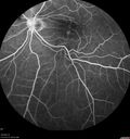
Elevated Episcleral Venous Pressure and Retinal Hemorrhages in both eyes355 views78 year old female with chronic red eye. The eyes are red all the time now. She is on Xeralto because of a history of blood clots in her lungs (2005 about). She has not had problems since. She has had floaters for many years. Ever since her eyes got funny she sees more floaters in both eyes. She had a brain scan over 6 months ago which was OK. She also had double vision when this first started which went away. She could not drive for a few months.
VA OD: Dcc20/32-1
VA OS: Dcc20/125
IOP: TP: OD:17 OS:16
Neuroimaging did not show a CC fistula
|
|
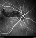
Cilioretinal Artery Occlusion - non central354 views81 year old female She noticed sudden vision loss in the right eye just before Christmas while watching television. This was about one week ago. She noticed when she closed her left eye and looked out of her right eye there was a darkness in the vision. The left eye is OK. She has no history of stroke and no neurological problems. She had a few headaches recently but they went away. Â
PMHx: HTN, Breast Cancer
Meds: HCTZ, Atorvastatin, Atenolol, Anastrozole
VA 20/80 OD, 20/32 OS
VA improved to 20/40 in one month in the involved eye
|
|

Branch Retinal Vein Occlusion and worsened cystoid macular edema following cataract surgery354 views51 year old female The patient noticed gradual vision loss in both eyes starting possibly after her last cataract surgery in May of 2022 (3 months ago). She had ECCE 3/2022 OD and 5/2022 OS. She does not have diabetes. She had glaucoma shunts placed about 2017. She has been on latanoprost and timolol. She is now getting ready to start Combigan and Bromfenac. She is also staring xidra for dry eye. She stopped the latanorpost on 8/15/22. She has had macular edema before for which she had intraocular injections which worked. She has had no injections lately. When she was getting treated for macular edema in the past she had an IOP of 60.
Medical Hx: Hypertension, Systemic. Glaucoma. Arthritis. Vertigo. Stroke.
Systemic Meds: Trimfya. NAPROXEN. LISINOPRIL.
VA OD: sc20/63-1
VA OS: sc20/50
IOP: TP: OD:12 OS:12
|
|
| 1845 files on 19 page(s) |
 |
|
|
|
|
14 | |
|
|
|
|


