am meisten angesehen - Age-Related Macular Degeneration - Wet
|
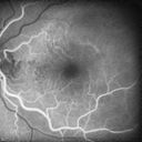
Polypoidal Choroidal Vasculopathy with Pulsating ICG polyp left eye560 x angesehen
|
|

Asymptomatic Juxtapapillary Choroidal Neovascular Membrane - Reticular Macular Disease (Wet AMD) 559 x angesehen
|
|

Asymptomatic Juxtapapillary Choroidal Neovascular Membrane - Reticular Macular Disease (Wet AMD) 558 x angesehen
|
|

Polypoidal Choroidal Vasculopathy with Pulsating ICG polyp left eye558 x angesehen
|
|

Juxtapapillary CNVM and Serous Macular Detachment Wet AMD Rx Laser557 x angesehen85-year-old man OD is 20/25, OS is 20/100. IOP: OD 21, OS 22.
OS: Vertical C/D ratio is 0.1. There is a posterior vitreous separation. There is a hemorrhagic pigment epithelial detachment on the superior pole of the optic nerve, extending a disc-and-a-half diameter off the nerve, with adjacent exudates. There is a serous macular detachment involving the center of the fovea.
VA improved from 20/100 to 20/50 in four months from laser.
|
|

Macular Degeneration - Choroidal Laser Left eye - New Subfoveal CNVM left eye - Did well with Avastin557 x angesehen69-year-old woman new vision loss right eye and old macular scar left eye. OD 20/40, OS 20/200
|
|

New Subfoveal Classic CNVM OS - Wet AMD - Macular Scar Congenital Toxoplasmosis Right Eye 556 x angesehen77-year-old woman as had poor vision in the right eye since birth from congenital toxoplasmosis of her macula in the right eye. Two to three weeks ago when she noticed vision loss in the left eye. VISUAL ACUITY: OD 2/200, OS 20/200.
|
|

Polypoidal Choroidal Vasculopathy Right Eye - Disciform Scar Left Eye556 x angesehen84-year-old woman has polypoidal choroidal vasculopathy in both eyes with macular degeneration. She has macular scars in both eyes and she remains with remarkably good vision in the right eye despite the scarring.
VISUAL ACUITY: OD 20/30, OS 1/200
Avastin Rx OD
|
|

Reticular Macular Disease (Pseudo-drusen) Both Eyes - Occult CNVM and Macular Edema Right Eye 556 x angesehenFluorescein Angiogram
|
|
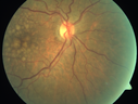
Polypoidal Choroidal Vasculopathy with Pulsating ICG polyp left eye556 x angesehen
|
|

Outer Retinal Tubulation555 x angesehen78 year old man chronic wet AMD - 20/200
|
|
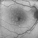
Polypoidal Choroidal Vasculopathy with Pulsating ICG polyp left eye553 x angesehen
|
|

Juxtapapillary CNVM - Hemorrhage553 x angesehen83 year old female VA 20/200 improved to 20/32 within 2 months with Lucentis
|
|
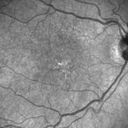
Polypoidal Choroidal Vasculopathy with Pulsating ICG polyp left eye552 x angesehen
|
|

Wet AMD OD and Lamellar Macular hole OS551 x angesehenWet AMD in right and better eye failed extend to 2 months but was able to stop therapy once he started on systemic Avastin for cancer. The last image shows the macula dry 9 months without local therapy. The left eye has a lamellar macular hole.
|
|

Reticular Macular Disease (Pseudo-drusen) Both Eyes - Occult CNVM and Macular Edema Right Eye 550 x angesehenFundus AutofluorescenceFundus Autofluorescence
|
|

Asymptomatic wet AMD - Subfoveal CNM both eyes550 x angesehen82 year old woman - 20/25 OU. Images show occult subfoveal CNVM both eyes best seen on late ICG. The left eye also has probable polypoidal choroidal vasculopathy. No treatment was recommended on the first visit - follow-up at 1 month.
|
|

Small Retinal Pigment Epithelial Rip and Poor Vision (20/400)549 x angesehen80-year-old woman vision in the right eye has substantially declined.
OD: 20/400; OS: 20/50
|
|
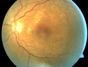
Polypoidal Choroidal Vasculopathy with Pulsating ICG polyp left eye549 x angesehen
|
|

Small Retinal Pigment Epithelial Rip and Poor Vision (20/400)548 x angesehen80-year-old woman vision in the right eye has substantially declined.
OD: 20/400; OS: 20/5080-year-old woman vision in the right eye has substantially declined.
OD: 20/400; OS: 20/50
|
|

IPCV - idiopathic polypoidal choroidal vasculopathy - juxtapapillary CNVM with hemorrhage both eyes (also choroidal nevus right eye) - Subretinal hemorrhage both eyes548 x angesehen86 year old woman with shadow in vision OD and new hemmorhage OU.
|
|

Reticular Macular Disease (Pseudo-drusen) Both Eyes - Occult CNVM and Macular Edema Right Eye 547 x angesehenFluorescein Angiogram
|
|

Retinal Angiomatous Proliferation - Type III choroidal neovascular membrane547 x angesehen83 year old female with wet AMD - 20/160 VA- Vision improved to 20/40 with 3 injections of anti-VEGF drugs. OCT shows break in RPE and FA and ICG show PED with hot spot on ICG
|
|
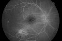
Macular Scar Left Eye from Age-Related Macular Degeneration546 x angesehen81-year-old woman has a macular scar in the left eye and then unfortunately developed a central retinal artery occlusion in the right eye OD 20/100, OS 6/200.
|
|

Asymptomatic Juxtapapillary Choroidal Neovascular Membrane - Reticular Macular Disease (Wet AMD) 546 x angesehen
|
|
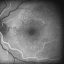
Polypoidal Choroidal Vasculopathy with Pulsating ICG polyp left eye546 x angesehen
|
|

Serous Macular Detachment Both Eyes - Wet AMD vs. CSR - 62 Year Old Man - Color Photo545 x angesehen
|
|
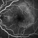
Polypoidal Choroidal Vasculopathy with Pulsating ICG polyp left eye545 x angesehen
|
|

Outer Retinal Tubulation545 x angesehen78 year old man chronic wet AMD - 20/200
|
|

IPCV - idiopathic polypoidal choroidal vasculopathy - juxtapapillary CNVM with hemorrhage both eyes (also choroidal nevus right eye) - Subretinal hemorrhage both eyes544 x angesehen86 year old woman with shadow in vision OD and new hemmorhage OU.FA shows juxtapapillary CNVM
|
|

Serous Macular Detachment Both Eyes - Wet AMD vs. CSR - 62 Year Old Man - IR543 x angesehen
|
|
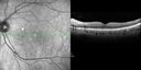
Cartwheel CNVM - Classic - wet AMD - evidence of possible old CSR543 x angesehen75 y.o Decreased Vision right eye for one year. VA OD: sc20/125+2 OS 20/25. Autofluorescence and SD OCT show evidence of old serous retinal detachment superior to the fovea suggesting a history of CSR. The macular lesion on FA shows a classic cartwheel appearance
|
|

Cartwheel CNVM - Classic - wet AMD - evidence of possible old CSR - ICG is normal - shows nothing543 x angesehen75 y.o Decreased Vision right eye for one year. VA OD: sc20/125+2 OS 20/25. Autofluorescence and SD OCT show evidence of old serous retinal detachment superior to the fovea suggesting a history of CSR. The macular lesion on FA shows a classic cartwheel appearance
|
|

Juxtapapillary CNVM - Hemorrhage542 x angesehen83 year old female VA 20/200 improved to 20/32 within 2 months with Lucentis
|
|

Small Retinal Pigment Epithelial Rip and Poor Vision (20/400)541 x angesehen80-year-old woman vision in the right eye has substantially declined.
OD: 20/400; OS: 20/50
|
|
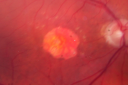
Wet Macular Degeneration Left Eye with Gutter (atypical) and Geographic Atrophy Right Eye540 x angesehen80-year-old man has wet age-related macular degeneration in his left eye. He has an atypical angiogram and that there is a gutter under the choroidal neovascular membrane suggesting still leakage of subretinal fluid. OS 20/400.
|
|

Serous Macular Detachment Both Eyes - Wet AMD vs. CSR - 62 Year Old Man - IR539 x angesehen
|
|

New Subfoveal Classic CNVM OS - Wet AMD - Macular Scar Congenital Toxoplasmosis Right Eye 538 x angesehen77-year-old woman as had poor vision in the right eye since birth from congenital toxoplasmosis of her macula in the right eye. Two to three weeks ago when she noticed vision loss in the left eye. VISUAL ACUITY: OD 2/200, OS 20/200.
|
|

Asymptomatic Juxtapapillary Choroidal Neovascular Membrane - Reticular Macular Disease (Wet AMD) 538 x angesehen
|
|

Idiopathic Polypoidal Choroidal Vasculopathy - IR538 x angesehen
|
|

New wet AMD OS with subretinal hemorrhage and recent vision loss, Scar OD538 x angesehen
|
|

New Subfoveal Classic CNVM OS - Wet AMD - Macular Scar Congenital Toxoplasmosis Right Eye 537 x angesehen77-year-old woman as had poor vision in the right eye since birth from congenital toxoplasmosis of her macula in the right eye. Two to three weeks ago when she noticed vision loss in the left eye. VISUAL ACUITY: OD 2/200, OS 20/200.
|
|

Macular Degeneration with Hemorrhage537 x angesehenPatient comes in with active leaking of AMD in the left eye. VA is 20/40 in the left eye. Patient was treated with Eylea and will return in 1-month for follow up.
|
|

Cartwheel CNVM - Classic - wet AMD - evidence of possible old CSR - ICG is normal - shows nothing537 x angesehen75 y.o Decreased Vision right eye for one year. VA OD: sc20/125+2 OS 20/25. Autofluorescence and SD OCT show evidence of old serous retinal detachment superior to the fovea suggesting a history of CSR. The macular lesion on FA shows a classic cartwheel appearance
|
|

Small Retinal Pigment Epithelial Rip and Poor Vision (20/400)536 x angesehen80-year-old woman vision in the right eye has substantially declined.
OD: 20/400; OS: 20/50
|
|

IPCV - idiopathic polypoidal choroidal vasculopathy - juxtapapillary CNVM with hemorrhage both eyes (also choroidal nevus right eye) - Subretinal hemorrhage both eyes536 x angesehen86 year old woman with shadow in vision OD and new hemmorhage OU.
|
|

IPCV - idiopathic polypoidal choroidal vasculopathy - juxtapapillary CNVM with hemorrhage both eyes (also choroidal nevus right eye) - Subretinal hemorrhage both eyes536 x angesehen86 year old woman with shadow in vision OD and new hemmorhage OU. ICG shows no plaque
|
|

Reticular Macular Disease (Pseudo-drusen) Both Eyes - Occult CNVM and Macular Edema Right Eye 535 x angesehenIndocyanine Green Angiogram
|
|

New Subfoveal Classic CNVM OS - Wet AMD - Macular Scar Congenital Toxoplasmosis Right Eye 534 x angesehen77-year-old woman as had poor vision in the right eye since birth from congenital toxoplasmosis of her macula in the right eye. Two to three weeks ago when she noticed vision loss in the left eye. VISUAL ACUITY: OD 2/200, OS 20/200.
|
|

Polypoidal Choroidal Vasculopathy - Right Eye534 x angesehen83 year old female presented in June 2013 with vision loss for 3 months. She had 2 injections elsewhere with no improvement (avastin). The first images show a peripapillary CNVM with polyps. I treated her once with PDT and she stayed dry with good vision for 5 years (2019). VA on presentation 20/80 OD. Improved to 20/40
|
|
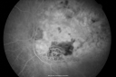
Disciform Macular Scar Both Eyes from Wet Macular Degeneration533 x angesehen86-year-old woman has disciform scars OD: 20/400; OS: 20/100
|
|

Macular Degeneration - Choroidal Laser Left eye - New Subfoveal CNVM left eye - Did well with Avastin532 x angesehen69-year-old woman new vision loss right eye and old macular scar left eye. OD 20/40, OS 20/200
|
|

Outer Retinal Tubulation532 x angesehen78 year old man chronic wet AMD - 20/200
|
|

Pigment Epithelial Detachment with Subretinal Fluid and Exudate 20/400 Vision531 x angesehen87-year-old woman has wet age-related macular degeneration in the left eye. She had treatment up north but there has been a gap in therapy and the vision in the left eye is poor. Vision OD is 20/25, OS is 20/400
|
|

Reticular Macular Disease (Pseudo-drusen) Both Eyes - Occult CNVM and Macular Edema Right Eye 531 x angesehenFluorescein Angiogram
|
|

Small Retinal Pigment Epithelial Rip and Poor Vision (20/400)530 x angesehen80-year-old woman vision in the right eye has substantially declined.
OD: 20/400; OS: 20/50
|
|

Wet AMD OD and Lamellar Macular hole OS530 x angesehenWet AMD in right and better eye failed extend to 2 months but was able to stop therapy once he started on systemic Avastin for cancer. The last image shows the macula dry 9 months without local therapy. The left eye has a lamellar macular hole.
|
|

IPCV - idiopathic polypoidal choroidal vasculopathy - juxtapapillary CNVM with hemorrhage both eyes (also choroidal nevus right eye) - Subretinal hemorrhage both eyes530 x angesehen86 year old woman with shadow in vision OD and new hemmorhage OU.FA shows juxtapapillary CNVM
|
|

IPCV - idiopathic polypoidal choroidal vasculopathy - juxtapapillary CNVM with hemorrhage both eyes (also choroidal nevus right eye) - Subretinal hemorrhage both eyes529 x angesehen86 year old woman with shadow in vision OD and new hemmorhage OU. OCT map shows progression of jpcnvm
|
|

Asymptomatic wet AMD - Subfoveal CNM both eyes528 x angesehen82 year old woman - 20/25 OU. Images show occult subfoveal CNVM both eyes best seen on late ICG. The left eye also has probable polypoidal choroidal vasculopathy. No treatment was recommended on the first visit - follow-up at 1 month.
|
|

Chorioretinal Scar with Radiating Chorioretinal Folds527 x angesehen79-year-old woman has wet age-related macular degeneration in both eyes. The left eye had intravitreal injections of Avastin starting in May of 2009. Her vision remains good in the left eye and poor in the right eye.
VISUAL ACUITY: OD 8/200, OS 20/60.
|
|

Serous Macular Detachment Both Eyes - Wet AMD vs. CSR - 62 Year Old Man - Fluorescein angiogram525 x angesehen
|
|

Asymptomatic wet AMD - Subfoveal CNM both eyes525 x angesehen82 year old woman - 20/25 OU. Images show occult subfoveal CNVM both eyes best seen on late ICG. The left eye also has probable polypoidal choroidal vasculopathy. No treatment was recommended on the first visit - follow-up at 1 month.
|
|

Outer Retinal Tubulation525 x angesehen78 year old man chronic wet AMD - 20/200
|
|

Asymptomatic wet AMD - Subfoveal CNM both eyes524 x angesehen82 year old woman - 20/25 OU. Images show occult subfoveal CNVM both eyes best seen on late ICG. The left eye also has probable polypoidal choroidal vasculopathy. No treatment was recommended on the first visit - follow-up at 1 month.
|
|

Asymptomatic wet AMD - Subfoveal CNM both eyes524 x angesehen82 year old woman - 20/25 OU. Images show occult subfoveal CNVM both eyes best seen on late ICG. The left eye also has probable polypoidal choroidal vasculopathy. No treatment was recommended on the first visit - follow-up at 1 month.
|
|

Small Retinal Pigment Epithelial Rip and Poor Vision (20/400)523 x angesehen80-year-old woman vision in the right eye has substantially declined.
OD: 20/400; OS: 20/50
|
|

New Subfoveal Classic CNVM OS - Wet AMD - Macular Scar Congenital Toxoplasmosis Right Eye 523 x angesehen77-year-old woman as had poor vision in the right eye since birth from congenital toxoplasmosis of her macula in the right eye. Two to three weeks ago when she noticed vision loss in the left eye. VISUAL ACUITY: OD 2/200, OS 20/200.
|
|

Asymptomatic wet AMD - Subfoveal CNM both eyes523 x angesehen82 year old woman - 20/25 OU. Images show occult subfoveal CNVM both eyes best seen on late ICG. The left eye also has probable polypoidal choroidal vasculopathy. No treatment was recommended on the first visit - follow-up at 1 month.
|
|

Serous Macular Detachment Both Eyes - Wet AMD vs. CSR - 62 Year Old Man - Fluorescein angiogram521 x angesehen
|
|

IPCV - idiopathic polypoidal choroidal vasculopathy - juxtapapillary CNVM with hemorrhage both eyes (also choroidal nevus right eye) - Subretinal hemorrhage both eyes521 x angesehen86 year old woman with shadow in vision OD and new hemmorhage OU. ICG angiogram
|
|

Submacular Hemorrhage - wet AMD521 x angesehen84 year old man with 20/200 from submacular hemorrhage. Vision improved to 20/32 with only Avastin therapy (no surgical intervention). The initial FAF is interesting because parts of the blood hyper auto fluoresce.
|
|

Submacular Hemorrhage - wet AMD521 x angesehen84 year old man with 20/200 from submacular hemorrhage. Vision improved to 20/32 with only Avastin therapy (no surgical intervention). The initial FAF is interesting because parts of the blood hyper auto fluoresce.
|
|

Polypoidal Choroidal Vasculopathy Left Eye521 x angesehen74 year old man with vision loss to 20/100 over a few months. He improved to 20/20 with Lucentis. The color and ICG show the polyps.
|
|

Small Retinal Pigment Epithelial Rip and Poor Vision (20/400)520 x angesehen80-year-old woman vision in the right eye has substantially declined.
OD: 20/400; OS: 20/50
|
|

Wet AMD controlled with Avastin 20/60 Vision520 x angesehen79-year-old woman has wet age-related macular degeneration in both eyes. The left eye had intravitreal injections of Avastin starting in May of 2009. Her vision remains good in the left eye and poor in the right eye.
VISUAL ACUITY: OD 8/200, OS 20/60.
|
|

Polypoidal Choroidal Vasculopathy Left Eye518 x angesehen74 year old man with vision loss to 20/100 over a few months. He improved to 20/20 with Lucentis. The color and ICG show the polyps.
|
|

Small Retinal Pigment Epithelial Rip and Poor Vision (20/400)517 x angesehen80-year-old woman vision in the right eye has substantially declined.
OD: 20/400; OS: 20/50
|
|

Small Retinal Pigment Epithelial Rip and Poor Vision (20/400)517 x angesehen80-year-old woman vision in the right eye has substantially declined.
OD: 20/400; OS: 20/50
|
|

IPCV - idiopathic polypoidal choroidal vasculopathy - juxtapapillary CNVM with hemorrhage both eyes (also choroidal nevus right eye) - Subretinal hemorrhage both eyes517 x angesehen86 year old woman with shadow in vision OD and new hemmorhage OU.FA shows juxtapapillary CNVM
|
|

Wet AMD controlled with Avastin 20/60 Vision516 x angesehen79-year-old woman has wet age-related macular degeneration in both eyes. The left eye had intravitreal injections of Avastin starting in May of 2009. Her vision remains good in the left eye and poor in the right eye.
VISUAL ACUITY: OD 8/200, OS 20/60.
|
|

Reticular Macular Disease (Pseudo-drusen) Both Eyes - Occult CNVM and Macular Edema Right Eye 516 x angesehen
|
|
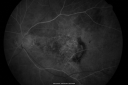
Wet Macular Degeneration Left Eye with Gutter (atypical) and Geographic Atrophy Right Eye515 x angesehen80-year-old man has wet age-related macular degeneration in his left eye. He has an atypical angiogram and that there is a gutter under the choroidal neovascular membrane suggesting still leakage of subretinal fluid. OS 20/400.
|
|

Wet Macular Degeneration Left Eye with Gutter (atypical) and Geographic Atrophy Right Eye515 x angesehen80-year-old man has wet age-related macular degeneration in his left eye. He has an atypical angiogram and that there is a gutter under the choroidal neovascular membrane suggesting still leakage of subretinal fluid. OS 20/400.
|
|

Asymptomatic Juxtapapillary Choroidal Neovascular Membrane - Reticular Macular Disease (Wet AMD) 515 x angesehen
|
|
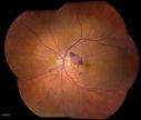
Macular Degeneration with Hemorrhage515 x angesehenPatient comes in with active leaking of AMD in the left eye. VA is 20/40 in the left eye. Patient was treated with Eylea and will return in 1-month for follow up.
|
|

OCT shows lamellar macular hole left eye515 x angesehenWet AMD in right and better eye failed extend to 2 months but was able to stop therapy once he started on systemic Avastin for cancer. The last image shows the macula dry 9 months without local therapy. The left eye has a lamellar macular hole.
|
|

IPCV - idiopathic polypoidal choroidal vasculopathy - juxtapapillary CNVM with hemorrhage both eyes (also choroidal nevus right eye) - Subretinal hemorrhage both eyes515 x angesehen86 year old woman with shadow in vision OD and new hemmorhage OU.
|
|

Asymptomatic Juxtapapillary Choroidal Neovascular Membrane - Reticular Macular Disease (Wet AMD) 514 x angesehen
|
|

Wet AMD - Subfoveal CNVM - Excellent Response to Avastin513 x angesehen76-year-old woman two to three months of vision loss OD
Vision started at 20/200 and with Avastin improved to 20/25. She has required monthly injections and as of October 2010 has been treated about 16 times. Her left eye remains good at 20/20.
The OCT from post-treatment as well as pretreatment images are in this album.
|
|

Small Retinal Pigment Epithelial Rip and Poor Vision (20/400)513 x angesehen80-year-old woman vision in the right eye has substantially declined.
OD: 20/400; OS: 20/50
|
|
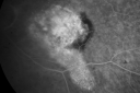
Wet Macular Degeneration Left Eye with Gutter (atypical) and Geographic Atrophy Right Eye513 x angesehen80-year-old man has wet age-related macular degeneration in his left eye. He has an atypical angiogram and that there is a gutter under the choroidal neovascular membrane suggesting still leakage of subretinal fluid. OS 20/400.
|
|

Wet Macular Degeneration Left Eye with Gutter (atypical) and Geographic Atrophy Right Eye513 x angesehen80-year-old man has wet age-related macular degeneration in his left eye. He has an atypical angiogram and that there is a gutter under the choroidal neovascular membrane suggesting still leakage of subretinal fluid. OS 20/400.
|
|

Asymptomatic Juxtapapillary Choroidal Neovascular Membrane - Reticular Macular Disease (Wet AMD) 513 x angesehen
|
|
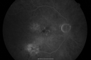
Macular Scar Left Eye from Age-Related Macular Degeneration512 x angesehen81-year-old woman has a macular scar in the left eye and then unfortunately developed a central retinal artery occlusion in the right eye OD 20/100, OS 6/200.
|
|

Wet AMD controlled with Avastin 20/60 Vision511 x angesehen79-year-old woman has wet age-related macular degeneration in both eyes. The left eye had intravitreal injections of Avastin starting in May of 2009. Her vision remains good in the left eye and poor in the right eye.
VISUAL ACUITY: OD 8/200, OS 20/60.
|
|
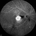
Cartwheel CNVM - Classic - wet AMD - evidence of possible old CSR - FA shows cartwheel shaped CNVM511 x angesehen75 y.o Decreased Vision right eye for one year. VA OD: sc20/125+2 OS 20/25. Autofluorescence and SD OCT show evidence of old serous retinal detachment superior to the fovea suggesting a history of CSR. The macular lesion on FA shows a classic cartwheel appearance
|
|

Reticular Macular Disease (Pseudo-drusen) Both Eyes - Occult CNVM and Macular Edema Right Eye 510 x angesehenFluorescein Angiogram
|
|
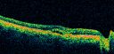
Macular Scar Left Eye from Age-Related Macular Degeneration509 x angesehen81-year-old woman has a macular scar in the left eye and then unfortunately developed a central retinal artery occlusion in the right eye OD 20/100, OS 6/200.
|
|

80 Year Old Woman Macular Degeneration Disciform Scar - Fibrosis509 x angesehenVA 20/100
|
|
| 1175 Dateien auf 12 Seite(n) |
 |
|
4 | |
|
|
|
|
|
|
|


