Les plus populaires - Macular Degeneration - Age Related, Myopic, Choroidal Neovascular Membranes
|

Reticular Macular Disease - Reticular Pseudodrusen and Soft Macular Drusen - 57 Year Old Womanvu 3067 fois57-year-old woman was seen in the office on 2/22/2011. She has macular drusen in both eyes. I saw her back in 2006. Since then she does notice slight increased distortion. OD is 20/25, OS is 20/25
|
|

Reticular Macular Disease - Reticular Pseudodrusen and Soft Macular Drusen - 57 Year Old Womanvu 2237 fois57-year-old woman was seen in the office on 2/22/2011. She has macular drusen in both eyes. I saw her back in 2006. Since then she does notice slight increased distortion. OD is 20/25, OS is 20/25
|
|

ANGIOID STREAKSvu 2073 foisA 29 year old woman was referred to the ophthalmologist for an eye exam since the dermatologist had noted “chicken-skin†papules in the neck skin. PAD showed for pseudoxathoma elasticum typical pathology. The eye exam disclosed angioid streaks, asymptomatic so far.
Magnus Gjötterberg MD
International membrer 1440
S:t Erik Eye Hospital
SE-11282
Stockholm
Sweden
magnusgjotterberg@hem.utfors.se
magnus.gjotterberg@sankterik.se.
Photographer/technician
Malin Langhals
|
|

Reticular Macular Disease - Reticular Pseudodrusen and Soft Macular Drusen - 57 Year Old Womanvu 2040 fois57-year-old woman was seen in the office on 2/22/2011. She has macular drusen in both eyes. I saw her back in 2006. Since then she does notice slight increased distortion. OD is 20/25, OS is 20/25
|
|

Reticular Macular Disease (Pseudo-drusen) Both Eyes - Wet AMD OS - Dry AMD OD SD-OCTvu 2021 fois84-year-old woman has wet age-related macular degeneration in the left eye and dry macular degeneration in the right eye. She takes the eye vitamins and her vision is stable since she was treated three months ago with Avastin. OD 20/50, OS 20/32
|
|

Pigment migration in dry age-related macular degenerationvu 1989 fois80 year old female. Dry AMD with GA in the left eye and pigment migration visible on OCT scan.
VA 20/40 OD, 20/160 OS
|
|

Reticular Macular Disease (Pseudo-drusen) Both Eyes - Wet AMD OS - Dry AMD OD SD-OCTvu 1882 fois84-year-old woman has wet age-related macular degeneration in the left eye and dry macular degeneration in the right eye. She takes the eye vitamins and her vision is stable since she was treated three months ago with Avastin. OD 20/50, OS 20/32
|
|

Basal Laminar Drusen - Cuticular Drusen - Equatorial Drusen - 70 Year Old Woman - Macular Photo Right Eyevu 1650 fois70 year old woman has had macular drusen since her thirties and she said her sister has a similar problem. Recently the vision in the left eye has substantially declined. OD 20/16, OS 10/200.
|
|

Reticular Macular Disease - Reticular Pseudodrusen and Soft Macular Drusen - 57 Year Old Womanvu 1634 fois57-year-old woman was seen in the office on 2/22/2011. She has macular drusen in both eyes. I saw her back in 2006. Since then she does notice slight increased distortion. OD is 20/25, OS is 20/25
|
|

80 year old woman with Reticular Drusen (pseudodrusen) 20/30 Visionvu 1576 fois80 year old woman with asteroid hyalosis and macular degeneration.
OPHTHALMIC EXAMINATION: Visual acuity with correction 20/30-3 OD and 20/30-2
|
|

Drusen Maculopathy - Confluent Subfoveal Soft Hydrophobic Drusen - Yellow Drusen in Center of the Maculavu 1557 fois
|
|

Equatorial Drusen Fundus Photo Left Eyevu 1501 fois
|
|

Reticular Macular Disease (Pseudo-drusen) Both Eyes - Wet AMD OS - Dry AMD OD SD-OCTvu 1495 fois84-year-old woman has wet age-related macular degeneration in the left eye and dry macular degeneration in the right eye. She takes the eye vitamins and her vision is stable since she was treated three months ago with Avastin. OD 20/50, OS 20/32
|
|

Basal Laminar Drusen - Cuticular Drusen - Equatorial Drusen - 70 Year Old Woman vu 1484 fois70 year old woman has had macular drusen since her thirties and she said her sister has a similar problem. Recently the vision in the left eye has substantially declined. OD 20/16, OS 10/200.
|
|

Posterior Staphyloma and Geographic Atrophy Myopic Degenerationvu 1412 fois54-year-old man . He is a high myope. He has posterior staphylomas. He has had gradually changing vision for the last month or two. OD is 20/40, OS is 20/50.
|
|

Reticular Macular Disease (Pseudo-drusen) Both Eyes - Wet AMD OS - Dry AMD OD SD-OCTvu 1342 fois84-year-old woman has wet age-related macular degeneration in the left eye and dry macular degeneration in the right eye. She takes the eye vitamins and her vision is stable since she was treated three months ago with Avastin. OD 20/50, OS 20/32
|
|

Reticular Macular Disease (Pseudo-drusen) Both Eyes - Occult CNVM and Macular Edema Right Eye vu 1342 foisSpectral Domain Optical Coherence Tomography
|
|

Hyperpigmentation of the Retinal Pigment Epithelium in Dry AMDvu 1332 fois87-year-old woman has wet age-related macular degeneration in the left eye. Vision OD is 20/25, OS is 20/400
|
|

Basal Laminar Drusen - Cuticular Drusen - 19 Year Old with Lupus Nephritisvu 1316 fois19-year-old. She has a history of developing severe systemic lupus erythematosus in 2001. She was in renal failure and had biopsy proven Class 2 lupus nephritis. She is currently on Plaquenil 400 mg for six years, as well as Prednisolone orally, every other day. OD 20/20, OS 20/25.
|
|

New Subfoveal Classic CNVM OS - Wet AMD - Macular Scar Congenital Toxoplasmosis Right Eye vu 1313 fois77-year-old woman as had poor vision in the right eye since birth from congenital toxoplasmosis of her macula in the right eye. Two to three weeks ago when she noticed vision loss in the left eye. VISUAL ACUITY: OD 2/200, OS 20/200.
|
|
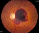
Retinal Hemorrhage vu 1298 foisFundus photography shows central retinal hemorrhage. Patient was treated with Avastin Injection and will be followed up in 4-weeks.
|
|

Hyperpigmentation of the Retinal Pigment Epithelium Right Eye (Treated wet AMD OS)vu 1287 fois85-year-old man has wet age-related macular degeneration in the left eye treated for one year most recently one year ago. OD is 20/30, OS is 20/60
|
|

Posterior Staphyloma and Geographic Atrophy Myopic Degenerationvu 1283 fois54-year-old man . He is a high myope. He has posterior staphylomas. He has had gradually changing vision for the last month or two. OD is 20/40, OS is 20/50.
|
|

AMD - Wet - Fibrovascular Hemorrhagic PED - Occult CNVMvu 1279 fois87-year-old woman for one month OS: 20/60 Fibrovascular PED and Hemorrhage
|
|

Wet AMD Fresh Submacular Hemorrhage - Displaced with Vitrectomy and Subretinal TPAvu 1279 fois85-year-old woman has wet age-related macular degeneration in her better eye and she was reading with it until two days ago when she had sudden severe vision loss in the eye. Her last injection with Avastin 2 weeks ago 20/160 (Pre-Surgical Displacement)
|
|

Basal Laminar Drusen - Cuticular Drusen - Equatorial Drusen - 70 Year Old Woman - Photo Montage Right Eyevu 1272 fois70 year old woman has had macular drusen since her thirties and she said her sister has a similar problem. Recently the vision in the left eye has substantially declined. OD 20/16, OS 10/200.
|
|

Macular Degeneration with Hemorrhagevu 1250 foisPatient comes in for follow up exam for wet AMD. Fundus photography reveals retinal hemorrhage temporally to the macula. Patient was not treated. Wait to see if the blood will absorb on its own.
|
|
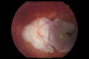
Posterior Staphyloma and Geographic Atrophy Myopic Degenerationvu 1244 fois54-year-old man . He is a high myope. He has posterior staphylomas. He has had gradually changing vision for the last month or two. OD is 20/40, OS is 20/50.
|
|

80 year old woman with Reticular Drusen (pseudodrusen) 20/30 Visionvu 1238 fois80 year old woman with asteroid hyalosis and macular degeneration.
OPHTHALMIC EXAMINATION: Visual acuity with correction 20/30-3 OD and 20/30-2
|
|

Angioid Streaks - Peau D'Orange - Pseudoxanthoma Elasticum - Asymptomatic Extrafoveal Choroidal Neovascular Membranevu 1237 fois60-year-old woman has pseudoxanthoma elasticum since 2008. Her sister has the same problem and lost vision in both eyes. She had a problem with blood vessel growth in the left eye for which she had injections of Avastin in August, September, and October of 2011 (5 months ago). Subsequent to that, the macula has been dry and you have just been watching her carefully. She is in Florida for a short time but will ultimately be spending nine months out of the year here. Both of her eyes have good vision at
|
|

Patterned Pigment Dystrophy of the Fovea 2 year later VA still goodvu 1226 fois
|
|

Reticular Macular Disease (Pseudo-drusen) Both Eyes - Wet AMD OS - Dry AMD OD SD-OCTvu 1221 fois84-year-old woman has wet age-related macular degeneration in the left eye and dry macular degeneration in the right eye. She takes the eye vitamins and her vision is stable since she was treated three months ago with Avastin. OD 20/50, OS 20/32
|
|

Patterned Pigment Dystrophy of the Fovea 79 YO Woman VA 20/20 Right 20/30 Leftvu 1217 fois
|
|

Angioid Streaks and Treated CNVMvu 1212 foisMaculopathy associated to angioid streaks in a 45 years old patient, man,
treated for related CNV
|
|

Cuticular Drusen (Basal Laminar Drusen) and Bilateral Chronic Subfoveal Fluid (Serous Retinal Detachment) unresponsive to monthly Avastin OCT OS EDIvu 1210 fois66-year-old man with bilateral subfoveal fluid unresponsive to one year of monthly avastin therapy.
OD 20/160, Pinhole 20/80. OS 20/63, Pinhole 20/50. OCT scan shows subretinal fluid with normal choroidal thickness. Fluorescein angiogram shows cuticular/basal laminar drusen. ICG shows no foci of choriocapillaris leakage.
|
|

Hyperpigmentation of Retinal Pigment Epithlium in Dry AMD (hyperplasia)vu 1197 fois87-year-old woman has wet age-related macular degeneration in the left eye. She had treatment up north but there has been a gap in therapy and the vision in the left eye is poor. Vision OD is 20/25, OS is 20/400
|
|

Equatorial Drusen Fundus Photo Left Eyevu 1194 fois
|
|

Calcified Drusen - Dry Age-related Macular Degeneration - Geographic Atrophyvu 1190 fois87 Year old woman with dry age-related macular degeneration in both eyes.With both eyes open she is fine, but when she closes the left eye, things are wavier.
VISUAL ACUITY: Vision OD is 20/100, OS is 20/40.
|
|

Hyperpigmentation of the Retinal Pigment Epithelium Right Eye (Treated wet AMD OS)vu 1186 fois85-year-old man has wet age-related macular degeneration in the left eye treated for one year most recently one year ago. OD is 20/30, OS is 20/60
|
|

Patterned Pigment Dystrophy of the Fovea 79 YO Woman VA 20/20 Right 20/30 Leftvu 1184 fois
|
|

PXE with Angioid Streaksvu 1179 foisPXE with angioid streaks OS (12/09/2011)
|
|

CNV secondary to Angioid Streaks, PXEvu 1176 fois
|
|

Basal Laminar Drusen - Cuticular Drusen - Equatorial Drusen - 70 Year Old Woman vu 1161 fois70 year old woman has had macular drusen since her thirties and she said her sister has a similar problem. Recently the vision in the left eye has substantially declined. OD 20/16, OS 10/200.
|
|

Myopic Macular Degeneration - Dry vu 1160 fois55-year-old woman has myopic macular degeneration in both eyes with vision and an Amsler grid change starting a few days ago in the left eye.
VISUAL ACUITY: OD: 20/60. PH: 20/25. OS: 20/200. PH: 20/30.
|
|

Posterior Staphyloma and Geographic Atrophy Myopic Degenerationvu 1158 fois54-year-old man . He is a high myope. He has posterior staphylomas. He has had gradually changing vision for the last month or two. OD is 20/40, OS is 20/50.
|
|

Calcified Drusen - Dry Age-related Macular Degeneration - Geographic Atrophyvu 1154 fois87 Year old woman with dry age-related macular degeneration in both eyes.With both eyes open she is fine, but when she closes the left eye, things are wavier.
VISUAL ACUITY: Vision OD is 20/100, OS is 20/40.
|
|

Patterned Pigment Dystrophy of the Fovea 79 YO Woman VA 20/20 Right 20/30 Leftvu 1146 fois
|
|

Adult Vitelliform Macular Dystrophy - Pattern Dystrophyvu 1142 fois69-year-old woman sees a dark spot centrally. When she looks at things she loses letters and numbers and is having more and more difficulty balancing her checkbook. OD is 20/80, OS is 20/40
|
|

AMD - Wet - Fibrovascular Hemorrhagic PED - Occult CNVMvu 1137 fois87-year-old woman for one month OS: 20/60 Fibrovascular PED and Hemorrhage
|
|

Myopic Degeneration with RPE Lossvu 1137 foisPatient comes in for follow up for glaucoma. VA is 20/30 in both eyes. Fundus photography shows the RPE loss due to myopic degeneration, nasally, inferiority in the left eye.
|
|

New Subfoveal Classic CNVM OS - Wet AMD - Macular Scar Congenital Toxoplasmosis Right Eye vu 1135 fois77-year-old woman as had poor vision in the right eye since birth from congenital toxoplasmosis of her macula in the right eye. Two to three weeks ago when she noticed vision loss in the left eye. VISUAL ACUITY: OD 2/200, OS 20/200.
|
|

Hyperpigmentation of Retinal Pigment Epithlium in Dry AMD (hyperplasia)vu 1131 fois87-year-old woman has wet age-related macular degeneration in the left eye. She had treatment up north but there has been a gap in therapy and the vision in the left eye is poor. Vision OD is 20/25, OS is 20/400
|
|

Calcified Drusen - Dry Age-related Macular Degeneration - Geographic Atrophyvu 1123 fois87 Year old woman with dry age-related macular degeneration in both eyes.With both eyes open she is fine, but when she closes the left eye, things are wavier.
VISUAL ACUITY: Vision OD is 20/100, OS is 20/40.
|
|

Equatorial Drusen Fundus Photo Right Eyevu 1119 fois
|
|
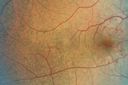
Angioid Streaks - Peau D'Orange - Pseudoxanthoma Elasticum - Asymptomatic Extrafoveal Choroidal Neovascular Membranevu 1119 fois60-year-old woman has pseudoxanthoma elasticum since 2008. Her sister has the same problem and lost vision in both eyes. She had a problem with blood vessel growth in the left eye for which she had injections of Avastin in August, September, and October of 2011 (5 months ago). Subsequent to that, the macula has been dry and you have just been watching her carefully. She is in Florida for a short time but will ultimately be spending nine months out of the year here. Both of her eyes have good vision at
|
|

AMD - Wet - Fibrovascular Hemorrhagic PED - Occult CNVMvu 1116 fois87-year-old woman for one month OS: 20/60 Fibrovascular PED and Hemorrhage
|
|

Large serous PEDvu 1116 fois63-year-old female with acute onset large retinal pigment epithelium detachment from age-related macular degeneration
|
|

Macular Scars - Laser (1980s) Ocular Histoplasmosisvu 1105 fois70 year old man who had laser at Wills, Wilmer and the Mayo clinic before better therapies were available for ocular histoplasmosis related CNVM
|
|

Posterior Staphyloma and Geographic Atrophy Myopic Degenerationvu 1096 fois54-year-old man . He is a high myope. He has posterior staphylomas. He has had gradually changing vision for the last month or two. OD is 20/40, OS is 20/50.
|
|
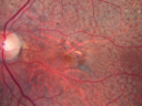
80 year old woman with Reticular Drusen (pseudodrusen) 20/30 Visionvu 1095 fois80 year old woman with asteroid hyalosis and macular degeneration.
OPHTHALMIC EXAMINATION: Visual acuity with correction 20/30-3 OD and 20/30-2
|
|

Myopic CNVM - Wet - rx Avastin for 1 yearvu 1089 fois55-year-old woman OD is 20/40, OS is 20/20. There is a hyperpigmented disc diameter choroidal neovascular membrane with fluid touching the fovea.
|
|

Macular Scar with Retinal Pigment Epithelial Hyperplasia - Hyperpigmentation - Wet Macular Degenerationvu 1087 foisMacular Scar in age related macular degeneration with pathology showing retinal pigment epithelial hyperplasia at the edge of the scar. Histopathogy, microscopic slide, pathology.
|
|

Myopic Macular Degeneration - Dry vu 1060 fois55-year-old woman has myopic macular degeneration in both eyes with vision and an Amsler grid change starting a few days ago in the left eye.
VISUAL ACUITY: OD: 20/60. PH: 20/25. OS: 20/200. PH: 20/30.
|
|

Equatorial Drusen Fundus Photo Right Eyevu 1059 fois
|
|

Macular Scars - Laser (1980s) Ocular Histoplasmosisvu 1058 fois70 year old man who had laser at Wills, Wilmer and the Mayo clinic before better therapies were available for ocular histoplasmosis related CNVM
|
|

Angioid Streaks - Peau D'Orange - Pseudoxanthoma Elasticum - Asymptomatic Extrafoveal Choroidal Neovascular Membranevu 1058 fois60-year-old woman has pseudoxanthoma elasticum since 2008. Her sister has the same problem and lost vision in both eyes. She had a problem with blood vessel growth in the left eye for which she had injections of Avastin in August, September, and October of 2011 (5 months ago). Subsequent to that, the macula has been dry and you have just been watching her carefully. She is in Florida for a short time but will ultimately be spending nine months out of the year here. Both of her eyes have good vision at
|
|

Drusen Maculopathy - Confluent Subfoveal Soft Hydrophobic Drusen - Yellow Drusen in Center of the Maculavu 1055 fois73-year-old woman. She is diabetic for thirteen years. She is also on Coumadin for her heart disease.
VISUAL ACUITY: OD 20/25, OS 20/50.
|
|

Myopic Macular Degeneration - Dry vu 1048 fois55-year-old woman has myopic macular degeneration in both eyes with vision and an Amsler grid change starting a few days ago in the left eye.
VISUAL ACUITY: OD: 20/60. PH: 20/25. OS: 20/200. PH: 20/30.
|
|

vu 1046 fois
|
|

Drusen Maculopathy - Confluent Subfoveal Soft Hydrophobic Drusen - Red Free Image shows Confluent Drusen vu 1042 fois73-year-old woman. She is diabetic for thirteen years. She is also on Coumadin for her heart disease.
VISUAL ACUITY: OD 20/25, OS 20/50.
|
|

Equatorial Drusen Fundus Photo Right Eyevu 1036 fois
|
|

Drusen Maculopathy - Confluent Subfoveal Soft Hydrophobic Drusen - Early Fluorescein Angiogram Drusen do not yet stainvu 1035 fois73-year-old woman. She is diabetic for thirteen years. She is also on Coumadin for her heart disease.
VISUAL ACUITY: OD 20/25, OS 20/50.
|
|
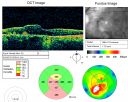
AMD - Wet - Fibrovascular Hemorrhagic PED - Occult CNVMvu 1033 fois87-year-old woman for one month OS: 20/60 Fibrovascular PED and Hemorrhage
|
|
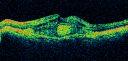
Subfoveal CNVM wet AMD oct line scanvu 1032 foisShe has noticed decreased vision in the left eye. It is hard to say for how long, maybe a few months. She had her last eye exam about a year ago. She thoughts perhaps it was cataracts, but you saw a problem with the macula and suggested she come here for evaluation.
VISUAL ACUITY: Vision OD is 20/25, OS is 2/200
|
|

Angioid Streaks - Peau D'Orange - Pseudoxanthoma Elasticum - Asymptomatic Extrafoveal Choroidal Neovascular Membranevu 1032 fois60-year-old woman has pseudoxanthoma elasticum since 2008. Her sister has the same problem and lost vision in both eyes. She had a problem with blood vessel growth in the left eye for which she had injections of Avastin in August, September, and October of 2011 (5 months ago). Subsequent to that, the macula has been dry and you have just been watching her carefully. She is in Florida for a short time but will ultimately be spending nine months out of the year here. Both of her eyes have good vision at
|
|

Cartwheel CNVM - Classic - wet AMD - evidence of possible old CSR - Subretinal hemorrhage next to subfoveal lesionvu 1031 fois75 y.o Decreased Vision right eye for one year. VA OD: sc20/125+2 OS 20/25. Autofluorescence and SD OCT show evidence of old serous retinal detachment superior to the fovea suggesting a history of CSR. The macular lesion on FA shows a classic cartwheel appearance
|
|

vu 1028 fois
|
|

Equatorial Drusen Fundus Photo Left Eyevu 1027 fois60-year-old woman OD 20/30, OS 20/20
|
|

Cuticular Drusen (Basal Laminar Drusen) and Bilateral Chronic Subfoveal Fluid (Serous Retinal Detachment) unresponsive to monthly Avastin OCT OD EDIvu 1027 fois66-year-old man with bilateral subfoveal fluid unresponsive to one year of monthly avastin therapy.
OD 20/160, Pinhole 20/80. OS 20/63, Pinhole 20/50. OCT scan shows subretinal fluid with normal choroidal thickness. Fluorescein angiogram shows cuticular/basal laminar drusen. ICG shows no foci of choriocapillaris leakage.
|
|

Drusen Maculopathy - Confluent Subfoveal Soft Hydrophobic Drusen - Yellow Drusen in the Center of the Maculavu 1023 fois73-year-old woman. She is diabetic for thirteen years. She is also on Coumadin for her heart disease.
VISUAL ACUITY: OD 20/25, OS 20/50.
|
|

Angioid Streaks - Peau D'Orange - Pseudoxanthoma Elasticum - Asymptomatic Extrafoveal Choroidal Neovascular Membranevu 1023 fois60-year-old woman has pseudoxanthoma elasticum since 2008. Her sister has the same problem and lost vision in both eyes. She had a problem with blood vessel growth in the left eye for which she had injections of Avastin in August, September, and October of 2011 (5 months ago). Subsequent to that, the macula has been dry and you have just been watching her carefully. She is in Florida for a short time but will ultimately be spending nine months out of the year here. Both of her eyes have good vision at
|
|

Myopic Macular Degeneration - Dry OCT OSvu 1017 fois55-year-old woman has myopic macular degeneration in both eyes with vision and an Amsler grid change starting a few days ago in the left eye.
VISUAL ACUITY: OD: 20/60. PH: 20/25. OS: 20/200. PH: 20/30.
|
|
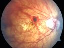
Myopic Choroidal Neovascular Membrane 54 year old woman 20/20vu 1013 fois54-year-old woman has had problems in her vision in the right eye for about the last three months. Inferotemporally she sees a gray area, which is distorted. OD is 20/20, OS is 20/20
|
|

Reticular Macular Disease (Pseudo-drusen) Both Eyes - Wet AMD OS - Dry AMD OD FAFvu 1013 fois84-year-old woman has wet age-related macular degeneration in the left eye and dry macular degeneration in the right eye. She takes the eye vitamins and her vision is stable since she was treated three months ago with Avastin. OD 20/50, OS 20/32
|
|

Hyperpigmentation of the Retinal Pigment Epithelium in Dry AMDvu 1001 fois87-year-old woman has wet age-related macular degeneration in the left eye. Vision OD is 20/25, OS is 20/400
|
|

Wet AMD Fresh Submacular Hemorrhage - Displaced with Vitrectomy and Subretinal TPAvu 1000 fois85-year-old woman has wet age-related macular degeneration in her better eye and she was reading with it until two days ago when she had sudden severe vision loss in the eye. Post-surgical displacment vision was 20/120.
|
|
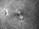
Non-Foveal Geographic Atrophy Both Eyesvu 991 fois81-year-old man has age-related macular degeneration in both eyes. Vision OD is 20/40, OS is 20/80.
|
|

Macular Scars - Laser (1980s) Ocular Histoplasmosisvu 990 fois70 year old man who had laser at Wills, Wilmer and the Mayo clinic before better therapies were available for ocular histoplasmosis related CNVM
|
|

Calcified Drusen - Dry Age-related Macular Degeneration - Geographic Atrophyvu 988 fois87 Year old woman with dry age-related macular degeneration in both eyes.With both eyes open she is fine, but when she closes the left eye, things are wavier.
VISUAL ACUITY: Vision OD is 20/100, OS is 20/40.
|
|

Macular Scars - Laser (1980s) Ocular Histoplasmosisvu 982 fois70 year old man who had laser at Wills, Wilmer and the Mayo clinic before better therapies were available for ocular histoplasmosis related CNVM
|
|
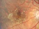
Hyperpigmentation of the Retinal Pigment Epithelium in Dry AMDvu 980 fois87-year-old woman has wet age-related macular degeneration in the left eye. Vision OD is 20/25, OS is 20/400
|
|

Basal Laminar Drusen - Cuticular Drusen - Equatorial Drusen - 70 Year Old Woman - Photo Equatorvu 980 fois70 year old woman has had macular drusen since her thirties and she said her sister has a similar problem. Recently the vision in the left eye has substantially declined. OD 20/16, OS 10/200.
|
|

Angioid Streaks - Pseudoxanthoma Elasticum - Supressed CNVMvu 978 fois54-year-old woman had angioid streaks and macular degeneration in both eyes. She has had intravitreal Avastin in the right eye about every three months and her vision in that eye is good.
VISUAL ACUITY: Vision OD is 20/30, OS is 2/200
|
|

80 year old woman with Reticular Drusen (pseudodrusen) 20/30 Vision - Before Vitrectomyvu 974 fois80 year old woman with asteroid hyalosis and macular degeneration.
OPHTHALMIC EXAMINATION: Visual acuity with correction 20/30-3 OD and 20/30-2
|
|
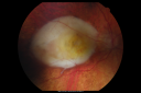
Disciform Scar after Photodynamic Laser - Macular Hemorrhage and Vitrectomyvu 973 fois84-year-old man has age-related macular degeneration in both eyes, with a disciform scar in the right eye. He had a subfoveal hemorrhage in that eye treated with vitrectomy and displacement on May 3, 2006. He previous to that had photodynamic laser. These photos are from October 2010. VA is 1/200.
|
|

Subfoveal CNVM wet AMD color photovu 971 foisShe has noticed decreased vision in the left eye. It is hard to say for how long, maybe a few months. She had her last eye exam about a year ago. She thoughts perhaps it was cataracts, but you saw a problem with the macula and suggested she come here for evaluation.
VISUAL ACUITY: Vision OD is 20/25, OS is 2/200
|
|

Multifocal Pattern Dystrophyvu 971 fois51 year old with normal vision - images look a lot like fundus flavimaculatus
|
|

Outer Retinal Tubulationvu 965 foisPatient with a history of wet macular degeneration and glaucoma in both eyes. VA is 20/50, right eye, 20/80, left eye. Patient is treated with Eylea in both eyes. Enhanced Depth Imaging OCT reveals a small like form of a cyst which in fact isn't a cyst at all. This is called Outer Retinal Tubulation in which degenerating photo-receptors may become arranged in a circular or ovoid fashion. This is sometimes misdiagnosed as cystic changes in the retinal pigment epithelium or sub-retinal fluid.
|
|

Equatorial Drusen Right Eyevu 959 fois
|
|

Hyperpigmentation of the Retinal Pigment Epithelium in Dry AMDvu 959 fois87-year-old woman has wet age-related macular degeneration in the left eye. Vision OD is 20/25, OS is 20/400
|
|
| 2694 photos sur 27 page(s) |
1 |
 |
|
|
|
|
|
|
|
|
|


