Les plus populaires - Macular Degeneration - Age Related, Myopic, Choroidal Neovascular Membranes
|

Post-Retinal Detachment Repair Occult Subfoveal CNVMvu 474 foisThis pleasant 56-year-old man has had a complex retinal detachment repair in the right eye. I most recently removed his silicone oil 3 months ago. His vision now is the best it has ever been.
VISUAL ACUITY: OD 4/200
|
|

Wet AMD OD and Lamellar Macular hole OSvu 474 foisWet AMD in right and better eye failed extend to 2 months but was able to stop therapy once he started on systemic Avastin for cancer. The last image shows the macula dry 9 months without local therapy. The left eye has a lamellar macular hole.
|
|

Dry AMD - Confluent Drusen Sparing Center of Macula which has Atrophyvu 474 fois65 year old woman, VA 20/40 OD; 20/80 OS. The center of the macula has few or no drusen with predominantly non-geographic atrophy
|
|

Dry AMD - Confluent Drusen Sparing Center of Macula which has Atrophyvu 474 fois65 year old woman, VA 20/40 OD; 20/80 OS. The center of the macula has few or no drusen with predominantly non-geographic atrophy
|
|

Wet AMD 1 month after Eylea - macula is dry and vision is bettervu 473 fois
|
|

Adult Vitelliform Macular Dystrophy - Pattern Dystrophyvu 473 foisBright subfoveal FAF from lipofuscin
|
|

Cuticular Drusen - 26 year old female - Glomerulonephritisvu 473 foisVA 20/25 OU. Images are over 4 years. FA shows starry sky early images - This is because of loss of RPE over the drusen creating very small window defects.
|
|

Juxtapapillary CNVM - Hemorrhagevu 473 fois83 year old female VA 20/200 improved to 20/32 within 2 months with Lucentis
|
|
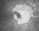
Geographic Atrophy and Age-Related Macular Degeneration - Choriocapillaris is Largely Still Intactvu 472 fois85-year-old woman has age-related macular degeneration in both eyes with geographic atrophy. She has noticed her vision getting slightly worse in the past 6 months. VISUAL ACUITY: OD 20/70, OS 20/400
|
|

Pattern Dystrophy - Adult Vitelliform Dystrophy - Macular Degenerationvu 472 fois 78 Year Old Woman with vision has diminished in the right eye over the past couple of years, but the diminishment has been slow.
20/60+1 OD, 20/25 OS
|
|
vu 472 fois
|
|

Wet AMD OD and Lamellar Macular hole OSvu 472 foisWet AMD in right and better eye failed extend to 2 months but was able to stop therapy once he started on systemic Avastin for cancer. The last image shows the macula dry 9 months without local therapy. The left eye has a lamellar macular hole.
|
|

OHS CNVM post treatment with Lucentis - VA now 20/25vu 472 fois
|
|

New wet AMD OS with subretinal hemorrhage and recent vision loss, Scar ODvu 472 fois
|
|

Juxtapapillary CNVM - Hemorrhagevu 472 fois83 year old female VA 20/200 improved to 20/32 within 2 months with Lucentis
|
|

Concentric Geograophic Atrophy - Age-related Macular Degeneration - Charles Bonnet Syndrome - Bulls Eyevu 471 fois88-year-old woman has age-related with vision gradually declining over the last year. She sees spots in her vision, like red spots and dark spots and she even notices during the daytime, for example when she was looking at her minister, it looked like there were little flowering pots coming out of his head.
VISUAL ACUITY: OD 20/200, OS 20/400.
|
|

Wet AMD controlled with Avastin 20/60 Visionvu 471 fois79-year-old woman has wet age-related macular degeneration in both eyes. The left eye had intravitreal injections of Avastin starting in May of 2009. Her vision remains good in the left eye and poor in the right eye.
VISUAL ACUITY: OD 8/200, OS 20/60.
|
|

Cuticular Drusen - 26 year old female - Glomerulonephritisvu 471 foisVA 20/25 OU. Images are over 4 years
|
|

Multifocal Pattern Dystrophyvu 471 fois51 year old with normal vision - images look a lot like fundus flavimaculatus
|
|

Wet AMD - Subfoveal CNVM - Excellent Response to Avastinvu 470 fois76-year-old woman two to three months of vision loss OD
Vision started at 20/200 and with Avastin improved to 20/25. She has required monthly injections and as of October 2010 has been treated about 16 times. Her left eye remains good at 20/20.
The OCT from post-treatment as well as pretreatment images are in this album.
|
|

Small Retinal Pigment Epithelial Rip and Poor Vision (20/400)vu 470 fois80-year-old woman vision in the right eye has substantially declined.
OD: 20/400; OS: 20/50
|
|

Small Retinal Pigment Epithelial Rip and Poor Vision (20/400)vu 470 fois80-year-old woman vision in the right eye has substantially declined.
OD: 20/400; OS: 20/50
|
|

Polypoidal Choroidal Vasculopathy Right Eye - Disciform Scar Left Eyevu 470 fois84-year-old woman has polypoidal choroidal vasculopathy in both eyes with macular degeneration. She has macular scars in both eyes and she remains with remarkably good vision in the right eye despite the scarring.
VISUAL ACUITY: OD 20/30, OS 1/200
Avastin Rx OD
|
|

Dry AMD - Confluent Drusen Sparing Center of Macula which has Atrophyvu 470 fois65 year old woman, VA 20/40 OD; 20/80 OS. The center of the macula has few or no drusen with predominantly non-geographic atrophy
|
|

Outer Retinal Tubulationvu 470 fois78 year old man chronic wet AMD - 20/200
|
|

Myopic CNVM in the left eye. New Classic subfoveal lesionvu 470 fois64 year old female. She had retinal detachment repair in the right eye in 1996 (25 years ago). The left eye has new distortion for a week or two.
VA 20/40 OU.
Left eye has a very small subfoveal CNVM
|
|
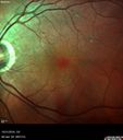
Submacular Hemorrhage - wet AMDvu 469 fois84 year old man with 20/200 from submacular hemorrhage. Vision improved to 20/32 with only Avastin therapy (no surgical intervention). The initial FAF is interesting because parts of the blood hyper auto fluoresce.
|
|

Non-exudative wet AMD right eye (treatment naive quiescent)vu 469 fois82 year old man. ICG shows CNVM in the right eye. This stayed quiet for 3 years and then started leaking and responded well to Avastin.
|
|

Myopic Degeneration - Inferior staphyloma with severe atrophyvu 469 fois76 year old with gradual vision loss. 20/32 OD; 20/40 OS
|
|

Myopic Degeneration - Inferior staphyloma with severe atrophyvu 469 fois76 year old with gradual vision loss. 20/32 OD; 20/40 OS
|
|

Adult Pseudovitelliform Macular Dystrophyvu 469 fois89 year old female states dark smudge on ceiling during night time came and went. Decreased vision in the left eye
VA OD: Dcc20/32
VA OS: Dcc20/63
|
|

Basal Laminar Drusen - Cuticular Drusen - Equatorial Drusen - 70 Year Old Woman - OCT Line Scan Right Eyevu 468 fois70 year old woman has had macular drusen since her thirties and she said her sister has a similar problem. Recently the vision in the left eye has substantially declined. OD 20/16, OS 10/200.
|
|

Wet AMD OD and Lamellar Macular hole OSvu 468 foisWet AMD in right and better eye failed extend to 2 months but was able to stop therapy once he started on systemic Avastin for cancer. The last image shows the macula dry 9 months without local therapy. The left eye has a lamellar macular hole.
|
|

Cuticular Drusen - 26 year old female - Glomerulonephritisvu 468 foisVA 20/25 OU. Images are over 4 years
|
|

Adult Pseudovitelliform Macular Dystrophyvu 468 fois89 year old female states dark smudge on ceiling during night time came and went. Decreased vision in the left eye
VA OD: Dcc20/32
VA OS: Dcc20/63
|
|

Myopic CNVM in the left eye. New Classic subfoveal lesionvu 468 fois64 year old female. She had retinal detachment repair in the right eye in 1996 (25 years ago). The left eye has new distortion for a week or two.
VA 20/40 OU.
Left eye has a very small subfoveal CNVM
|
|

Myopic CNVM in the left eye. New Classic subfoveal lesionvu 468 fois64 year old female. She had retinal detachment repair in the right eye in 1996 (25 years ago). The left eye has new distortion for a week or two.
VA 20/40 OU.
Left eye has a very small subfoveal CNVM
|
|

Small Retinal Pigment Epithelial Rip and Poor Vision (20/400)vu 467 fois80-year-old woman vision in the right eye has substantially declined.
OD: 20/400; OS: 20/50
|
|

Pattern Dystrophy - Adult Vitellifrom (Best)vu 467 fois71 year old female - lesions nasal to the fovea in both eyes. (20/40 OU)
|
|
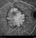
Asymptomatic wet AMD - Subfoveal CNM both eyesvu 466 fois82 year old woman - 20/25 OU. Images show occult subfoveal CNVM both eyes best seen on late ICG. The left eye also has probable polypoidal choroidal vasculopathy. No treatment was recommended on the first visit - follow-up at 1 month.
|
|

Submacular Hemorrhage - wet AMDvu 466 fois84 year old man with 20/200 from submacular hemorrhage. Vision improved to 20/32 with only Avastin therapy (no surgical intervention). The initial FAF is interesting because parts of the blood hyper auto fluoresce.
|
|

Myopic CNVM in the left eye. New Classic subfoveal lesionvu 466 fois64 year old female. She had retinal detachment repair in the right eye in 1996 (25 years ago). The left eye has new distortion for a week or two.
VA 20/40 OU.
Left eye has a very small subfoveal CNVM
|
|

Post-Retinal Detachment Repair Occult Subfoveal CNVMvu 465 foisThis pleasant 56-year-old man has had a complex retinal detachment repair in the right eye. I most recently removed his silicone oil 3 months ago. His vision now is the best it has ever been.
VISUAL ACUITY: OD 4/200
|
|

Serous Macular Detachment Both Eyes - Wet AMD vs. CSR - 62 Year Old Man - Fluorescein angiogramvu 465 fois
|
|
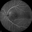
Serous Macular Detachment Both Eyes - Wet AMD vs. CSR - 62 Year Old Man - Fluorescein angiogramvu 465 fois
|
|

Serous Macular Detachment Both Eyes - Wet AMD vs. CSR - 62 Year Old Man ICG vu 465 fois
|
|

Cuticular Drusen - 26 year old female - Glomerulonephritisvu 465 foisVA 20/25 OU. Images are over 4 years FA shows starry sky early images - This is because of loss of RPE over the drusen creating very small window defects.
|
|

Pattern Dystrophy - Adult Vitellifrom (Best)vu 465 fois71 year old female - lesions nasal to the fovea in both eyes. (20/40 OU)
|
|

Polypoidal Choroidal Vasculopathy Left Eyevu 465 fois74 year old man with vision loss to 20/100 over a few months. He improved to 20/20 with Lucentis. The color and ICG show the polyps.
|
|

Pattern Dystrophy - Foveal Atrophyvu 464 fois73 year old female with vision loss - looks like macular hole
|
|

Chorioretinal Scar with Radiating Chorioretinal Foldsvu 463 fois79-year-old woman has wet age-related macular degeneration in both eyes. The left eye had intravitreal injections of Avastin starting in May of 2009. Her vision remains good in the left eye and poor in the right eye.
VISUAL ACUITY: OD 8/200, OS 20/60.
|
|

Post-Retinal Detachment Repair Occult Subfoveal CNVMvu 463 foisThis pleasant 56-year-old man has had a complex retinal detachment repair in the right eye. I most recently removed his silicone oil 3 months ago. His vision now is the best it has ever been.
VISUAL ACUITY: OD 4/200
|
|

Angioid Streaks and Macular Degeneration - CNVM OS - Band Keratopathy vu 463 fois79-year-old man has wet age-related macular degeneration in the left eye, for which he is having Lucentis treatment. OD 20/40, Pinhole 20/32. OS 20/160, Pinhole 20/50. He has band keratopathy sparing the visual axis in both eyes. ICG shows angioid streaks both eye.
|
|

Polypoidal Choroidal Vasculopathy Left Eyevu 463 fois74 year old man with vision loss to 20/100 over a few months. He improved to 20/20 with Lucentis. The color and ICG show the polyps.
|
|
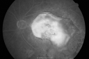
Macular Scar Left Eye from Age-Related Macular Degenerationvu 462 fois81-year-old woman has a macular scar in the left eye and then unfortunately developed a central retinal artery occlusion in the right eye OD 20/100, OS 6/200.
|
|

Patterned Pigment Dystrophy of the Fovea 79 YO Woman VA 20/20 Right 20/30 Leftvu 462 fois
|
|

Polypoidal Choroidal Vasculopathy Right Eye - Disciform Scar Left Eyevu 462 fois84-year-old woman has polypoidal choroidal vasculopathy in both eyes with macular degeneration. She has macular scars in both eyes and she remains with remarkably good vision in the right eye despite the scarring.
VISUAL ACUITY: OD 20/30, OS 1/200
Avastin Rx OD
|
|

Wet AMD OD and Lamellar Macular hole OSvu 462 foisWet AMD in right and better eye failed extend to 2 months but was able to stop therapy once he started on systemic Avastin for cancer. The last image shows the macula dry 9 months without local therapy. The left eye has a lamellar macular hole.
|
|

IPCV - idiopathic polypoidal choroidal vasculopathy - juxtapapillary CNVM with hemorrhage both eyes (also choroidal nevus right eye) - Subretinal hemorrhage both eyesvu 462 fois86 year old woman with shadow in vision OD and new hemmorhage OU.
|
|

Submacular Hemorrhage - wet AMDvu 462 fois84 year old man with 20/200 from submacular hemorrhage. Vision improved to 20/32 with only Avastin therapy (no surgical intervention). The initial FAF is interesting because parts of the blood hyper auto fluoresce.
|
|

Outer Retinal Tubulationvu 462 fois78 year old man chronic wet AMD - 20/200
|
|

Pattern Dystrophy - Foveal Atrophyvu 462 fois73 year old female with vision loss - looks like macular hole
|
|

Non-exudative wet AMD right eye (treatment naive quiescent)vu 462 fois82 year old man. ICG shows CNVM in the right eye. This stayed quiet for 3 years and then started leaking and responded well to Avastin.
|
|

Basal Laminar Drusen - Cuticular Drusen - Equatorial Drusen - 70 Year Old Woman vu 461 fois70 year old woman has had macular drusen since her thirties and she said her sister has a similar problem. Recently the vision in the left eye has substantially declined. OD 20/16, OS 10/200.
|
|

Serous Macular Detachment Both Eyes - Wet AMD vs. CSR - 62 Year Old Man - Fluorescein angiogramvu 461 fois
|
|

Outer Retinal Tubulationvu 461 fois78 year old man chronic wet AMD - 20/200
|
|

Non-exudative wet AMD right eye (treatment naive quiescent)vu 461 fois82 year old man. ICG shows CNVM in the right eye. This stayed quiet for 3 years and then started leaking and responded well to Avastin.
|
|

Polypoidal Choroidal Vasculopathy - Right Eyevu 461 fois83 year old female presented in June 2013 with vision loss for 3 months. She had 2 injections elsewhere with no improvement (avastin). The first images show a peripapillary CNVM with polyps. I treated her once with PDT and she stayed dry with good vision for 5 years (2019). VA on presentation 20/80 OD. Improved to 20/40
|
|
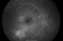
Macular Scar Left Eye from Age-Related Macular Degenerationvu 460 fois81-year-old woman has a macular scar in the left eye and then unfortunately developed a central retinal artery occlusion in the right eye OD 20/100, OS 6/200.
|
|

Cuticular Drusen (Basal Laminar Drusen) and Bilateral Chronic Subfoveal Fluid (Serous Retinal Detachment) unresponsive to monthly Avastin - Fill ICG FA ODvu 460 fois66-year-old man with bilateral subfoveal fluid unresponsive to one year of monthly avastin therapy.
OD 20/160, Pinhole 20/80. OS 20/63, Pinhole 20/50. OCT scan shows subretinal fluid with normal choroidal thickness. Fluorescein angiogram shows cuticular/basal laminar drusen. ICG shows no foci of choriocapillaris leakage.
|
|

Confluent Soft Drusen with Subetinal Fluid - Right Eyevu 460 foisAsymptomatic 85 year old man. OCT shows pocket of subfoveal fluid in the center of the macula. This could be mechanical from the drusen preventing the retina from settling down. Testing did not reveal a CNVM and subsequent OCT 6 weeks later showed no change in structure.
|
|

Submacular Hemorrhage - wet AMDvu 460 fois84 year old man with 20/200 from submacular hemorrhage. Vision improved to 20/32 with only Avastin therapy (no surgical intervention). The initial FAF is interesting because parts of the blood hyper auto fluoresce.
|
|

Myopic Macular Degeneration - Staphyloma - Recent Vision Loss Right Eye - Possible CNVMvu 459 fois83-year-old woman is a high myope with myopic macular degeneration. She had a retinal detachment repair in the right eye in 1968. I saw her in June of 2003. She had posterior colobomas and areas of pigment epithelial atrophy. She has noticed over the last six weeks substantial declining vision in the right eye. Prior to that, the vision in the two eyes was about the same. She uses Xalatan in the left eye and Alphagan in both eyes.
VISUAL ACUITY: Vision OD is 20/160, OS is 20/40
|
|

Basal Laminar Drusen - Cuticular Drusen - Equatorial Drusen - 70 Year Old Woman vu 459 fois70 year old woman has had macular drusen since her thirties and she said her sister has a similar problem. Recently the vision in the left eye has substantially declined. OD 20/16, OS 10/200.
|
|

New wet AMD OS with subretinal hemorrhage and recent vision loss, Scar ODvu 459 fois
|
|

Pattern Dystrophy - Adult Vitellifrom (Best)vu 459 fois71 year old female - lesions nasal to the fovea in both eyes. (20/40 OU)
|
|

Pattern Dystrophy - Foveal Atrophyvu 459 fois73 year old female with vision loss - looks like macular hole
|
|

Myopic CNVM in the left eye. New Classic subfoveal lesionvu 459 fois64 year old female. She had retinal detachment repair in the right eye in 1996 (25 years ago). The left eye has new distortion for a week or two.
VA 20/40 OU.
Left eye has a very small subfoveal CNVM
|
|

Myopic Macular Degeneration - Staphyloma - Recent Vision Loss Right Eye - Possible CNVMvu 458 fois83-year-old woman is a high myope with myopic macular degeneration. She had a retinal detachment repair in the right eye in 1968. I saw her in June of 2003. She had posterior colobomas and areas of pigment epithelial atrophy. She has noticed over the last six weeks substantial declining vision in the right eye. Prior to that, the vision in the two eyes was about the same. She uses Xalatan in the left eye and Alphagan in both eyes.
VISUAL ACUITY: Vision OD is 20/160, OS is 20/40
|
|

vu 458 fois
|
|

Serous Macular Detachment Both Eyes - Wet AMD vs. CSR - 62 Year Old Man ICGvu 458 fois
|
|

Wet AMD OD and Lamellar Macular hole OSvu 458 foisWet AMD in right and better eye failed extend to 2 months but was able to stop therapy once he started on systemic Avastin for cancer. The last image shows the macula dry 9 months without local therapy. The left eye has a lamellar macular hole.
|
|

IPCV - idiopathic polypoidal choroidal vasculopathy - juxtapapillary CNVM with hemorrhage both eyes (also choroidal nevus right eye) - Subretinal hemorrhage both eyesvu 458 fois86 year old woman with shadow in vision OD and new hemmorhage OU. ICG angiogram
|
|

Asymptomatic wet AMD - Subfoveal CNM both eyesvu 458 fois82 year old woman - 20/25 OU. Images show occult subfoveal CNVM both eyes best seen on late ICG. The left eye also has probable polypoidal choroidal vasculopathy. No treatment was recommended on the first visit - follow-up at 1 month.
|
|

Submacular Hemorrhage - wet AMDvu 458 fois84 year old man with 20/200 from submacular hemorrhage. Vision improved to 20/32 with only Avastin therapy (no surgical intervention). The initial FAF is interesting because parts of the blood hyper auto fluoresce.
|
|

Outer Retinal Tubulationvu 458 fois78 year old man chronic wet AMD - 20/200
|
|

Concentric Geographic Atrophyvu 458 fois76 year old man Gradual vision loss
20/32 OD; 20/40 OS
No medicines, Non-smoker
Working and Driving
|
|

Small Retinal Pigment Epithelial Rip and Poor Vision (20/400)vu 457 fois80-year-old woman vision in the right eye has substantially declined.
OD: 20/400; OS: 20/50
|
|
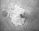
Geographic Atrophy and Age-Related Macular Degeneration - Choriocapillaris is Largely Still Intactvu 457 fois85-year-old woman has age-related macular degeneration in both eyes with geographic atrophy. She has noticed her vision getting slightly worse in the past 6 months. VISUAL ACUITY: OD 20/70, OS 20/400
|
|

Submacular Hemorrhage - wet AMDvu 457 fois84 year old man with 20/200 from submacular hemorrhage. Vision improved to 20/32 with only Avastin therapy (no surgical intervention). The initial FAF is interesting because parts of the blood hyper auto fluoresce.
|
|

Adult Pseudovitelliform Macular Dystrophyvu 457 fois89 year old female states dark smudge on ceiling during night time came and went. Decreased vision in the left eye
VA OD: Dcc20/32
VA OS: Dcc20/63
|
|

Adult Pseudovitelliform Macular Dystrophyvu 457 fois89 year old female states dark smudge on ceiling during night time came and went. Decreased vision in the left eye
VA OD: Dcc20/32
VA OS: Dcc20/63
|
|

Cuticular Drusen (Basal Laminar Drusen) and Bilateral Chronic Subfoveal Fluid (Serous Retinal Detachment) unresponsive to monthly Avastin Very Early ICG OSvu 456 fois66-year-old man with bilateral subfoveal fluid unresponsive to one year of monthly avastin therapy.
OD 20/160, Pinhole 20/80. OS 20/63, Pinhole 20/50. OCT scan shows subretinal fluid with normal choroidal thickness. Fluorescein angiogram shows cuticular/basal laminar drusen. ICG shows no foci of choriocapillaris leakage.
|
|

Post-Retinal Detachment Repair Occult Subfoveal CNVMvu 456 foisThis pleasant 56-year-old man has had a complex retinal detachment repair in the right eye. I most recently removed his silicone oil 3 months ago. His vision now is the best it has ever been.
VISUAL ACUITY: OD 4/200
|
|

IPCV - idiopathic polypoidal choroidal vasculopathy - juxtapapillary CNVM with hemorrhage both eyes (also choroidal nevus right eye) - Subretinal hemorrhage both eyesvu 455 fois86 year old woman with shadow in vision OD and new hemmorhage OU. ICG angiogram - polyp shows in first 6 minutes of test with branching vascular network.
|
|
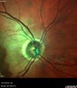
Submacular Hemorrhage - wet AMDvu 455 fois84 year old man with 20/200 from submacular hemorrhage. Vision improved to 20/32 with only Avastin therapy (no surgical intervention). The initial FAF is interesting because parts of the blood hyper auto fluoresce.
|
|
Pattern Dystrophy - Adult Vitellifrom (Best)vu 455 fois71 year old female - lesions nasal to the fovea in both eyes. (20/40 OU)
|
|

Pattern Dystrophy - Adult Vitellifrom (Best)vu 455 fois71 year old female - lesions nasal to the fovea in both eyes. (20/40 OU)
|
|

Outer Retinal Tubulationvu 455 fois78 year old man chronic wet AMD - 20/200
|
|

Non-exudative wet AMD right eye (treatment naive quiescent)vu 455 fois82 year old man. ICG shows CNVM in the right eye. This stayed quiet for 3 years and then started leaking and responded well to Avastin.
|
|
| 2694 photos sur 27 page(s) |
 |
|
|
|
|
12 | |
|
|
|
|


