Most viewed - Macular Degeneration - Age Related, Myopic, Choroidal Neovascular Membranes
|

Post-Retinal Detachment Repair Occult Subfoveal CNVM475 viewsThis pleasant 56-year-old man has had a complex retinal detachment repair in the right eye. I most recently removed his silicone oil 3 months ago. His vision now is the best it has ever been.
VISUAL ACUITY: OD 4/200
|
|

Submacular Hemorrhage - wet AMD475 views84 year old man with 20/200 from submacular hemorrhage. Vision improved to 20/32 with only Avastin therapy (no surgical intervention). The initial FAF is interesting because parts of the blood hyper auto fluoresce.
|
|

Outer Retinal Tubulation475 views78 year old man chronic wet AMD - 20/200
|
|

Outer Retinal Tubulation475 views78 year old man chronic wet AMD - 20/200
|
|

Pattern Dystrophy - Foveal Atrophy475 views73 year old female with vision loss - looks like macular hole
|
|
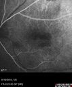
New wet AMD OS with subretinal hemorrhage and recent vision loss, Scar OD474 views
|
|

Polypoidal Choroidal Vasculopathy Left Eye474 views74 year old man with vision loss to 20/100 over a few months. He improved to 20/20 with Lucentis. The color and ICG show the polyps.
|
|

Pattern Dystrophy - Foveal Atrophy474 views73 year old female with vision loss - looks like macular hole
|
|

Myopic Degeneration - Inferior staphyloma with severe atrophy474 views76 year old with gradual vision loss. 20/32 OD; 20/40 OS
|
|

Cuticular Drusen (Basal Laminar Drusen) and Bilateral Chronic Subfoveal Fluid (Serous Retinal Detachment) unresponsive to monthly Avastin Very Early ICG OS473 views66-year-old man with bilateral subfoveal fluid unresponsive to one year of monthly avastin therapy.
OD 20/160, Pinhole 20/80. OS 20/63, Pinhole 20/50. OCT scan shows subretinal fluid with normal choroidal thickness. Fluorescein angiogram shows cuticular/basal laminar drusen. ICG shows no foci of choriocapillaris leakage.
|
|

Dry AMD - Confluent Drusen Sparing Center of Macula which has Atrophy473 views65 year old woman, VA 20/40 OD; 20/80 OS. The center of the macula has few or no drusen with predominantly non-geographic atrophy
|
|

Non-exudative wet AMD right eye (treatment naive quiescent)473 views82 year old man. ICG shows CNVM in the right eye. This stayed quiet for 3 years and then started leaking and responded well to Avastin.
|
|

Concentric Geographic Atrophy473 views76 year old man Gradual vision loss
20/32 OD; 20/40 OS
No medicines, Non-smoker
Working and Driving
|
|

Adult Pseudovitelliform Macular Dystrophy473 views89 year old female states dark smudge on ceiling during night time came and went. Decreased vision in the left eye
VA OD: Dcc20/32
VA OS: Dcc20/63
|
|

Wet AMD OD and Lamellar Macular hole OS472 viewsWet AMD in right and better eye failed extend to 2 months but was able to stop therapy once he started on systemic Avastin for cancer. The last image shows the macula dry 9 months without local therapy. The left eye has a lamellar macular hole.
|
|

IPCV - idiopathic polypoidal choroidal vasculopathy - juxtapapillary CNVM with hemorrhage both eyes (also choroidal nevus right eye) - Subretinal hemorrhage both eyes472 views86 year old woman with shadow in vision OD and new hemmorhage OU.FA shows juxtapapillary CNVM
|
|

Dry AMD - Confluent Drusen Sparing Center of Macula which has Atrophy472 views65 year old woman, VA 20/40 OD; 20/80 OS. The center of the macula has few or no drusen with predominantly non-geographic atrophy
|
|

Submacular Hemorrhage - wet AMD472 views84 year old man with 20/200 from submacular hemorrhage. Vision improved to 20/32 with only Avastin therapy (no surgical intervention). The initial FAF is interesting because parts of the blood hyper auto fluoresce.
|
|

Pattern Dystrophy - Foveal Atrophy472 views73 year old female with vision loss - looks like macular hole
|
|
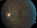
Angioid Streaks - CNVM OS with vision loss472 views48 year old soccer player. Prior trauma OS (with soccer ball). Vision loss for 3 weeks to 20/60 - with Lucentis vision improved to 20/30
|
|

Angioid Streaks - CNVM OS with vision loss472 views48 year old soccer player. Prior trauma OS (with soccer ball). Vision loss for 3 weeks to 20/60 - with Lucentis vision improved to 20/30
|
|

Myopic Degeneration - Inferior staphyloma with severe atrophy472 views76 year old with gradual vision loss. 20/32 OD; 20/40 OS
|
|

Concentric Geographic Atrophy472 views76 year old man Gradual vision loss
20/32 OD; 20/40 OS
No medicines, Non-smoker
Working and Driving
|
|

Adult Pseudovitelliform Macular Dystrophy472 views89 year old female states dark smudge on ceiling during night time came and went. Decreased vision in the left eye
VA OD: Dcc20/32
VA OS: Dcc20/63
|
|

Myopic CNVM in the left eye. New Classic subfoveal lesion472 views64 year old female. She had retinal detachment repair in the right eye in 1996 (25 years ago). The left eye has new distortion for a week or two.
VA 20/40 OU.
Left eye has a very small subfoveal CNVM
|
|

Adult Vitelliform Macular Dystrophy - Pattern Dystrophy471 views
|
|

Asymptomatic wet AMD - Subfoveal CNM both eyes471 views82 year old woman - 20/25 OU. Images show occult subfoveal CNVM both eyes best seen on late ICG. The left eye also has probable polypoidal choroidal vasculopathy. No treatment was recommended on the first visit - follow-up at 1 month.
|
|

Outer Retinal Tubulation471 views78 year old man chronic wet AMD - 20/200
|
|

Non-exudative wet AMD right eye (treatment naive quiescent)471 views82 year old man. ICG shows CNVM in the right eye. This stayed quiet for 3 years and then started leaking and responded well to Avastin.
|
|

Cuticular Drusen (Basal Laminar Drusen) and Bilateral Chronic Subfoveal Fluid (Serous Retinal Detachment) unresponsive to monthly Avastin FA OS470 views66-year-old man with bilateral subfoveal fluid unresponsive to one year of monthly avastin therapy.
OD 20/160, Pinhole 20/80. OS 20/63, Pinhole 20/50. OCT scan shows subretinal fluid with normal choroidal thickness. Fluorescein angiogram shows cuticular/basal laminar drusen. ICG shows no foci of choriocapillaris leakage.
|
|
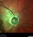
Submacular Hemorrhage - wet AMD470 views84 year old man with 20/200 from submacular hemorrhage. Vision improved to 20/32 with only Avastin therapy (no surgical intervention). The initial FAF is interesting because parts of the blood hyper auto fluoresce.
|
|

Pattern Dystrophy - Foveal Atrophy470 views73 year old female with vision loss - looks like macular hole
|
|

Basal Laminar Drusen - Cuticular Drusen - Equatorial Drusen - 70 Year Old Woman 469 views70 year old woman has had macular drusen since her thirties and she said her sister has a similar problem. Recently the vision in the left eye has substantially declined. OD 20/16, OS 10/200.
|
|
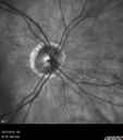
Submacular Hemorrhage - wet AMD469 views84 year old man with 20/200 from submacular hemorrhage. Vision improved to 20/32 with only Avastin therapy (no surgical intervention). The initial FAF is interesting because parts of the blood hyper auto fluoresce.
|
|

Chorioretinal Scar with Radiating Chorioretinal Folds468 views79-year-old woman has wet age-related macular degeneration in both eyes. The left eye had intravitreal injections of Avastin starting in May of 2009. Her vision remains good in the left eye and poor in the right eye.
VISUAL ACUITY: OD 8/200, OS 20/60.
|
|

Cuticular Drusen - 26 year old female - Glomerulonephritis468 viewsVA 20/25 OU. Images are over 4 years FA shows starry sky early images - This is because of loss of RPE over the drusen creating very small window defects.
|
|

Myopic CNVM in the left eye. New Classic subfoveal lesion468 views64 year old female. She had retinal detachment repair in the right eye in 1996 (25 years ago). The left eye has new distortion for a week or two.
VA 20/40 OU.
Left eye has a very small subfoveal CNVM
|
|
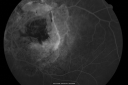
Angioid Streaks New Vision Change in Good Right Eye Treated with Lucentis467 views53-year-old man has angioid streaks. He has a macular scar in the left eye from choroidal neovasculari-zation that was treated with photodynamic laser 5-8 years ago. His vision in the right eye had been fine until about a week ago when he noticed temporally in the right eye an area that does not look normal and looks a little distorted.
VISUAL ACUITY: Vision OD is 20/30, OS is 20/400. Treatment was started within a month and patient responded well and is now 3 years out requiring every 3 month Lucentis
|
|
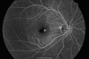
Macular Hemorrahge and Probable CNVM Right eye for 2 weeks VA 20/400 - 20 Year old467 views20-year-old man noticed vision loss in the right eye about two weeks ago and if anything it seems to be getting worse. He is a high myope. VISUAL ACUITY: OD 20/400
|
|

Myopic Macular Degeneration - Staphyloma - Recent Vision Loss Right Eye - Possible CNVM467 views83-year-old woman is a high myope with myopic macular degeneration. She had a retinal detachment repair in the right eye in 1968. I saw her in June of 2003. She had posterior colobomas and areas of pigment epithelial atrophy. She has noticed over the last six weeks substantial declining vision in the right eye. Prior to that, the vision in the two eyes was about the same. She uses Xalatan in the left eye and Alphagan in both eyes.
VISUAL ACUITY: Vision OD is 20/160, OS is 20/40
|
|

Basal Laminar Drusen - Cuticular Drusen - Equatorial Drusen - 70 Year Old Woman 467 views70 year old woman has had macular drusen since her thirties and she said her sister has a similar problem. Recently the vision in the left eye has substantially declined. OD 20/16, OS 10/200.
|
|

Cuticular Drusen (Basal Laminar Drusen) and Bilateral Chronic Subfoveal Fluid (Serous Retinal Detachment) unresponsive to monthly Avastin ICG OS467 views66-year-old man with bilateral subfoveal fluid unresponsive to one year of monthly avastin therapy.
OD 20/160, Pinhole 20/80. OS 20/63, Pinhole 20/50. OCT scan shows subretinal fluid with normal choroidal thickness. Fluorescein angiogram shows cuticular/basal laminar drusen. ICG shows no foci of choriocapillaris leakage.
|
|

Polypoidal Choroidal Vasculopathy Left Eye467 views74 year old man with vision loss to 20/100 over a few months. He improved to 20/20 with Lucentis. The color and ICG show the polyps.
|
|

Non-exudative wet AMD right eye (treatment naive quiescent)467 views82 year old man. ICG shows CNVM in the right eye. This stayed quiet for 3 years and then started leaking and responded well to Avastin.
|
|

Non-exudative wet AMD right eye (treatment naive quiescent)467 views82 year old man. ICG shows CNVM in the right eye. This stayed quiet for 3 years and then started leaking and responded well to Avastin.
|
|

Large and Extensive Macular Drusen467 views64 year old female with 20/25 vision taking vitamins in for a checkup.
|
|

Macula is Dry466 viewsWet AMD in right and better eye failed extend to 2 months but was able to stop therapy once he started on systemic Avastin for cancer. The last image shows the macula dry 9 months without local therapy. The left eye has a lamellar macular hole.
|
|
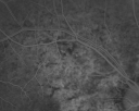
Angioid Streaks New Vision Change in Good Right Eye Treated with Lucentis465 views53-year-old man has angioid streaks. He has a macular scar in the left eye from choroidal neovasculari-zation that was treated with photodynamic laser 5-8 years ago. His vision in the right eye had been fine until about a week ago when he noticed temporally in the right eye an area that does not look normal and looks a little distorted.
VISUAL ACUITY: Vision OD is 20/30, OS is 20/400. Treatment was started within a month and patient responded well and is now 3 years out requiring every 3 month Lucentis
|
|

Basal Laminar Drusen - Cuticular Drusen - Equatorial Drusen - 70 Year Old Woman Line Scan Left Eye465 views70 year old woman has had macular drusen since her thirties and she said her sister has a similar problem. Recently the vision in the left eye has substantially declined. OD 20/16, OS 10/200.
|
|

465 viewsIndocyanine Green Angiogram
|
|

Angioid Streaks - CNVM OS with vision loss465 views48 year old soccer player. Prior trauma OS (with soccer ball). Vision loss for 3 weeks to 20/60 - with Lucentis vision improved to 20/30
|
|

Concentric Geographic Atrophy465 views76 year old man Gradual vision loss
20/32 OD; 20/40 OS
No medicines, Non-smoker
Working and Driving
|
|

Choroidal Neovascular Membrane following Prior Central Serous Chorioretinopathy - OCT-A465 views67 year old female with CSR in the left eye first 20 years ago. She was managed with out therapy and has had a few episodes since. Now ther eis distortion in the left eye for a few days. She had a steroid shot in her shoulder 5 months ago.
VA 20/20 OD, 20/50 OS- ICG and OCT-A confirm CNVM in the left eye
|
|
464 views
|
|

Submacular Hemorrhage - wet AMD464 views84 year old man with 20/200 from submacular hemorrhage. Vision improved to 20/32 with only Avastin therapy (no surgical intervention). The initial FAF is interesting because parts of the blood hyper auto fluoresce.
|
|

Submacular Hemorrhage - wet AMD464 views84 year old man with 20/200 from submacular hemorrhage. Vision improved to 20/32 with only Avastin therapy (no surgical intervention). The initial FAF is interesting because parts of the blood hyper auto fluoresce.
|
|

Pattern Dystrophy - Adult Vitellifrom (Best)464 views71 year old female - lesions nasal to the fovea in both eyes. (20/40 OU)
|
|

Outer Retinal Tubulation464 views78 year old man chronic wet AMD - 20/200
|
|

Outer Retinal Tubulation464 views78 year old man chronic wet AMD - 20/200
|
|

Adult Pseudovitelliform Macular Dystrophy464 views89 year old female states dark smudge on ceiling during night time came and went. Decreased vision in the left eye
VA OD: Dcc20/32
VA OS: Dcc20/63
|
|
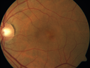
Choroidal Neovascular Membrane following Prior Central Serous Chorioretinopathy - OCT-A464 views67 year old female with CSR in the left eye first 20 years ago. She was managed with out therapy and has had a few episodes since. Now ther eis distortion in the left eye for a few days. She had a steroid shot in her shoulder 5 months ago.
VA 20/20 OD, 20/50 OS- ICG and OCT-A confirm CNVM in the left eye
|
|

Macular Hemorrahge and Probable CNVM Right eye for 2 weeks VA 20/400 - 20 Year old463 views20-year-old man noticed vision loss in the right eye about two weeks ago and if anything it seems to be getting worse. He is a high myope. VISUAL ACUITY: OD 20/400
|
|

Wet AMD OD and Lamellar Macular hole OS463 viewsWet AMD in right and better eye failed extend to 2 months but was able to stop therapy once he started on systemic Avastin for cancer. The last image shows the macula dry 9 months without local therapy. The left eye has a lamellar macular hole.
|
|

Asymptomatic wet AMD - Subfoveal CNM both eyes463 views82 year old woman - 20/25 OU. Images show occult subfoveal CNVM both eyes best seen on late ICG. The left eye also has probable polypoidal choroidal vasculopathy. No treatment was recommended on the first visit - follow-up at 1 month.
|
|

Outer Retinal Tubulation463 views78 year old man chronic wet AMD - 20/200
|
|

Concentric Geographic Atrophy - 76 year old man463 viewsGradual vision loss, worse at night. VA 20/40 OU.
|
|

Choriodal Neovascular Membrane - Subfoveal - Pseudo-Retinitis Pigmentosa - 50 years post Retinal Detachment Repair462 views86-year-old with a history of decreased vision in the right eye. The patient reports she has lost vision in the right eye over the past two months or so. She reports the vision was always compromised in that eye since retinal detachment surgery 50 years ago with a scleral buckler. Vision dropped from 20/50 to 20/200 right eye in 2 months.
|
|

New wet AMD OS with subretinal hemorrhage and recent vision loss, Scar OD462 views
|
|

Submacular Hemorrhage - wet AMD462 views84 year old man with 20/200 from submacular hemorrhage. Vision improved to 20/32 with only Avastin therapy (no surgical intervention). The initial FAF is interesting because parts of the blood hyper auto fluoresce.
|
|

Polypoidal Choroidal Vasculopathy Left Eye462 views74 year old man with vision loss to 20/100 over a few months. He improved to 20/20 with Lucentis. The color and ICG show the polyps.
|
|

Adult Pseudovitelliform Macular Dystrophy462 views89 year old female states dark smudge on ceiling during night time came and went. Decreased vision in the left eye
VA OD: Dcc20/32
VA OS: Dcc20/63
|
|

Wet Macular Degeneration Left Eye with Gutter (atypical) and Geographic Atrophy Right Eye461 views80-year-old man has wet age-related macular degeneration in his left eye. He has an atypical angiogram and that there is a gutter under the choroidal neovascular membrane suggesting still leakage of subretinal fluid. OS 20/400.
|
|

New wet AMD OS with subretinal hemorrhage and recent vision loss, Scar OD461 views
|
|

Outer Retinal Tubulation461 views78 year old man chronic wet AMD - 20/200
|
|
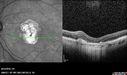
Outer Retinal Tubulation461 views78 year old man chronic wet AMD - 20/200
|
|
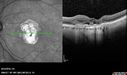
Outer Retinal Tubulation461 views78 year old man chronic wet AMD - 20/200
|
|

Myopic CNVM in the left eye. New Classic subfoveal lesion461 views64 year old female. She had retinal detachment repair in the right eye in 1996 (25 years ago). The left eye has new distortion for a week or two.
VA 20/40 OU.
Left eye has a very small subfoveal CNVM
|
|

Polypoidal Choroidal Vasculopathy Right Eye - Disciform Scar Left Eye460 views84-year-old woman has polypoidal choroidal vasculopathy in both eyes with macular degeneration. She has macular scars in both eyes and she remains with remarkably good vision in the right eye despite the scarring.
VISUAL ACUITY: OD 20/30, OS 1/200
Avastin Rx OD
|
|

Polypoidal Choroidal Vasculopathy Right Eye - Disciform Scar Left Eye460 views84-year-old woman has polypoidal choroidal vasculopathy in both eyes with macular degeneration. She has macular scars in both eyes and she remains with remarkably good vision in the right eye despite the scarring.
VISUAL ACUITY: OD 20/30, OS 1/200
Avastin Rx OD
|
|

New wet AMD OS with subretinal hemorrhage and recent vision loss, Scar OD460 views
|
|
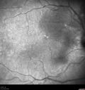
IPCV - idiopathic polypoidal choroidal vasculopathy - juxtapapillary CNVM with hemorrhage both eyes (also choroidal nevus right eye) - Subretinal hemorrhage both eyes460 views86 year old woman with shadow in vision OD and new hemmorhage OU.
|
|
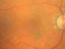
Non-exudative wet AMD right eye (treatment naive quiescent)460 views82 year old man. ICG shows CNVM in the right eye. This stayed quiet for 3 years and then started leaking and responded well to Avastin.
|
|

Concentric Geographic Atrophy460 views76 year old man Gradual vision loss
20/32 OD; 20/40 OS
No medicines, Non-smoker
Working and Driving
|
|

Myopic CNVM in the left eye. New Classic subfoveal lesion460 views64 year old female. She had retinal detachment repair in the right eye in 1996 (25 years ago). The left eye has new distortion for a week or two.
VA 20/40 OU.
Left eye has a very small subfoveal CNVM
|
|

Choriodal Neovascular Membrane - Subfoveal - Pseudo-Retinitis Pigmentosa - 50 years post Retinal Detachment Repair459 views86-year-old with a history of decreased vision in the right eye. The patient reports she has lost vision in the right eye over the past two months or so. She reports the vision was always compromised in that eye since retinal detachment surgery 50 years ago with a scleral buckler. Vision dropped from 20/50 to 20/200 right eye in 2 months.
|
|

Submacular Hemorrhage - wet AMD459 views84 year old man with 20/200 from submacular hemorrhage. Vision improved to 20/32 with only Avastin therapy (no surgical intervention). The initial FAF is interesting because parts of the blood hyper auto fluoresce.
|
|

Outer Retinal Tubulation459 views78 year old man chronic wet AMD - 20/200
|
|

Non-exudative wet AMD right eye (treatment naive quiescent)459 views82 year old man. ICG shows CNVM in the right eye. This stayed quiet for 3 years and then started leaking and responded well to Avastin.
|
|

Choroidal Neovascular Membrane following Prior Central Serous Chorioretinopathy - OCT-A459 views67 year old female with CSR in the left eye first 20 years ago. She was managed with out therapy and has had a few episodes since. Now ther eis distortion in the left eye for a few days. She had a steroid shot in her shoulder 5 months ago.
VA 20/20 OD, 20/50 OS- ICG and OCT-A confirm CNVM in the left eye
|
|

Angioid Streaks - CNVM OS with vision loss458 views48 year old soccer player. Prior trauma OS (with soccer ball). Vision loss for 3 weeks to 20/60 - with Lucentis vision improved to 20/30
|
|
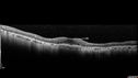
Concentric Geographic Atrophy458 views76 year old man Gradual vision loss
20/32 OD; 20/40 OS
No medicines, Non-smoker
Working and Driving
|
|

Concentric Geographic Atrophy - 76 year old man458 viewsGradual vision loss, worse at night. VA 20/40 OU.
|
|

Choroidal Neovascular Membrane following Prior Central Serous Chorioretinopathy - OCT-A458 views67 year old female with CSR in the left eye first 20 years ago. She was managed with out therapy and has had a few episodes since. Now ther eis distortion in the left eye for a few days. She had a steroid shot in her shoulder 5 months ago.
VA 20/20 OD, 20/50 OS- ICG and OCT-A confirm CNVM in the left eye
|
|

Wet AMD controlled with Avastin 20/60 Vision457 views79-year-old woman has wet age-related macular degeneration in both eyes. The left eye had intravitreal injections of Avastin starting in May of 2009. Her vision remains good in the left eye and poor in the right eye.
VISUAL ACUITY: OD 8/200, OS 20/60.
|
|

Asymptomatic wet AMD - Subfoveal CNM both eyes457 views82 year old woman - 20/25 OU. Images show occult subfoveal CNVM both eyes best seen on late ICG. The left eye also has probable polypoidal choroidal vasculopathy. No treatment was recommended on the first visit - follow-up at 1 month.
|
|

IPCV - idiopathic polypoidal choroidal vasculopathy - juxtapapillary CNVM with hemorrhage both eyes (also choroidal nevus right eye) - Subretinal hemorrhage both eyes456 views86 year old woman with shadow in vision OD and new hemmorhage OU. ICG angiogram
|
|

IPCV - idiopathic polypoidal choroidal vasculopathy - juxtapapillary CNVM with hemorrhage both eyes (also choroidal nevus right eye) - Subretinal hemorrhage both eyes456 views86 year old woman with shadow in vision OD and new hemmorhage OU.
|
|

Pattern Dystrophy - Adult Vitellifrom (Best)456 views71 year old female - lesions nasal to the fovea in both eyes. (20/40 OU)
|
|

Rapid macular atrophy with anti-VEGF therapy for myopic CNVM456 views64 year old female with 20/32 vision and distortion in the left and better eye. She was treated with monthly lucentis and her vision declined to 20/100. Medicine was stopped and she improved a little to 20/80.
|
|

Choriodal Neovascular Membrane - Subfoveal - Pseudo-Retinitis Pigmentosa - 50 years post Retinal Detachment Repair455 views86-year-old with a history of decreased vision in the right eye. The patient reports she has lost vision in the right eye over the past two months or so. She reports the vision was always compromised in that eye since retinal detachment surgery 50 years ago with a scleral buckler. Vision dropped from 20/50 to 20/200 right eye in 2 months.
|
|
| 2694 files on 27 page(s) |
 |
|
|
|
|
13 | |
|
|
|
|


