Les plus populaires - Branch Retinal Artery Occlusion
|

Asymptomatic Hollenhorst Plaque - Cholesterol Embolisvu 2703 fois71-year-old woman is not noticing any vision change. She does take pressure-lowering drops for glaucoma, both Timolol and Xalatan.
VISUAL ACUITY: Vision OD is 20/30, OS is 20/16
|
|

Inferotemporal and Inferonasal BRAOvu 1640 foisBranch Retinal Artery Occlusions - Multiple
|
|

Asymptomatic Hollenhorst Plaque - Cholesterol Embolisvu 1359 fois71-year-old woman is not noticing any vision change. She does take pressure-lowering drops for glaucoma, both Timolol and Xalatan.
VISUAL ACUITY: Vision OD is 20/30, OS is 20/16
|
|

Asymptomatic Hollenhorst Plaque - Cholesterol Embolisvu 1290 fois71-year-old woman is not noticing any vision change. She does take pressure-lowering drops for glaucoma, both Timolol and Xalatan.
VISUAL ACUITY: Vision OD is 20/30, OS is 20/16
|
|
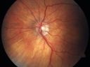
Cilioretinal artery occlusion and Ophthalmic Artery Occlusion with Relative Sparing of the Central Retinal Arteryvu 1268 fois80-year-old woman who says she was looking in the mirror, covered her right eye and noticed she could not see well out of the left eye. That just happened yesterday. Her last eye check was a few years ago. She also had a carotid endarterectomy on the right side in 2006 shortly after coronary bypass surgery. OD is 20/40, OS is 20/200.
|
|

Old BRAO twig with plaquevu 1068 fois81-year-old woman Her left eye is amblyopic. Her right eye was doing fine until about last Sunday, which is five days ago. She had a change in vision where for five minutes she saw a shadow in her side vision out of the right eye. She describes it as being more up into the left. It lasted for just a brief time and then went away. She has had atrial fibrillation since January of 2008. She is on Coumadin for that. She is diabetic for eleven years. OD is 20/25, OS is 20/60. IOP: OD 15, OS 18.
|
|
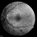
14.5 secondsvu 1057 foisCorresponding FA for Cilioretinal artery occlusion with early venous stasis retinopathy
|
|

BRAO and PAMMvu 1024 foisLayers of retina are thickened showing ischemia to the middle retina (deep retinal capillary plexus)
|
|

Sectoral Optic Atrophy from an old Branch Retinal Artery Occlusion vu 949 fois74-year-old woman had cataract surgery 3 months ago. Her vision in the right eye has been abnormal since. She notices a shadow in the inferior vision in the right eye. OD is 20/60, OS is 20/50
|
|

Cilioretinal artery occlusion and Ophthalmic Artery Occlusion with Relative Sparing of the Central Retinal Arteryvu 924 fois80-year-old woman who says she was looking in the mirror, covered her right eye and noticed she could not see well out of the left eye. That just happened yesterday. Her last eye check was a few years ago. She also had a carotid endarterectomy on the right side in 2006 shortly after coronary bypass surgery. OD is 20/40, OS is 20/200.
|
|

Branch Retinal Arterial Occlusion 20/20 Vision - Symptoms for 1 Dayvu 890 fois
|
|

Cilioretinal artery occlusion and Ophthalmic Artery Occlusion with Relative Sparing of the Central Retinal Arteryvu 886 fois80-year-old woman who says she was looking in the mirror, covered her right eye and noticed she could not see well out of the left eye. That just happened yesterday. Her last eye check was a few years ago. She also had a carotid endarterectomy on the right side in 2006 shortly after coronary bypass surgery. OD is 20/40, OS is 20/200.
|
|

Sectoral Optic Atrophy from an old Branch Retinal Artery Occlusion vu 880 fois74-year-old woman had cataract surgery 3 months ago. Her vision in the right eye has been abnormal since. She notices a shadow in the inferior vision in the right eye. OD is 20/60, OS is 20/50
|
|

Cilioretinal artery occlusion and Ophthalmic Artery Occlusion with Relative Sparing of the Central Retinal Arteryvu 871 fois80-year-old woman who says she was looking in the mirror, covered her right eye and noticed she could not see well out of the left eye. That just happened yesterday. Her last eye check was a few years ago. She also had a carotid endarterectomy on the right side in 2006 shortly after coronary bypass surgery. OD is 20/40, OS is 20/200.
|
|

Sectoral Optic Atrophy from an old Branch Retinal Artery Occlusion vu 867 fois74-year-old woman had cataract surgery 3 months ago. Her vision in the right eye has been abnormal since. She notices a shadow in the inferior vision in the right eye. OD is 20/60, OS is 20/50
|
|

Cilioretinal artery occlusion and Ophthalmic Artery Occlusion with Relative Sparing of the Central Retinal Arteryvu 862 fois80-year-old woman who says she was looking in the mirror, covered her right eye and noticed she could not see well out of the left eye. That just happened yesterday. Her last eye check was a few years ago. She also had a carotid endarterectomy on the right side in 2006 shortly after coronary bypass surgery. OD is 20/40, OS is 20/200.
|
|

BRAO and PAMMvu 816 fois
|
|

Sectoral Optic Atrophy from an old Branch Retinal Artery Occlusion vu 812 fois74-year-old woman had cataract surgery 3 months ago. Her vision in the right eye has been abnormal since. She notices a shadow in the inferior vision in the right eye. OD is 20/60, OS is 20/50
|
|

Cilioretinal artery occlusion and Ophthalmic Artery Occlusion with Relative Sparing of the Central Retinal Arteryvu 805 fois80-year-old woman who says she was looking in the mirror, covered her right eye and noticed she could not see well out of the left eye. That just happened yesterday. Her last eye check was a few years ago. She also had a carotid endarterectomy on the right side in 2006 shortly after coronary bypass surgery. OD is 20/40, OS is 20/200.
|
|

Cilioretinal artery occlusion and Ophthalmic Artery Occlusion with Relative Sparing of the Central Retinal Arteryvu 796 fois80-year-old woman who says she was looking in the mirror, covered her right eye and noticed she could not see well out of the left eye. That just happened yesterday. Her last eye check was a few years ago. She also had a carotid endarterectomy on the right side in 2006 shortly after coronary bypass surgery. OD is 20/40, OS is 20/200.
|
|

Branch Retinal Arterial Occlusion 20/20 Vision - Symptoms for 1 Dayvu 775 fois
|
|

Cilioretinal artery occlusion and Ophthalmic Artery Occlusion with Relative Sparing of the Central Retinal Arteryvu 772 fois80-year-old woman who says she was looking in the mirror, covered her right eye and noticed she could not see well out of the left eye. That just happened yesterday. Her last eye check was a few years ago. She also had a carotid endarterectomy on the right side in 2006 shortly after coronary bypass surgery. OD is 20/40, OS is 20/200.
|
|
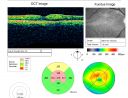
Branch Retinal Arterial Occlusion 20/20 Vision - Symptoms for 1 Dayvu 764 fois75-year-old man was seen in the office on 4/6/2010. He noticed a sudden vision change in the right eye yesterday morning. 20/20 Vision both Eyes
|
|

Old BRAO twig with plaquevu 754 fois81-year-old woman Her left eye is amblyopic. Her right eye was doing fine until about last Sunday, which is five days ago. She had a change in vision where for five minutes she saw a shadow in her side vision out of the right eye. She describes it as being more up into the left. It lasted for just a brief time and then went away. She has had atrial fibrillation since January of 2008. She is on Coumadin for that. She is diabetic for eleven years. OD is 20/25, OS is 20/60. IOP: OD 15, OS 18.
|
|

Old BRAO twig with plaquevu 754 fois81-year-old woman Her left eye is amblyopic. Her right eye was doing fine until about last Sunday, which is five days ago. She had a change in vision where for five minutes she saw a shadow in her side vision out of the right eye. She describes it as being more up into the left. It lasted for just a brief time and then went away. She has had atrial fibrillation since January of 2008. She is on Coumadin for that. She is diabetic for eleven years. OD is 20/25, OS is 20/60. IOP: OD 15, OS 18.
|
|

Acute Branch Retinal Artery Occlusion Left Eyevu 740 foisThis 84 year old woman had an old BRVO in the right eye and saw a shadow for 2 days in the left eye. She has a superotemporal BRAO. The photos show retinal whitening, the OCT shows inner retinal edema and the FA shows poor circulation.
|
|

Cilioretinal artery occlusion and Ophthalmic Artery Occlusion with Relative Sparing of the Central Retinal Arteryvu 734 fois80-year-old woman who says she was looking in the mirror, covered her right eye and noticed she could not see well out of the left eye. That just happened yesterday. Her last eye check was a few years ago. She also had a carotid endarterectomy on the right side in 2006 shortly after coronary bypass surgery. OD is 20/40, OS is 20/200.
|
|

Cilioretinal artery occlusion and Ophthalmic Artery Occlusion with Relative Sparing of the Central Retinal Arteryvu 732 fois80-year-old woman who says she was looking in the mirror, covered her right eye and noticed she could not see well out of the left eye. That just happened yesterday. Her last eye check was a few years ago. She also had a carotid endarterectomy on the right side in 2006 shortly after coronary bypass surgery. OD is 20/40, OS is 20/200.
|
|

Cilioretinal artery occlusion and Ophthalmic Artery Occlusion with Relative Sparing of the Central Retinal Arteryvu 731 fois80-year-old woman who says she was looking in the mirror, covered her right eye and noticed she could not see well out of the left eye. That just happened yesterday. Her last eye check was a few years ago. She also had a carotid endarterectomy on the right side in 2006 shortly after coronary bypass surgery. OD is 20/40, OS is 20/200.
|
|

Cilioretinal artery occlusion and Ophthalmic Artery Occlusion with Relative Sparing of the Central Retinal Arteryvu 729 fois80-year-old woman who says she was looking in the mirror, covered her right eye and noticed she could not see well out of the left eye. That just happened yesterday. Her last eye check was a few years ago. She also had a carotid endarterectomy on the right side in 2006 shortly after coronary bypass surgery. OD is 20/40, OS is 20/200.
|
|

Sectoral Optic Atrophy from an old Branch Retinal Artery Occlusion vu 726 fois74-year-old woman had cataract surgery 3 months ago. Her vision in the right eye has been abnormal since. She notices a shadow in the inferior vision in the right eye. OD is 20/60, OS is 20/50
|
|

Sectoral Optic Atrophy from an old Branch Retinal Artery Occlusion vu 710 fois74-year-old woman had cataract surgery 3 months ago. Her vision in the right eye has been abnormal since. She notices a shadow in the inferior vision in the right eye. OD is 20/60, OS is 20/50
|
|

Sectoral Optic Atrophy from an old Branch Retinal Artery Occlusion vu 705 fois74-year-old woman had cataract surgery 3 months ago. Her vision in the right eye has been abnormal since. She notices a shadow in the inferior vision in the right eye. OD is 20/60, OS is 20/50
|
|

Cilioretinal artery occlusionvu 699 foisCilioretinal artery occlusion with early venous stasis retinopathy
|
|

BRAO and PAMMvu 697 fois
|
|

Branch Retinal Arterial Occlusion 20/20 Vision - Symptoms for 1 Dayvu 695 fois
|
|

Cilioretinal artery occlusion and Ophthalmic Artery Occlusion with Relative Sparing of the Central Retinal Arteryvu 695 fois80-year-old woman who says she was looking in the mirror, covered her right eye and noticed she could not see well out of the left eye. That just happened yesterday. Her last eye check was a few years ago. She also had a carotid endarterectomy on the right side in 2006 shortly after coronary bypass surgery. OD is 20/40, OS is 20/200.
|
|
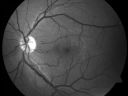
Branch Retinal Arterial Occlusion 20/20 Vision - Symptoms for 1 Dayvu 689 fois
|
|

Sectoral Optic Atrophy from an old Branch Retinal Artery Occlusion vu 686 fois74-year-old woman had cataract surgery 3 months ago. Her vision in the right eye has been abnormal since. She notices a shadow in the inferior vision in the right eye. OD is 20/60, OS is 20/50
|
|

Acute Branch Retinal Artery Occlusion Left Eyevu 684 foisThis 84 year old woman had an old BRVO in the right eye and saw a shadow for 2 days in the left eye. She has a superotemporal BRAO. The photos show retinal whitening, the OCT shows inner retinal edema and the FA shows poor circulation.
|
|

BRAO and PAMMvu 677 fois
|
|

Branch Retinal Arterial Occlusion Right Eye - Atheromata - Both Eyesvu 677 fois83 year old woman with no symptoms - VA 20/40 in both eyes. Right eye has ST BRAO with silver wiring. Left eye has superotemporal atheromata
|
|

Branch Retinal Arterial Occlusion 20/20 Vision - Symptoms for 1 Dayvu 676 fois75-year-old man was seen in the office on 4/6/2010. He noticed a sudden vision change in the right eye yesterday morning. 20/20 Vision both Eyes
|
|

Latevu 675 foisCorresponding FA for Cilioretinal artery occlusion with early venous stasis retinopathy
|
|

Acute Branch Retinal Artery Occlusion Left Eyevu 669 foisThis 84 year old woman had an old BRVO in the right eye and saw a shadow for 2 days in the left eye. She has a superotemporal BRAO. The photos show retinal whitening, the OCT shows inner retinal edema and the FA shows poor circulation.
|
|

Acute Branch Retinal Artery Occlusion Left Eyevu 669 foisThis 84 year old woman had an old BRVO in the right eye and saw a shadow for 2 days in the left eye. She has a superotemporal BRAO. The photos show retinal whitening, the OCT shows inner retinal edema and the FA shows poor circulation.
|
|

BRAO and PAMMvu 668 fois
|
|

BRAO and PAMMvu 668 fois
|
|

Acute Branch Retinal Artery Occlusion Left Eyevu 668 foisThis 84 year old woman had an old BRVO in the right eye and saw a shadow for 2 days in the left eye. She has a superotemporal BRAO. The photos show retinal whitening, the OCT shows inner retinal edema and the FA shows poor circulation.
|
|

Branch Retinal Arterial Occlusion 20/20 Vision - Symptoms for 1 Dayvu 667 fois
|
|

BRAO and PAMMvu 667 foisGray paracentral lesion from BRAO is a PAMM lesion
|
|
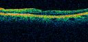
Sectoral Optic Atrophy from an old Branch Retinal Artery Occlusion vu 665 fois74-year-old woman had cataract surgery 3 months ago. Her vision in the right eye has been abnormal since. She notices a shadow in the inferior vision in the right eye. OD is 20/60, OS is 20/50
|
|

Acute Branch Retinal Artery Occlusion Left Eyevu 659 foisThis 84 year old woman had an old BRVO in the right eye and saw a shadow for 2 days in the left eye. She has a superotemporal BRAO. The photos show retinal whitening, the OCT shows inner retinal edema and the FA shows poor circulation.
|
|

Acute Branch Retinal Artery Occlusion Left Eyevu 652 foisThis 84 year old woman had an old BRVO in the right eye and saw a shadow for 2 days in the left eye. She has a superotemporal BRAO. The photos show retinal whitening, the OCT shows inner retinal edema and the FA shows poor circulation.
|
|

Bilateral Branch Retinal Artery Occlusion - Intraarterial (hollenhorst) plaques visiblevu 651 fois67 year old with acute visual field loss in each eye. Vision is 20/25 right eye 20/32 left eye. Images show intra-arterial plaques in each eye. The right eye on the optic nerve and the left eye in a branch retinal artery.
|
|
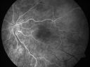
Cilioretinal artery occlusion and Ophthalmic Artery Occlusion with Relative Sparing of the Central Retinal Arteryvu 649 fois80-year-old woman who says she was looking in the mirror, covered her right eye and noticed she could not see well out of the left eye. That just happened yesterday. Her last eye check was a few years ago. She also had a carotid endarterectomy on the right side in 2006 shortly after coronary bypass surgery. OD is 20/40, OS is 20/200.
|
|

Acute Branch Retinal Artery Occlusion Left Eyevu 649 foisThis 84 year old woman had an old BRVO in the right eye and saw a shadow for 2 days in the left eye. She has a superotemporal BRAO. The photos show retinal whitening, the OCT shows inner retinal edema and the FA shows poor circulation.
|
|

Choroidal Nevusvu 647 fois
|
|

BRAO and PAMMvu 646 fois
|
|

Acute Branch Retinal Artery Occlusion Left Eyevu 645 foisThis 84 year old woman had an old BRVO in the right eye and saw a shadow for 2 days in the left eye. She has a superotemporal BRAO. The photos show retinal whitening, the OCT shows inner retinal edema and the FA shows poor circulation.
|
|

Branch Retinal Arterial Occlusion 20/20 Vision - Symptoms for 1 Dayvu 643 fois75-year-old man was seen in the office on 4/6/2010. He noticed a sudden vision change in the right eye yesterday morning. 20/20 Vision both Eyes
|
|

Holenhorst Plaque - Small and asymptomaticvu 640 fois72 year old woman with mild dry ARMD. VA 20/40 OU. Asymptomatic plaque noted in the right eye.
|
|

Branch Retinal Arterial Occlusion 20/20 Vision - Symptoms for 1 Dayvu 637 fois
|
|

Intraluminal Plaque Retinal Vessel vu 637 fois69 year old female referred with atypical plaque in the right retina. Her vision is normal. The plaque looks to be adjacent to the blood column visible on the images and the SC OCT shows it to be in the vessel wall.
|
|

Acute Branch Retinal Artery Occlusion Left Eyevu 632 foisThis 84 year old woman had an old BRVO in the right eye and saw a shadow for 2 days in the left eye. She has a superotemporal BRAO. The photos show retinal whitening, the OCT shows inner retinal edema and the FA shows poor circulation.
|
|

BRAO and PAMMvu 630 fois
|
|

BRAO and PAMMvu 625 fois
|
|

BRAO and PAMMvu 623 fois
|
|

Chronic BRAO with Hemorrhages - Sclerosed Vesselvu 623 fois95 year old female with blurred vision in both eyes in for a checkup. History of perpapillary CNVM in the left eye 2 years prior not treated. History of stroke with multiple emboli 1 year ago.
Vision 20/32 OD, 20/40 OS
|
|

Branch Retinal Arterial Occlusion 20/20 Vision - Symptoms for 1 Dayvu 622 fois75-year-old man was seen in the office on 4/6/2010. He noticed a sudden vision change in the right eye yesterday morning. 20/20 Vision both Eyes
|
|

Acute Branch Retinal Artery Occlusion Left Eyevu 620 foisThis 84 year old woman had an old BRVO in the right eye and saw a shadow for 2 days in the left eye. She has a superotemporal BRAO. The photos show retinal whitening, the OCT shows inner retinal edema and the FA shows poor circulation.
|
|

BRAO and PAMMvu 619 fois
|
|

Acute Branch Retinal Artery Occlusion Left Eyevu 616 foisThis 84 year old woman had an old BRVO in the right eye and saw a shadow for 2 days in the left eye. She has a superotemporal BRAO. The photos show retinal whitening, the OCT shows inner retinal edema and the FA shows poor circulation.
|
|

Cilioretinal artery occlusion and Ophthalmic Artery Occlusion with Relative Sparing of the Central Retinal Arteryvu 614 fois80-year-old woman who says she was looking in the mirror, covered her right eye and noticed she could not see well out of the left eye. That just happened yesterday. Her last eye check was a few years ago. She also had a carotid endarterectomy on the right side in 2006 shortly after coronary bypass surgery. OD is 20/40, OS is 20/200.
|
|

Subtle Branch Retinal Artery Occlusionvu 613 fois68 year old man with shadow in the left eye yesterday. IR and OCT show subtle retinal swelling. FA is normal
|
|

Branch Retinal Artery Occlusionvu 611 foisAcute Inferotemporal Occlusion Right Eye - FA shows delayed circulation, OCT shows edema of superficial layers of retina
|
|

Branch Retinal Arterial Occlusion Right Eye - Atheromata - Both Eyesvu 611 fois83 year old woman with no symptoms - VA 20/40 in both eyes. Right eye has ST BRAO with silver wiring. Left eye has superotemporal atheromata
|
|

BRAO and PAMMvu 608 fois
|
|
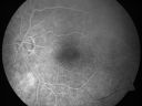
Cilioretinal artery occlusion and Ophthalmic Artery Occlusion with Relative Sparing of the Central Retinal Arteryvu 606 fois80-year-old woman who says she was looking in the mirror, covered her right eye and noticed she could not see well out of the left eye. That just happened yesterday. Her last eye check was a few years ago. She also had a carotid endarterectomy on the right side in 2006 shortly after coronary bypass surgery. OD is 20/40, OS is 20/200.
|
|

Branch Retinal Arterial Occlusion Right Eye - Atheromata - Both Eyesvu 604 fois83 year old woman with no symptoms - VA 20/40 in both eyes. Right eye has ST BRAO with silver wiring. Left eye has superotemporal atheromata
|
|
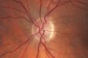
Sectoral Optic Atrophy from an old Branch Retinal Artery Occlusion vu 598 fois74-year-old woman had cataract surgery 3 months ago. Her vision in the right eye has been abnormal since. She notices a shadow in the inferior vision in the right eye. OD is 20/60, OS is 20/50
|
|

Cilioretinal artery occlusion and Ophthalmic Artery Occlusion with Relative Sparing of the Central Retinal Arteryvu 597 fois80-year-old woman who says she was looking in the mirror, covered her right eye and noticed she could not see well out of the left eye. That just happened yesterday. Her last eye check was a few years ago. She also had a carotid endarterectomy on the right side in 2006 shortly after coronary bypass surgery. OD is 20/40, OS is 20/200.
|
|

Branch Retinal Arterial Occlusion Right Eye - Atheromata - Both Eyesvu 597 fois83 year old woman with no symptoms - VA 20/40 in both eyes. Right eye has ST BRAO with silver wiring. Left eye has superotemporal atheromata
|
|

Branch Retinal Arterial Occlusion Right Eye - Atheromata - Both Eyesvu 597 fois83 year old woman with no symptoms - VA 20/40 in both eyes. Right eye has ST BRAO with silver wiring. Left eye has superotemporal atheromata
|
|

BRAO and PAMMvu 596 fois
|
|
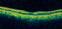
Cilioretinal artery occlusion and Ophthalmic Artery Occlusion with Relative Sparing of the Central Retinal Arteryvu 595 fois80-year-old woman who says she was looking in the mirror, covered her right eye and noticed she could not see well out of the left eye. That just happened yesterday. Her last eye check was a few years ago. She also had a carotid endarterectomy on the right side in 2006 shortly after coronary bypass surgery. OD is 20/40, OS is 20/200.
|
|

Bilateral Branch Retinal Artery Occlusion - Intraarterial (hollenhorst) plaques visiblevu 592 fois67 year old with acute visual field loss in each eye. Vision is 20/25 right eye 20/32 left eye. Images show intra-arterial plaques in each eye. The right eye on the optic nerve and the left eye in a branch retinal artery.
|
|
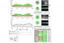
Sectoral Optic Atrophy from an old Branch Retinal Artery Occlusion vu 591 fois74-year-old woman had cataract surgery 3 months ago. Her vision in the right eye has been abnormal since. She notices a shadow in the inferior vision in the right eye. OD is 20/60, OS is 20/50
|
|

Bilateral Branch Retinal Artery Occlusion - Intraarterial (hollenhorst) plaques visiblevu 590 fois67 year old with acute visual field loss in each eye. Vision is 20/25 right eye 20/32 left eye. Images show intra-arterial plaques in each eye. The right eye on the optic nerve and the left eye in a branch retinal artery.
|
|

Branch Retinal Arterial Occlusion Right Eye - Atheromata - Both Eyesvu 586 fois83 year old woman with no symptoms - VA 20/40 in both eyes. Right eye has ST BRAO with silver wiring. Left eye has superotemporal atheromata
|
|
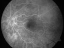
Cilioretinal artery occlusion and Ophthalmic Artery Occlusion with Relative Sparing of the Central Retinal Arteryvu 585 fois80-year-old woman who says she was looking in the mirror, covered her right eye and noticed she could not see well out of the left eye. That just happened yesterday. Her last eye check was a few years ago. She also had a carotid endarterectomy on the right side in 2006 shortly after coronary bypass surgery. OD is 20/40, OS is 20/200.
|
|

Branch Retinal Artery Occlusionvu 585 foisAcute Inferotemporal Occlusion Right Eye - FA shows delayed circulation, OCT shows edema of superficial layers of retina
|
|

Cardiac Catheterization induced Cilioretinal Artery Occlusion - Paracentral Acute Middle Maculopathyvu 585 fois69 year old female with sudden vision loss on completing cardiac catheterization. The SD OCT shows a PAMM lesion and the lesion looks gray on color imaging. There is delayed filling of the cilioretinal artery.
|
|

Branch Retinal Artery Occlusionvu 584 foisAcute Inferotemporal Occlusion Right Eye - FA shows delayed circulation, OCT shows edema of superficial layers of retina
|
|

Branch Retinal Arterial Occlusion 20/20 Vision - Symptoms for 1 Dayvu 582 fois75-year-old man was seen in the office on 4/6/2010. He noticed a sudden vision change in the right eye yesterday morning. 20/20 Vision both Eyes
|
|

Subtle Branch Retinal Artery Occlusionvu 581 fois68 year old man with shadow in the left eye yesterday. IR and OCT show subtle retinal swelling. FA is normal
|
|

Branch Retinal Arterial Occlusion 20/20 Vision - Symptoms for 1 Dayvu 580 fois
|
|

Branch Retinal Artery Occlusionvu 580 foisAcute Inferotemporal Occlusion Right Eye - FA shows delayed circulation, OCT shows edema of superficial layers of retina
|
|

PAMM with Follow-upvu 579 fois
|
|

Chronic BRAO with Hemorrhages - Sclerosed Vesselvu 578 fois95 year old female with blurred vision in both eyes in for a checkup. History of perpapillary CNVM in the left eye 2 years prior not treated. History of stroke with multiple emboli 1 year ago.
Vision 20/32 OD, 20/40 OS
|
|
| 467 photos sur 5 page(s) |
1 |
 |
|
|


