Most viewed - Branch Retinal Artery Occlusion
|

Branch Retinal Arterial Occlusion Right Eye - Atheromata - Both Eyes501 views83 year old woman with no symptoms - VA 20/40 in both eyes. Right eye has ST BRAO with silver wiring. Left eye has superotemporal atheromata
|
|

Chronic BRAO with Hemorrhages - Sclerosed Vessel501 views95 year old female with blurred vision in both eyes in for a checkup. History of perpapillary CNVM in the left eye 2 years prior not treated. History of stroke with multiple emboli 1 year ago.
Vision 20/32 OD, 20/40 OS
|
|

Branch Retinal Arterial Occlusion Right Eye - Atheromata - Both Eyes500 views83 year old woman with no symptoms - VA 20/40 in both eyes. Right eye has ST BRAO with silver wiring. Left eye has superotemporal atheromata
|
|

Intraluminal Plaque Retinal Vessel 500 views69 year old female referred with atypical plaque in the right retina. Her vision is normal. The plaque looks to be adjacent to the blood column visible on the images and the SC OCT shows it to be in the vessel wall.
|
|

Branch Retinal Artery Occlusion - No Plaque500 views74 year old man with 2 days of vision loss. Vision OS is 20/200 but improved to 20/25 in 1 month. He has dysfibrinogen anemia.
|
|

Chronic BRAO with Hemorrhages - Sclerosed Vessel500 views95 year old female with blurred vision in both eyes in for a checkup. History of perpapillary CNVM in the left eye 2 years prior not treated. History of stroke with multiple emboli 1 year ago.
Vision 20/32 OD, 20/40 OS
|
|

Branch Retinal Arterial Occlusion 20/20 Vision - Symptoms for 1 Day499 views75-year-old man was seen in the office on 4/6/2010. He noticed a sudden vision change in the right eye yesterday morning. 20/20 Vision both Eyes
|
|

Bilateral Branch Retinal Artery Occlusion - Intraarterial (hollenhorst) plaques visible499 views67 year old with acute visual field loss in each eye. Vision is 20/25 right eye 20/32 left eye. Images show intra-arterial plaques in each eye. The right eye on the optic nerve and the left eye in a branch retinal artery.
|
|

Bilateral Branch Retinal Artery Occlusion - Intraarterial (hollenhorst) plaques visible499 views67 year old with acute visual field loss in each eye. Vision is 20/25 right eye 20/32 left eye. Images show intra-arterial plaques in each eye. The right eye on the optic nerve and the left eye in a branch retinal artery.
|
|

Branch Retinal Arterial Occlusion Right Eye - Atheromata - Both Eyes499 views83 year old woman with no symptoms - VA 20/40 in both eyes. Right eye has ST BRAO with silver wiring. Left eye has superotemporal atheromata
|
|

Subtle Branch Retinal Artery Occlusion498 views68 year old man with shadow in the left eye yesterday. IR and OCT show subtle retinal swelling. FA is normal
|
|

Cilioretinal Artery Occlusion - Good vision497 views67 year old female - 5 days ago she was getting ready for bed and closed one eye (she had worked for an optometrist for 20 years) and she noticed a spot in the vision in the right eye and came in. She has no focal neurological problems. She once in a while gets some weakness. She has a neurologist and there are some brain lesions. Father had stroke at age 52.
VA OD: Dcc20/20 NccJ1
VA OS: Dcc20/16-2 NccJ1-2
|
|

Cardiac Catheterization induced Cilioretinal Artery Occlusion - Paracentral Acute Middle Maculopathy496 views69 year old female with sudden vision loss on completing cardiac catheterization. The SD OCT shows a PAMM lesion and the lesion looks gray on color imaging. There is delayed filling of the cilioretinal artery.
|
|

Subtle Branch Retinal Artery Occlusion496 views68 year old man with shadow in the left eye yesterday. IR and OCT show subtle retinal swelling. FA is normal
|
|

Acute Branch Retinal Artery Occlusion Left Eye495 viewsThis 84 year old woman had an old BRVO in the right eye and saw a shadow for 2 days in the left eye. She has a superotemporal BRAO. The photos show retinal whitening, the OCT shows inner retinal edema and the FA shows poor circulation.
|
|

Branch Retinal Artery Occlusion495 viewsAcute Inferotemporal Occlusion Right Eye - FA shows delayed circulation, OCT shows edema of superficial layers of retina
|
|

Branch Retinal Arterial Occlusion Right Eye - Atheromata - Both Eyes495 views83 year old woman with no symptoms - VA 20/40 in both eyes. Right eye has ST BRAO with silver wiring. Left eye has superotemporal atheromata
|
|

Bilateral Branch Retinal Artery Occlusion - Intraarterial (hollenhorst) plaques visible494 views67 year old with acute visual field loss in each eye. Vision is 20/25 right eye 20/32 left eye. Images show intra-arterial plaques in each eye. The right eye on the optic nerve and the left eye in a branch retinal artery.
|
|

Cardiac Catheterization induced Cilioretinal Artery Occlusion - Paracentral Acute Middle Maculopathy494 views69 year old female with sudden vision loss on completing cardiac catheterization. The SD OCT shows a PAMM lesion and the lesion looks gray on color imaging. There is delayed filling of the cilioretinal artery.
|
|

Bilateral Branch Retinal Artery Occlusion - Intraarterial (hollenhorst) plaques visible493 views67 year old with acute visual field loss in each eye. Vision is 20/25 right eye 20/32 left eye. Images show intra-arterial plaques in each eye. The right eye on the optic nerve and the left eye in a branch retinal artery.
|
|

Bilateral Branch Retinal Artery Occlusion - Intraarterial (hollenhorst) plaques visible493 views67 year old with acute visual field loss in each eye. Vision is 20/25 right eye 20/32 left eye. Images show intra-arterial plaques in each eye. The right eye on the optic nerve and the left eye in a branch retinal artery.
|
|

Subtle Branch Retinal Artery Occlusion493 views68 year old man with shadow in the left eye yesterday. IR and OCT show subtle retinal swelling. FA is normal
|
|

Subtle Branch Retinal Artery Occlusion491 views68 year old man with shadow in the left eye yesterday. IR and OCT show subtle retinal swelling. FA is normal
|
|

Intraluminal Plaque Retinal Vessel 490 views69 year old female referred with atypical plaque in the right retina. Her vision is normal. The plaque looks to be adjacent to the blood column visible on the images and the SC OCT shows it to be in the vessel wall.
|
|
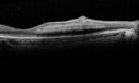
Cardiac Catheterization induced Cilioretinal Artery Occlusion - Paracentral Acute Middle Maculopathy490 views69 year old female with sudden vision loss on completing cardiac catheterization. The SD OCT shows a PAMM lesion and the lesion looks gray on color imaging. There is delayed filling of the cilioretinal artery.
|
|

Subtle Branch Retinal Artery Occlusion489 views68 year old man with shadow in the left eye yesterday. IR and OCT show subtle retinal swelling. FA is normal
|
|

Bilateral Branch Retinal Artery Occlusion - Intraarterial (hollenhorst) plaques visible488 views67 year old with acute visual field loss in each eye. Vision is 20/25 right eye 20/32 left eye. Images show intra-arterial plaques in each eye. The right eye on the optic nerve and the left eye in a branch retinal artery.
|
|

Subtle Branch Retinal Artery Occlusion488 views68 year old man with shadow in the left eye yesterday. IR and OCT show subtle retinal swelling. FA is normal
|
|

Branch Retinal Arterial Occlusion Right Eye - Atheromata - Both Eyes487 views83 year old woman with no symptoms - VA 20/40 in both eyes. Right eye has ST BRAO with silver wiring. Left eye has superotemporal atheromata
|
|

Cilioretinal Artery Occlusion - Good vision487 views67 year old female - 5 days ago she was getting ready for bed and closed one eye (she had worked for an optometrist for 20 years) and she noticed a spot in the vision in the right eye and came in. She has no focal neurological problems. She once in a while gets some weakness. She has a neurologist and there are some brain lesions. Father had stroke at age 52.
VA OD: Dcc20/20 NccJ1
VA OS: Dcc20/16-2 NccJ1-2
|
|

Acute Branch Retinal Artery Occlusion Left Eye485 viewsThis 84 year old woman had an old BRVO in the right eye and saw a shadow for 2 days in the left eye. She has a superotemporal BRAO. The photos show retinal whitening, the OCT shows inner retinal edema and the FA shows poor circulation.
|
|

Branch Retinal Arterial Occlusion Right Eye - Atheromata - Both Eyes485 views83 year old woman with no symptoms - VA 20/40 in both eyes. Right eye has ST BRAO with silver wiring. Left eye has superotemporal atheromata
|
|

Cardiac Catheterization induced Cilioretinal Artery Occlusion - Paracentral Acute Middle Maculopathy485 views69 year old female with sudden vision loss on completing cardiac catheterization. The SD OCT shows a PAMM lesion and the lesion looks gray on color imaging. There is delayed filling of the cilioretinal artery.
|
|

Subtle Branch Retinal Artery Occlusion485 views68 year old man with shadow in the left eye yesterday. IR and OCT show subtle retinal swelling. FA is normal
|
|

Cilioretinal Artery Occlusion - Good vision485 views67 year old female - 5 days ago she was getting ready for bed and closed one eye (she had worked for an optometrist for 20 years) and she noticed a spot in the vision in the right eye and came in. She has no focal neurological problems. She once in a while gets some weakness. She has a neurologist and there are some brain lesions. Father had stroke at age 52.
VA OD: Dcc20/20 NccJ1
VA OS: Dcc20/16-2 NccJ1-2
|
|

Cardiac Catheterization induced Cilioretinal Artery Occlusion - Paracentral Acute Middle Maculopathy484 views69 year old female with sudden vision loss on completing cardiac catheterization. The SD OCT shows a PAMM lesion and the lesion looks gray on color imaging. There is delayed filling of the cilioretinal artery.
|
|

Cardiac Catheterization induced Cilioretinal Artery Occlusion - Paracentral Acute Middle Maculopathy484 views69 year old female with sudden vision loss on completing cardiac catheterization. The SD OCT shows a PAMM lesion and the lesion looks gray on color imaging. There is delayed filling of the cilioretinal artery.
|
|

Branch Retinal Artery Occlusion - No Plaque482 views74 year old man with 2 days of vision loss. Vision OS is 20/200 but improved to 20/25 in 1 month. He has dysfibrinogen anemia.
|
|

Chronic BRAO with Hemorrhages - Sclerosed Vessel482 views95 year old female with blurred vision in both eyes in for a checkup. History of perpapillary CNVM in the left eye 2 years prior not treated. History of stroke with multiple emboli 1 year ago.
Vision 20/32 OD, 20/40 OS
|
|

Branch Retinal Arterial Occlusion 20/20 Vision - Symptoms for 1 Day480 views75-year-old man was seen in the office on 4/6/2010. He noticed a sudden vision change in the right eye yesterday morning. 20/20 Vision both Eyes
|
|

Branch Retinal Arterial Occlusion 20/20 Vision - Symptoms for 1 Day479 views75-year-old man was seen in the office on 4/6/2010. He noticed a sudden vision change in the right eye yesterday morning. 20/20 Vision both Eyes
|
|

Bilateral Branch Retinal Artery Occlusion - Intraarterial (hollenhorst) plaques visible479 views67 year old with acute visual field loss in each eye. Vision is 20/25 right eye 20/32 left eye. Images show intra-arterial plaques in each eye. The right eye on the optic nerve and the left eye in a branch retinal artery.
|
|

Subtle Branch Retinal Artery Occlusion479 views68 year old man with shadow in the left eye yesterday. IR and OCT show subtle retinal swelling. FA is normal
|
|

Bilateral Branch Retinal Artery Occlusion - Intraarterial (hollenhorst) plaques visible478 views67 year old with acute visual field loss in each eye. Vision is 20/25 right eye 20/32 left eye. Images show intra-arterial plaques in each eye. The right eye on the optic nerve and the left eye in a branch retinal artery.
|
|

Cardiac Catheterization induced Cilioretinal Artery Occlusion - Paracentral Acute Middle Maculopathy478 views69 year old female with sudden vision loss on completing cardiac catheterization. The SD OCT shows a PAMM lesion and the lesion looks gray on color imaging. There is delayed filling of the cilioretinal artery.
|
|

Branch Retinal Arterial Occlusion Right Eye - Atheromata - Both Eyes477 views83 year old woman with no symptoms - VA 20/40 in both eyes. Right eye has ST BRAO with silver wiring. Left eye has superotemporal atheromata
|
|

Subtle Branch Retinal Artery Occlusion475 views68 year old man with shadow in the left eye yesterday. IR and OCT show subtle retinal swelling. FA is normal
|
|

Cilioretinal Artery Occlusion - Good vision475 views67 year old female - 5 days ago she was getting ready for bed and closed one eye (she had worked for an optometrist for 20 years) and she noticed a spot in the vision in the right eye and came in. She has no focal neurological problems. She once in a while gets some weakness. She has a neurologist and there are some brain lesions. Father had stroke at age 52.
VA OD: Dcc20/20 NccJ1
VA OS: Dcc20/16-2 NccJ1-2
|
|

Branch Retinal Artery Occlusion - No Plaque473 views74 year old man with 2 days of vision loss. Vision OS is 20/200 but improved to 20/25 in 1 month. He has dysfibrinogen anemia.
|
|

Branch Retinal Artery Occlusion472 viewsAcute Inferotemporal Occlusion Right Eye - FA shows delayed circulation, OCT shows edema of superficial layers of retina
|
|

Branch Retinal Artery Occlusion - No Plaque472 views74 year old man with 2 days of vision loss. Vision OS is 20/200 but improved to 20/25 in 1 month. He has dysfibrinogen anemia.
|
|

Cardiac Catheterization induced Cilioretinal Artery Occlusion - Paracentral Acute Middle Maculopathy471 views69 year old female with sudden vision loss on completing cardiac catheterization. The SD OCT shows a PAMM lesion and the lesion looks gray on color imaging. There is delayed filling of the cilioretinal artery.
|
|

Chronic BRAO with Hemorrhages - Sclerosed Vessel471 views95 year old female with blurred vision in both eyes in for a checkup. History of perpapillary CNVM in the left eye 2 years prior not treated. History of stroke with multiple emboli 1 year ago.
Vision 20/32 OD, 20/40 OS
|
|

Subtle Branch Retinal Artery Occlusion471 views68 year old man with shadow in the left eye yesterday. IR and OCT show subtle retinal swelling. FA is normal
|
|

Branch Retinal Arterial Occlusion Right Eye - Atheromata - Both Eyes470 views83 year old woman with no symptoms - VA 20/40 in both eyes. Right eye has ST BRAO with silver wiring. Left eye has superotemporal atheromata
|
|

Branch Retinal Arterial Occlusion Right Eye - Atheromata - Both Eyes469 views83 year old woman with no symptoms - VA 20/40 in both eyes. Right eye has ST BRAO with silver wiring. Left eye has superotemporal atheromata
|
|

Branch Retinal Arterial Occlusion Right Eye - Atheromata - Both Eyes467 views83 year old woman with no symptoms - VA 20/40 in both eyes. Right eye has ST BRAO with silver wiring. Left eye has superotemporal atheromata
|
|

Chronic BRAO with Hemorrhages - Sclerosed Vessel467 views95 year old female with blurred vision in both eyes in for a checkup. History of perpapillary CNVM in the left eye 2 years prior not treated. History of stroke with multiple emboli 1 year ago.
Vision 20/32 OD, 20/40 OS
|
|
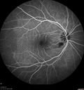
Cilioretinal Artery Occlusion - Good vision467 views67 year old female - 5 days ago she was getting ready for bed and closed one eye (she had worked for an optometrist for 20 years) and she noticed a spot in the vision in the right eye and came in. She has no focal neurological problems. She once in a while gets some weakness. She has a neurologist and there are some brain lesions. Father had stroke at age 52.
VA OD: Dcc20/20 NccJ1
VA OS: Dcc20/16-2 NccJ1-2
|
|

Cardiac Catheterization induced Cilioretinal Artery Occlusion - Paracentral Acute Middle Maculopathy466 views69 year old female with sudden vision loss on completing cardiac catheterization. The SD OCT shows a PAMM lesion and the lesion looks gray on color imaging. There is delayed filling of the cilioretinal artery.
|
|

Cardiac Catheterization induced Cilioretinal Artery Occlusion - Paracentral Acute Middle Maculopathy466 views69 year old female with sudden vision loss on completing cardiac catheterization. The SD OCT shows a PAMM lesion and the lesion looks gray on color imaging. There is delayed filling of the cilioretinal artery.
|
|

Subtle Branch Retinal Artery Occlusion465 views68 year old man with shadow in the left eye yesterday. IR and OCT show subtle retinal swelling. FA is normal
|
|

Cilioretinal Artery Occlusion - Good vision465 views67 year old female - 5 days ago she was getting ready for bed and closed one eye (she had worked for an optometrist for 20 years) and she noticed a spot in the vision in the right eye and came in. She has no focal neurological problems. She once in a while gets some weakness. She has a neurologist and there are some brain lesions. Father had stroke at age 52.
VA OD: Dcc20/20 NccJ1
VA OS: Dcc20/16-2 NccJ1-2
|
|

Bilateral Branch Retinal Artery Occlusion - Intraarterial (hollenhorst) plaques visible464 views67 year old with acute visual field loss in each eye. Vision is 20/25 right eye 20/32 left eye. Images show intra-arterial plaques in each eye. The right eye on the optic nerve and the left eye in a branch retinal artery.
|
|

BRAO and PAMM463 views
|
|

Cilioretinal Artery Occlusion - Good vision462 views67 year old female - 5 days ago she was getting ready for bed and closed one eye (she had worked for an optometrist for 20 years) and she noticed a spot in the vision in the right eye and came in. She has no focal neurological problems. She once in a while gets some weakness. She has a neurologist and there are some brain lesions. Father had stroke at age 52.
VA OD: Dcc20/20 NccJ1
VA OS: Dcc20/16-2 NccJ1-2
|
|

Bilateral Branch Retinal Artery Occlusion - Intraarterial (hollenhorst) plaques visible461 views67 year old with acute visual field loss in each eye. Vision is 20/25 right eye 20/32 left eye. Images show intra-arterial plaques in each eye. The right eye on the optic nerve and the left eye in a branch retinal artery.
|
|

Branch Retinal Arterial Occlusion Right Eye - Atheromata - Both Eyes460 views83 year old woman with no symptoms - VA 20/40 in both eyes. Right eye has ST BRAO with silver wiring. Left eye has superotemporal atheromata
|
|

Cardiac Catheterization induced Cilioretinal Artery Occlusion - Paracentral Acute Middle Maculopathy459 views69 year old female with sudden vision loss on completing cardiac catheterization. The SD OCT shows a PAMM lesion and the lesion looks gray on color imaging. There is delayed filling of the cilioretinal artery.
|
|
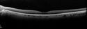
Cardiac Catheterization induced Cilioretinal Artery Occlusion - Paracentral Acute Middle Maculopathy459 views69 year old female with sudden vision loss on completing cardiac catheterization. The SD OCT shows a PAMM lesion and the lesion looks gray on color imaging. There is delayed filling of the cilioretinal artery.
|
|

Chronic BRAO with Hemorrhages - Sclerosed Vessel459 views95 year old female with blurred vision in both eyes in for a checkup. History of perpapillary CNVM in the left eye 2 years prior not treated. History of stroke with multiple emboli 1 year ago.
Vision 20/32 OD, 20/40 OS
|
|

Cilioretinal Artery Occlusion - Good vision459 views67 year old female - 5 days ago she was getting ready for bed and closed one eye (she had worked for an optometrist for 20 years) and she noticed a spot in the vision in the right eye and came in. She has no focal neurological problems. She once in a while gets some weakness. She has a neurologist and there are some brain lesions. Father had stroke at age 52.
VA OD: Dcc20/20 NccJ1
VA OS: Dcc20/16-2 NccJ1-2
|
|
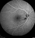
Cilioretinal Artery Occlusion - Good vision458 views67 year old female - 5 days ago she was getting ready for bed and closed one eye (she had worked for an optometrist for 20 years) and she noticed a spot in the vision in the right eye and came in. She has no focal neurological problems. She once in a while gets some weakness. She has a neurologist and there are some brain lesions. Father had stroke at age 52.
VA OD: Dcc20/20 NccJ1
VA OS: Dcc20/16-2 NccJ1-2
|
|

Cilioretinal Artery Occlusion - Good vision457 views67 year old female - 5 days ago she was getting ready for bed and closed one eye (she had worked for an optometrist for 20 years) and she noticed a spot in the vision in the right eye and came in. She has no focal neurological problems. She once in a while gets some weakness. She has a neurologist and there are some brain lesions. Father had stroke at age 52.
VA OD: Dcc20/20 NccJ1
VA OS: Dcc20/16-2 NccJ1-2
|
|

Cilioretinal Artery Occlusion - Good vision456 views67 year old female - 5 days ago she was getting ready for bed and closed one eye (she had worked for an optometrist for 20 years) and she noticed a spot in the vision in the right eye and came in. She has no focal neurological problems. She once in a while gets some weakness. She has a neurologist and there are some brain lesions. Father had stroke at age 52.
VA OD: Dcc20/20 NccJ1
VA OS: Dcc20/16-2 NccJ1-2
|
|

Cardiac Catheterization induced Cilioretinal Artery Occlusion - Paracentral Acute Middle Maculopathy454 views69 year old female with sudden vision loss on completing cardiac catheterization. The SD OCT shows a PAMM lesion and the lesion looks gray on color imaging. There is delayed filling of the cilioretinal artery.
|
|

Branch Retinal Artery Occlusion - No Plaque453 views74 year old man with 2 days of vision loss. Vision OS is 20/200 but improved to 20/25 in 1 month. He has dysfibrinogen anemia.
|
|

Chronic BRAO with Hemorrhages - Sclerosed Vessel453 views95 year old female with blurred vision in both eyes in for a checkup. History of perpapillary CNVM in the left eye 2 years prior not treated. History of stroke with multiple emboli 1 year ago.
Vision 20/32 OD, 20/40 OS
|
|

Acute Branch Retinal Arteriolar Occlusion453 views75 year old female treated for wet AMD with 2 days of inferior shadow in the right eye. VA 20/40. There is a plaque visible and retinal whitening from the arteriolar occlusion.
|
|

Branch Retinal Artery Occlusion - No Plaque451 views74 year old man with 2 days of vision loss. Vision OS is 20/200 but improved to 20/25 in 1 month. He has dysfibrinogen anemia.
|
|

Cilioretinal Artery Occlusion - Good vision451 views67 year old female - 5 days ago she was getting ready for bed and closed one eye (she had worked for an optometrist for 20 years) and she noticed a spot in the vision in the right eye and came in. She has no focal neurological problems. She once in a while gets some weakness. She has a neurologist and there are some brain lesions. Father had stroke at age 52.
VA OD: Dcc20/20 NccJ1
VA OS: Dcc20/16-2 NccJ1-2
|
|

Cardiac Catheterization induced Cilioretinal Artery Occlusion - Paracentral Acute Middle Maculopathy450 views69 year old female with sudden vision loss on completing cardiac catheterization. The SD OCT shows a PAMM lesion and the lesion looks gray on color imaging. There is delayed filling of the cilioretinal artery.
|
|

Branch Retinal Artery Occlusion448 viewsAcute Inferotemporal Occlusion Right Eye - FA shows delayed circulation, OCT shows edema of superficial layers of retina
|
|

Acute Branch Retinal Arteriolar Occlusion448 views75 year old female treated for wet AMD with 2 days of inferior shadow in the right eye. VA 20/40. There is a plaque visible and retinal whitening from the arteriolar occlusion.
|
|

Branch Retinal Arterial Occlusion Right Eye - Atheromata - Both Eyes447 views83 year old woman with no symptoms - VA 20/40 in both eyes. Right eye has ST BRAO with silver wiring. Left eye has superotemporal atheromata
|
|

Cardiac Catheterization induced Cilioretinal Artery Occlusion - Paracentral Acute Middle Maculopathy447 views69 year old female with sudden vision loss on completing cardiac catheterization. The SD OCT shows a PAMM lesion and the lesion looks gray on color imaging. There is delayed filling of the cilioretinal artery.
|
|

Cardiac Catheterization induced Cilioretinal Artery Occlusion - Paracentral Acute Middle Maculopathy447 views69 year old female with sudden vision loss on completing cardiac catheterization. The SD OCT shows a PAMM lesion and the lesion looks gray on color imaging. There is delayed filling of the cilioretinal artery.
|
|

Branch Retinal Artery Occlusion - No Plaque447 views74 year old man with 2 days of vision loss. Vision OS is 20/200 but improved to 20/25 in 1 month. He has dysfibrinogen anemia.
|
|

Cilioretinal Artery Occlusion - Good vision447 views67 year old female - 5 days ago she was getting ready for bed and closed one eye (she had worked for an optometrist for 20 years) and she noticed a spot in the vision in the right eye and came in. She has no focal neurological problems. She once in a while gets some weakness. She has a neurologist and there are some brain lesions. Father had stroke at age 52.
VA OD: Dcc20/20 NccJ1
VA OS: Dcc20/16-2 NccJ1-2
|
|

Bilateral Branch Retinal Artery Occlusion - Intraarterial (hollenhorst) plaques visible446 views67 year old with acute visual field loss in each eye. Vision is 20/25 right eye 20/32 left eye. Images show intra-arterial plaques in each eye. The right eye on the optic nerve and the left eye in a branch retinal artery.
|
|

Cilioretinal Artery Occlusion - Good vision446 views67 year old female - 5 days ago she was getting ready for bed and closed one eye (she had worked for an optometrist for 20 years) and she noticed a spot in the vision in the right eye and came in. She has no focal neurological problems. She once in a while gets some weakness. She has a neurologist and there are some brain lesions. Father had stroke at age 52.
VA OD: Dcc20/20 NccJ1
VA OS: Dcc20/16-2 NccJ1-2
|
|

Cilioretinal Artery Occlusion - Nasal (not macular)445 views77 year old female There is a shadow in the top half of her temporal vision in the left eye that came on suddenly yesterday morning. It has not changed since then and the right eye is OK. She has had sinus headaches. She had no focal neurological changes. Â
VA OD: Dcc20/32
VA OS: Dcc20/16-1
IOP: TP: OD:18 OS:19Â
|
|

Cilioretinal Artery Occlusion - Good vision444 views67 year old female - 5 days ago she was getting ready for bed and closed one eye (she had worked for an optometrist for 20 years) and she noticed a spot in the vision in the right eye and came in. She has no focal neurological problems. She once in a while gets some weakness. She has a neurologist and there are some brain lesions. Father had stroke at age 52.
VA OD: Dcc20/20 NccJ1
VA OS: Dcc20/16-2 NccJ1-2
|
|
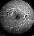
Chronic BRAO with Hemorrhages - Sclerosed Vessel442 views95 year old female with blurred vision in both eyes in for a checkup. History of perpapillary CNVM in the left eye 2 years prior not treated. History of stroke with multiple emboli 1 year ago.
Vision 20/32 OD, 20/40 OS
|
|

Branch Retinal Artery Occlusion - No Plaque441 views74 year old man with 2 days of vision loss. Vision OS is 20/200 but improved to 20/25 in 1 month. He has dysfibrinogen anemia.
|
|

Branch Retinal Arterial Occlusion Right Eye - Atheromata - Both Eyes440 views83 year old woman with no symptoms - VA 20/40 in both eyes. Right eye has ST BRAO with silver wiring. Left eye has superotemporal atheromata
|
|

Acute Branch Retinal Arteriolar Occlusion440 views75 year old female treated for wet AMD with 2 days of inferior shadow in the right eye. VA 20/40. There is a plaque visible and retinal whitening from the arteriolar occlusion.
|
|

Chronic BRAO with Hemorrhages - Sclerosed Vessel439 views95 year old female with blurred vision in both eyes in for a checkup. History of perpapillary CNVM in the left eye 2 years prior not treated. History of stroke with multiple emboli 1 year ago.
Vision 20/32 OD, 20/40 OS
|
|

Subtle Branch Retinal Artery Occlusion439 views68 year old man with shadow in the left eye yesterday. IR and OCT show subtle retinal swelling. FA is normal
|
|

Acute Branch Retinal Artery Occlusion Left Eye438 viewsThis 84 year old woman had an old BRVO in the right eye and saw a shadow for 2 days in the left eye. She has a superotemporal BRAO. The photos show retinal whitening, the OCT shows inner retinal edema and the FA shows poor circulation.
|
|
| 467 files on 5 page(s) |
 |
3 | |
|


