am meisten angesehen - Central Retinal Artery Occlusion
|

Central Retinal Artery Occlusion - 2 months old by history1833 x angesehen77-year-old man was doing fine. His left eye was a little worse than the right eye, and then in the 2 months ago he had sudden severe vision loss in the right eye. Vision OD is 3/200; OS is 20/60. IOP: OD 30, OS 15.
OD: Vertical C/D ratio is 0.4. The macula is white from ischemia. The arteries are narrow.
Photos confirm clinical findings.
OCT SCAN: OCT scan of the right eye shows retinal thickening, consistent with macular edema associated with a central retinal artery occlusion. The left eye has severe inferior retinal thinning, consistent with an old hemicentral retinal artery occlusion in the left eye. The fovea in the left eye is a little bit thin but not too bad.
IMPRESSION: 1. CENTRAL RETINAL ARTERY OCCLUSION RIGHT EYE
2 OLD HEMICENTRAL RETINAL ARTERY OCCLUSION LEFT EYE
3 DIABETES WITHOUT RETINOPATHY
4 ELEVATED INTRAOCULAR PRESSURE RIGHT EYE STATUS POST INJECTION RIGHT EYE A WEEK AGO
DISCUSSION: I explained to the patient that the right eye unfortunately has had a central retinal artery and unfortunately we do not have anything to treat that. His vision will probably improve some over time. He needs to be watched though. It is possible he is developing rubeotic glaucoma, although it is probable the intraocular pressure is high from the injection last week. I asked him to restart the Alphagan, to stop the other two drops, and to return here in three weeks to make sure his pressure is more acceptable.
At this point, having lost almost all the retinal circulation in the right eye and half of the retinal circulation in the left eye, he only has half a retina in the left eye he is working off of. I told him it is very important he keep seeing you and anything that can be done to keep his blood pressure low and his cholesterol reasonable would be helpful to what remains of his retinal circulation.
|
|

Central Retinal Artery Occlusion1777 x angesehen60 year old male with LP vision due to extensive blood flow loss.
|
|
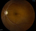
Retinal Artery Occlusion1709 x angesehenPatient comes in for eval on artery occlusions in both eyes. VA is 20/400, right eye and NLP, left eye. Fundus photos show paniretinal scars in the right eye with arterial narrowing and the left eye has arterial narrowing as well.
|
|
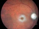
Central Retinal Artery Occlusion - 2 months old by history1607 x angesehen77-year-old man was doing fine. His left eye was a little worse than the right eye, and then in the 2 months ago he had sudden severe vision loss in the right eye. Vision OD is 3/200; OS is 20/60. IOP: OD 30, OS 15.
OD: Vertical C/D ratio is 0.4. The macula is white from ischemia. The arteries are narrow.
Photos confirm clinical findings.
OCT SCAN: OCT scan of the right eye shows retinal thickening, consistent with macular edema associated with a central retinal artery occlusion. The left eye has severe inferior retinal thinning, consistent with an old hemicentral retinal artery occlusion in the left eye. The fovea in the left eye is a little bit thin but not too bad.
IMPRESSION: 1. CENTRAL RETINAL ARTERY OCCLUSION RIGHT EYE
2 OLD HEMICENTRAL RETINAL ARTERY OCCLUSION LEFT EYE
3 DIABETES WITHOUT RETINOPATHY
4 ELEVATED INTRAOCULAR PRESSURE RIGHT EYE STATUS POST INJECTION RIGHT EYE A WEEK AGO
DISCUSSION: I explained to the patient that the right eye unfortunately has had a central retinal artery and unfortunately we do not have anything to treat that. His vision will probably improve some over time. He needs to be watched though. It is possible he is developing rubeotic glaucoma, although it is probable the intraocular pressure is high from the injection last week. I asked him to restart the Alphagan, to stop the other two drops, and to return here in three weeks to make sure his pressure is more acceptable.
At this point, having lost almost all the retinal circulation in the right eye and half of the retinal circulation in the left eye, he only has half a retina in the left eye he is working off of. I told him it is very important he keep seeing you and anything that can be done to keep his blood pressure low and his cholesterol reasonable would be helpful to what remains of his retinal circulation.
|
|

Central Retinal Artery Occlusion with Cilioretinal Sparing - Acute - SD-OCT Line Scan Inner retinal edema1532 x angesehen 77-year-old man was doing fine until midnight of last night when he noticed sudden severe vision loss in the left eye. VISUAL ACUITY: OD 20/20, OS 2/200.
|
|

Central Retinal Artery Occlusion with Cilioretinal Sparing - Acute1218 x angesehen 77-year-old man was doing fine until midnight of last night when he noticed sudden severe vision loss in the left eye. VISUAL ACUITY: OD 20/20, OS 2/200.
|
|

Retinal Artery Occlusion1200 x angesehenPatient comes in for eval on artery occlusions in both eyes. VA is 20/400, right eye and NLP, left eye. Fundus photos show paniretinal scars in the right eye with arterial narrowing and the left eye has arterial narrowing as well.
|
|
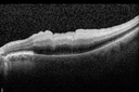
Central Retinal Artery Occlusion with Cilioretinal Sparing - Acute - SD-OCT Line Scan Inner retinal edema1182 x angesehen 77-year-old man was doing fine until midnight of last night when he noticed sudden severe vision loss in the left eye. VISUAL ACUITY: OD 20/20, OS 2/200.
|
|

Central Retinal Artery Occlusion1106 x angesehenCentral Retinal Artery Occlusion with cilio artery perfusion.
|
|

Central Retinal Artery Occlusion less than 24 hours - 69 year old man VA light perception1104 x angesehen
|
|

Central Retinal Artery Occlusion with Cilioretinal Sparing - Acute - FA Very Slow Filling CRA1048 x angesehen 77-year-old man was doing fine until midnight of last night when he noticed sudden severe vision loss in the left eye. VISUAL ACUITY: OD 20/20, OS 2/200.
|
|

Central Arterial Occlusion with Embolism Present1045 x angesehenPatient comes in with CRAO in the right eye. VA was hand motion. Fundus photo shows white veil over the retina with 2-emboli in a branch artery temporal in the right eye. She will be evaluated for emboli and followed up in a month
|
|

Retinal Artery Occlusion1034 x angesehenPatient comes in for eval on artery occlusions in both eyes. VA is 20/400, right eye and NLP, left eye. Fundus photos show paniretinal scars in the right eye with arterial narrowing and the left eye has arterial narrowing as well.
|
|

Central Retinal Artery Occlusion with Cilioretinal Sparing - Acute - Line scan good eye994 x angesehen 77-year-old man was doing fine until midnight of last night when he noticed sudden severe vision loss in the left eye. VISUAL ACUITY: OD 20/20, OS 2/200.
|
|

Central Retinal Artery Occlusion with Cilioretinal Sparing - Acute - FA Very Slow Filling CRA967 x angesehen 77-year-old man was doing fine until midnight of last night when he noticed sudden severe vision loss in the left eye. VISUAL ACUITY: OD 20/20, OS 2/200.
|
|

Central Retinal Artery Occlusion less than 24 hours - 69 year old man VA light perception939 x angesehen
|
|

Pathology - Central Retinal Artery Occlusion - 6 weeks old - Inner Retinal Atrophy - Macular Pucker932 x angesehenpatient died about 6 to 8 weeks after the artery occlusion, and he had no history of glaucoma. The inner retina atrophy, which looks very similar to that caused by COAG, is due to recent infarct.
|
|

Central Retinal Artery Occlusion with Cilioretinal Sparing - Acute924 x angesehen 77-year-old man was doing fine until midnight of last night when he noticed sudden severe vision loss in the left eye. VISUAL ACUITY: OD 20/20, OS 2/200.
|
|

Central Retinal Artery Occlusion with Cilioretinal Sparing - Acute - FA Very Slow Filling CRA922 x angesehen 77-year-old man was doing fine until midnight of last night when he noticed sudden severe vision loss in the left eye. VISUAL ACUITY: OD 20/20, OS 2/200.
|
|

Central Retinal Artery Occlusion with Cilioretinal Sparing - Acute - FA Very Slow Filling CRA921 x angesehen 77-year-old man was doing fine until midnight of last night when he noticed sudden severe vision loss in the left eye. VISUAL ACUITY: OD 20/20, OS 2/200.
|
|

Pathology - Central Retinal Artery Occlusion - 6 weeks old - Inner Retinal Atrophy - Macular Pucker907 x angesehenpatient died about 6 to 8 weeks after the artery occlusion, and he had no history of glaucoma. The inner retina atrophy, which looks very similar to that caused by COAG, is due to recent infarct.
|
|

Central Retinal Artery Occlusion - 2 months old by history876 x angesehen77-year-old man was doing fine. His left eye was a little worse than the right eye, and then in the 2 months ago he had sudden severe vision loss in the right eye. Vision OD is 3/200; OS is 20/60. IOP: OD 30, OS 15.
OD: Vertical C/D ratio is 0.4. The macula is white from ischemia. The arteries are narrow.
Photos confirm clinical findings.
OCT SCAN: OCT scan of the right eye shows retinal thickening, consistent with macular edema associated with a central retinal artery occlusion. The left eye has severe inferior retinal thinning, consistent with an old hemicentral retinal artery occlusion in the left eye. The fovea in the left eye is a little bit thin but not too bad.
IMPRESSION: 1. CENTRAL RETINAL ARTERY OCCLUSION RIGHT EYE
2 OLD HEMICENTRAL RETINAL ARTERY OCCLUSION LEFT EYE
3 DIABETES WITHOUT RETINOPATHY
4 ELEVATED INTRAOCULAR PRESSURE RIGHT EYE STATUS POST INJECTION RIGHT EYE A WEEK AGO
DISCUSSION: I explained to the patient that the right eye unfortunately has had a central retinal artery and unfortunately we do not have anything to treat that. His vision will probably improve some over time. He needs to be watched though. It is possible he is developing rubeotic glaucoma, although it is probable the intraocular pressure is high from the injection last week. I asked him to restart the Alphagan, to stop the other two drops, and to return here in three weeks to make sure his pressure is more acceptable.
At this point, having lost almost all the retinal circulation in the right eye and half of the retinal circulation in the left eye, he only has half a retina in the left eye he is working off of. I told him it is very important he keep seeing you and anything that can be done to keep his blood pressure low and his cholesterol reasonable would be helpful to what remains of his retinal circulation.
|
|

Pathology - Central Retinal Artery Occlusion - 6 weeks old - Inner Retinal Atrophy872 x angesehenpatient died about 6 to 8 weeks after the artery occlusion, and he had no history of glaucoma. The inner retina atrophy, which looks very similar to that caused by COAG, is due to recent infarct.
|
|

Embolic CRAO color850 x angesehen
|
|

846 x angesehen
|
|

Central Retinal Artery Occlusion less than 24 hours - 69 year old man VA light perception845 x angesehen
|
|

Central Retinal Artery Occlusion with Cilioretinal Sparing - Acute - FA Very Slow Filling CRA840 x angesehen 77-year-old man was doing fine until midnight of last night when he noticed sudden severe vision loss in the left eye. VISUAL ACUITY: OD 20/20, OS 2/200.
|
|

FA: CRAO840 x angesehenCRAO with cilio artery perfusion
|
|
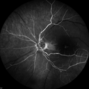
Central Retinal Artery Occlusion with Cilioretinal Sparing - Acute - FA Very Slow Filling CRA825 x angesehen 77-year-old man was doing fine until midnight of last night when he noticed sudden severe vision loss in the left eye. VISUAL ACUITY: OD 20/20, OS 2/200.
|
|

Central Retinal Artery Occlusion Left Eye 2 Weeks Old - Still Non-Perfusion818 x angesehen85-year-old woman who had sudden severevision loss in the left eye two weeks ago. she can see a little more centrally and much more in the periphery, since about the last four days.
VISUAL ACUITY: OD 20/30, OS: Hand motion
|
|

Central Retinal Artery Occlusion Left Eye 2 Weeks Old - Still Non-Perfusion811 x angesehen85-year-old woman who had sudden severevision loss in the left eye two weeks ago. she can see a little more centrally and much more in the periphery, since about the last four days.
VISUAL ACUITY: OD 20/30, OS: Hand motion
|
|

Central Retinal Artery Occlusion Left Eye 2 Weeks Old - Still Non-Perfusion793 x angesehen85-year-old woman who had sudden severevision loss in the left eye two weeks ago. she can see a little more centrally and much more in the periphery, since about the last four days.
VISUAL ACUITY: OD 20/30, OS: Hand motion
|
|

Central Retinal Artery Occlusion Left Eye 2 Weeks Old - Still Non-Perfusion791 x angesehen85-year-old woman who had sudden severevision loss in the left eye two weeks ago. she can see a little more centrally and much more in the periphery, since about the last four days.
VISUAL ACUITY: OD 20/30, OS: Hand motion
|
|

782 x angesehen
|
|

Central Retinal Artery Occlusion with Cilioretinal Sparing - Acute782 x angesehen 77-year-old man was doing fine until midnight of last night when he noticed sudden severe vision loss in the left eye. VISUAL ACUITY: OD 20/20, OS 2/200.
|
|

Pathology - Central Retinal Artery Occlusion - 6 weeks old - Inner Retinal Atrophy779 x angesehenpatient died about 6 to 8 weeks after the artery occlusion, and he had no history of glaucoma. The inner retina atrophy, which looks very similar to that caused by COAG, is due to recent infarct.
|
|

Central Retinal Artery Occlusion - MAP765 x angesehenProgression from acute to atrophy - decreasing retinal thickenss
|
|

753 x angesehen
|
|
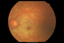
Central Retinal Artery Occlusion Left Eye 2 Weeks Old - Still Non-Perfusion735 x angesehen85-year-old woman who had sudden severevision loss in the left eye two weeks ago. she can see a little more centrally and much more in the periphery, since about the last four days.
VISUAL ACUITY: OD 20/30, OS: Hand motion
|
|

Central Retinal Artery Occlusion less than 24 hours - 69 year old man VA light perception734 x angesehen
|
|

Montage of Hemi-Central Retinal Artery Occlusion732 x angesehen
|
|

Central Retinal Artery Occlusion Left Eye 2 Weeks Old - Still Non-Perfusion704 x angesehen85-year-old woman who had sudden severevision loss in the left eye two weeks ago. she can see a little more centrally and much more in the periphery, since about the last four days.
VISUAL ACUITY: OD 20/30, OS: Hand motion
|
|

Central Retinal Artery Occlusion Left Eye 2 Weeks Old - Still Non-Perfusion703 x angesehen85-year-old woman who had sudden severevision loss in the left eye two weeks ago. she can see a little more centrally and much more in the periphery, since about the last four days.
VISUAL ACUITY: OD 20/30, OS: Hand motion
|
|

Montage of Hemi-Central Retinal Artery Occlusion701 x angesehen
|
|
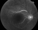
698 x angesehen60 year old male with LP vision due to extensive blood flow loss.
|
|

Central Retinal Artery Occlusion with Cilioretinal Sparing - Acute696 x angesehen 77-year-old man was doing fine until midnight of last night when he noticed sudden severe vision loss in the left eye. VISUAL ACUITY: OD 20/20, OS 2/200.
|
|

Central Retinal Artery Occlusion less than 24 hours - 69 year old man VA light perception694 x angesehen
|
|

Central Retinal Artery Occlusion Left Eye 2 Weeks Old - Still Non-Perfusion690 x angesehen85-year-old woman who had sudden severevision loss in the left eye two weeks ago. she can see a little more centrally and much more in the periphery, since about the last four days.
VISUAL ACUITY: OD 20/30, OS: Hand motion
|
|

Central Retinal Artery Occlusion Left Eye 2 Weeks Old - Still Non-Perfusion671 x angesehen85-year-old woman who had sudden severevision loss in the left eye two weeks ago. she can see a little more centrally and much more in the periphery, since about the last four days.
VISUAL ACUITY: OD 20/30, OS: Hand motion
|
|

Central Retinal Artery Occlusion less than 24 hours - 69 year old man VA light perception671 x angesehen
|
|

660 x angesehen
|
|

Central Retinal Artery Occlusion Left Eye 2 Weeks Old - Still Non-Perfusion657 x angesehen85-year-old woman who had sudden severevision loss in the left eye two weeks ago. she can see a little more centrally and much more in the periphery, since about the last four days.
VISUAL ACUITY: OD 20/30, OS: Hand motion
|
|

Central Retinal Artery Occlusion Left Eye 2 Weeks Old - Still Non-Perfusion651 x angesehen85-year-old woman who had sudden severevision loss in the left eye two weeks ago. she can see a little more centrally and much more in the periphery, since about the last four days.
VISUAL ACUITY: OD 20/30, OS: Hand motion
|
|

Central Retinal Artery Occlusion Left Eye 2 Weeks Old - Still Non-Perfusion650 x angesehen85-year-old woman who had sudden severevision loss in the left eye two weeks ago. she can see a little more centrally and much more in the periphery, since about the last four days.
VISUAL ACUITY: OD 20/30, OS: Hand motion
|
|

Central Retinal Artery Occlusion Left Eye 2 Weeks Old - Still Non-Perfusion645 x angesehen85-year-old woman who had sudden severevision loss in the left eye two weeks ago. she can see a little more centrally and much more in the periphery, since about the last four days.
VISUAL ACUITY: OD 20/30, OS: Hand motion
|
|

Central Retinal Artery Occlusion - Diabetic Retinopathy - NO cherry red spot624 x angesehen58 year old man with sudden vision loss in the right eye. The vision had been 20/40 and dropped to 20/200 the day prior to presentation.
VA 20/200 OD, 20/20 OS
FA shows CRAO. No cherry red spot.
|
|

Embolic CRAO Early621 x angesehen
|
|

Central Retinal Artery Occlusion less than 24 hours - 69 year old man VA light perception615 x angesehen
|
|

Central Retinal Artery Occlusion less than 24 hours - 69 year old man VA light perception615 x angesehen
|
|

Central Retinal Artery Occlusion less than 24 hours - 69 year old man VA light perception610 x angesehen
|
|

Central Retinal Artery Occlusion Left Eye 2 Weeks Old - Still Non-Perfusion597 x angesehen85-year-old woman who had sudden severevision loss in the left eye two weeks ago. she can see a little more centrally and much more in the periphery, since about the last four days.
VISUAL ACUITY: OD 20/30, OS: Hand motion
|
|

80 Year old man with 3 day history of vision loss right eye. Vision 4/200.595 x angesehen
|
|

Retinal Artery Occlusion595 x angesehenPatient comes in for eval on artery occlusions in both eyes. VA is 20/400, right eye and NLP, left eye. Fundus photos show paniretinal scars in the right eye with arterial narrowing and the left eye has arterial narrowing as well.
|
|

80 Year old man with 3 day history of vision loss right eye. Vision 4/200.593 x angesehen
|
|

80 Year old man with 3 day history of vision loss right eye. Vision 4/200.591 x angesehen
|
|

Central Retinal Artery Occlusion less than 24 hours - 69 year old man VA light perception589 x angesehen
|
|

80 Year old man with 3 day history of vision loss right eye. Vision 4/200.582 x angesehen
|
|

Central Retinal Artery Occlusion less than 24 hours - 69 year old man VA light perception577 x angesehen
|
|

Central Retinal Artery Occlusion - Diabetic Retinopathy - NO cherry red spot575 x angesehen58 year old man with sudden vision loss in the right eye. The vision had been 20/40 and dropped to 20/200 the day prior to presentation.
VA 20/200 OD, 20/20 OS
FA shows CRAO. No cherry red spot.
|
|

Central Retinal Artery Occlusion then Neovascular Glaucoma573 x angesehen81 year old male with sudden vision loss. 2 months later developed NVG
|
|

Central Retinal Artery Occlusion less than 24 hours - 69 year old man VA light perception571 x angesehen
|
|

Central Retinal Artery Occlusion less than 24 hours - 69 year old man VA light perception571 x angesehen
|
|

Central Retinal Artery Occlusion - Diabetic Retinopathy - NO cherry red spot567 x angesehen58 year old man with sudden vision loss in the right eye. The vision had been 20/40 and dropped to 20/200 the day prior to presentation.
VA 20/200 OD, 20/20 OS
FA shows CRAO. No cherry red spot.
|
|

Embolic CRAO Mid563 x angesehen
|
|

80 Year old man with 3 day history of vision loss right eye. Vision 4/200.562 x angesehen
|
|

Central Retinal Artery Occlusion Left Eye 2 Weeks Old - Still Non-Perfusion561 x angesehen85-year-old woman who had sudden severevision loss in the left eye two weeks ago. she can see a little more centrally and much more in the periphery, since about the last four days.
VISUAL ACUITY: OD 20/30, OS: Hand motion
|
|

Central Retinal Artery Occlusion less than 24 hours - 69 year old man VA light perception558 x angesehen
|
|

Central Retinal Artery Occlusion less than 24 hours - 69 year old man VA light perception558 x angesehen
|
|
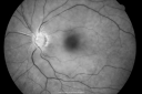
Central Retinal Artery Occlusion less than 24 hours - 69 year old man VA light perception557 x angesehen
|
|

Central Retinal Artery Occlusion less than 24 hours - 69 year old man VA light perception555 x angesehen
|
|

Central Retinal Artery Occlusion less than 24 hours - 69 year old man VA light perception554 x angesehen
|
|

Central Retinal Artery Occlusion - Diabetic Retinopathy - NO cherry red spot553 x angesehen58 year old man with sudden vision loss in the right eye. The vision had been 20/40 and dropped to 20/200 the day prior to presentation.
VA 20/200 OD, 20/20 OS
FA shows CRAO. No cherry red spot.
|
|

Central Retinal Artery Occlusion then Neovascular Glaucoma550 x angesehen81 year old male with sudden vision loss. 2 months later developed NVG
|
|

540 x angesehen
|
|

Central Retinal Artery Occlusion - Diabetic Retinopathy - NO cherry red spot540 x angesehen58 year old man with sudden vision loss in the right eye. The vision had been 20/40 and dropped to 20/200 the day prior to presentation.
VA 20/200 OD, 20/20 OS
FA shows CRAO. No cherry red spot.
|
|

Central Retinal Artery Occlusion less than 24 hours - 69 year old man VA light perception539 x angesehen
|
|

80 Year old man with 3 day history of vision loss right eye. Vision 4/200.537 x angesehen
|
|

Central Retinal Artery Occlusion - Diabetic Retinopathy - NO cherry red spot532 x angesehen58 year old man with sudden vision loss in the right eye. The vision had been 20/40 and dropped to 20/200 the day prior to presentation.
VA 20/200 OD, 20/20 OS
FA shows CRAO. No cherry red spot.
|
|

Embolic CRAO B&W531 x angesehen
|
|

Central Retinal Artery Occlusion - Diabetic Retinopathy - NO cherry red spot530 x angesehen58 year old man with sudden vision loss in the right eye. The vision had been 20/40 and dropped to 20/200 the day prior to presentation.
VA 20/200 OD, 20/20 OS
FA shows CRAO. No cherry red spot.
|
|

Central Retinal Artery Occlusion less than 24 hours - 69 year old man VA light perception523 x angesehen
|
|

80 Year old man with 3 day history of vision loss right eye. Vision 4/200.523 x angesehen
|
|

522 x angesehen
|
|

Central Retinal Artery Occlusion less than 24 hours - 69 year old man VA light perception515 x angesehen
|
|

Central Retinal Artery Occlusion then Neovascular Glaucoma514 x angesehen81 year old male with sudden vision loss. 2 months later developed NVG
|
|

Embolic CRAO Late511 x angesehen
|
|

Acute Central Retinal Artery Occlusion511 x angesehen1 day of vision loss 79 year old man to HM. NVG developed 2 months later.
|
|
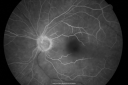
Central Retinal Artery Occlusion less than 24 hours - 69 year old man VA light perception508 x angesehen
|
|

Central Retinal Artery Occlusion then Neovascular Glaucoma506 x angesehen81 year old male with sudden vision loss. 2 months later developed NVG
|
|

Acute Central Retinal Artery Occlusion503 x angesehen1 day of vision loss 79 year old man to HM. NVG developed 2 months later.
|
|
| 239 Dateien auf 3 Seite(n) |
1 |
|


