am meisten angesehen - Anterior Segment
|

Correctopia - post laser iridoplasty - 20/20 vision2619 x angesehen1 month after the iridoplasty, her vision was 20/20. She began to improve on the drive home from the treatment. The laser setting were : green argon 500 microns, 500 mw, 500 msec.
|
|
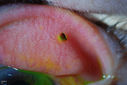
Corneal Abrasion with Foreign Body Present 2610 x angesehenER patient comes in with corneal abrasion in the left eye which was getting worst. VA 20/25. Slit lamp exam showed corneal abrasion superiorly at 2-o'clock. Flipped eyelid and foreign body appeared on the upper tarsal plate. Foreign body was removed.
|
|
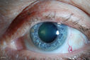
Dilated Conjunctival Blood Vessels2517 x angesehenPatient comes in for routine eye exam. VA 20/20 in both eyes. Fundus exam was normal. Dilated blood vessels are shown temporal inferiorly in the left eye.
|
|

"Jelly Bumps" Soft Contact Lens2433 x angesehenYoung female patient comes in wearing overnight contact lenses. Complains of irritation in both eyes. Denies sleeping in SCL. Slit lamp exam shows little jelly bumps underneath the contact lens in both eyes. Was issued a new pair and lectured on handling them.
|
|

Corneal Abrasion with Foreign Body Present 2414 x angesehenER patient comes in with corneal abrasion in the left eye which was getting worst. VA 20/25. Slit lamp exam showed corneal abrasion superiorly at 2-o'clock. Flipped eyelid and foreign body appeared on the upper tarsal plate. Foreign body was removed.
|
|

Dilated Conjunctival Blood Vessels2395 x angesehenPatient comes in for routine eye exam. VA 20/20 in both eyes. Fundus exam was normal. Dilated blood vessels are shown temporal inferiorly in the left eye.
|
|

Dilated Conjunctival Blood Vessels2363 x angesehenPatient comes in for routine eye exam. VA 20/20 in both eyes. Fundus exam was normal. Dilated blood vessels are shown temporal inferiorly in the left eye.
|
|

Uveitis - Iris Nodule - Koeppe Nodule2125 x angesehen29-year-old woman has had problems with uveitis in the right eye for about the last two years. Her symptoms are usually floaters. Occasionally the eye gets red, it has not been very uncomfortable, and it has always been in the right eye. Has had Iris Nodule like one in photo in left eye and Right eye. They clear with or without steroids after about a month.
|
|

Conjunctival Cyst2080 x angesehenPatient comes in with mechanical irritation in the right eye. No foreign body was found. Slit lamp photo shows conjunctival cyst was found temporal in the right eye. Patient will proceed with steroid/antibiotic drops and will stay out of contact lenses.
|
|

Corneal Foreign Body 2023 x angesehenPatient complains of foreign body sensation in the right eye. Slit lamp photos shows a piece of glass metal embedded into the inferior part of the cornea at 7-o'clock in the right eye. Foreign body was removed.
|
|

Correctopia - post laser iridoplasty - 20/20 vision2015 x angesehen1 month after the iridoplasty, her vision was 20/20. She began to improve on the drive home from the treatment. The laser setting were : green argon 500 microns, 500 mw, 500 msec.
|
|

Conjunctival Cyst1986 x angesehenPatient comes in with mechanical irritation in the right eye. No foreign body was found. Slit lamp photo shows conjunctival cyst was found temporal in the right eye. Patient will proceed with steroid/antibiotic drops and will stay out of contact lenses.
|
|

Ectopia Lentis - Marfan's Syndrome - Intermittent Pupillary Block Glaucoma Right Eye1975 x angesehen41-year-old woman decreased vision right eye and intermittent pupillary block glaucoma for 6 months from a dislocated natural lens in the eye, probably associated with Marfan syndrome.Vision OD is 20/160, PH 20/40; OS is 20/30, PH 20/25
|
|
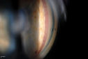
Gonioscopy, Neovascularization in the Angle1864 x angesehenNeovascularization in the right eye, temporally. VA is 20/40, right eye with IOP at 25. Patient will proceed with Avastin injection to the right eye.
|
|

Gonioscopy; Scattered Peripheral Anterior Synechiae1752 x angesehenPatient comes in for evaluation for glaucoma. Patient also has a history of Uveitis. Last flare up was back in 1990. Patient's VA was 20/30, Right eye and 20/40-1, Left eye. Slit Lamp Gonioscopy reveals iris bow with scattered PAS around the angles of the anterior chamber. You can also see pigmentation in the trabecular meshwork. Patient will follow up in 3-months.
|
|
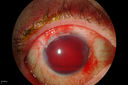
Hyphema1743 x angesehenHyphema
|
|
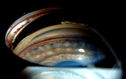
Gonioscopy- Pigmentary Glaucoma Suspect1723 x angesehenPatient with no family history of glaucoma, comes in with elevated IOP. During Gonioscopy exam. brown pigment overlying the trabecular meshwork. Also, trans-illumination defects on the iris.
|
|
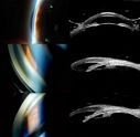
Angle Closure/ UBM & Gonioscopy1719 x angesehenPatient comes in a day after dilation with severe headache and nausea. Patient's IOP was 64 in the right eye. Patient ended up having angle closure attack. Promoted severe nausea and eye pain. Peripheral Iridotomy was performed and IOP dropped slowly. Patient was followed up the next morning and will be seen 2-days after that.
|
|

Ozurdex Implant in AC1699 x angesehen79 yr old male presents with Blurry vison with slight discomfort x 1 in OD. Pt is S/P Ozurdex Implant x 2 mos which has now surfaced in the Anterior Chamber.
|
|
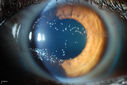
"Jelly Bumps" Soft Contact Lens1688 x angesehenYoung female patient comes in wearing overnight contact lenses. Complains of irritation in both eyes. Denies sleeping in SCL. Slit lamp exam shows little jelly bumps underneath the contact lens in both eyes. Was issued a new pair and lectured on handling them.
|
|

Gonioscopy; Scattered Peripheral Anterior Synechiae1664 x angesehenPatient comes in for evaluation for glaucoma. Patient also has a history of Uveitis. Last flare up was back in 1990. Patient's VA was 20/30, Right eye and 20/40-1, Left eye. Slit Lamp Gonioscopy reveals iris bow with scattered PAS around the angles of the anterior chamber. You can also see pigmentation in the trabecular meshwork. Patient will follow up in 3-months.
|
|

Ectopia Lentis - Marfan's Syndrome - Intermittent Pupillary Block Glaucoma Right Eye1663 x angesehen41-year-old woman decreased vision right eye and intermittent pupillary block glaucoma for 6 months from a dislocated natural lens in the eye, probably associated with Marfan syndrome.Vision OD is 20/160, PH 20/40; OS is 20/30, PH 20/25
|
|

Fuchs Heterochromic Iridocyclitis - Left eye has inflammation1647 x angesehenThe right eye is darker than the left eye which has chronic iritis
|
|

360-Degree's Pseudoexfoliation1604 x angesehenFollow-up on glaucoma. Patient has sever pseudoexfoliation in the right eye. VA is 20/25 with IOP at 27. Patient will return in 6-months for re-evaluation.
|
|

Gonioscopy; Scattered Peripheral Anterior Synechiae1574 x angesehenPatient comes in for evaluation for glaucoma. Patient also has a history of Uveitis. Last flare up was back in 1990. Patient's VA was 20/30, Right eye and 20/40-1, Left eye. Slit Lamp Gonioscopy reveals iris bow with scattered PAS around the angles of the anterior chamber. You can also see pigmentation in the trabecular meshwork. Patient will follow up in 3-months.
|
|

Correctopia - before laser iridoplasty - 20/60 vision1526 x angesehenIris is occluding visual axis
|
|

Flower Cataract1524 x angesehenPatient with ahmed tube and flower cataract
|
|

"Jelly Bumps" Soft Contact Lens1508 x angesehenYoung female patient comes in wearing overnight contact lenses. Complains of irritation in both eyes. Denies sleeping in SCL. Slit lamp exam shows little jelly bumps underneath the contact lens in both eyes. Was issued a new pair and lectured on handling them.
|
|

Ectopia Lentis - Marfan's Syndrome - Intermittent Pupillary Block Glaucoma Right Eye1499 x angesehen41-year-old woman decreased vision right eye and intermittent pupillary block glaucoma for 6 months from a dislocated natural lens in the eye, probably associated with Marfan syndrome.Vision OD is 20/160, PH 20/40; OS is 20/30, PH 20/25
|
|

Gonioscopy, Pigment Dispersion Syndrome1473 x angesehenPatient comes in with elevated pressures. Goinoscopy shows heavy pigment dusted off the iris into the trabecular meshwork. Laser procedure was done to break up the pigment for more aqueous flow.
|
|

Correctopia - before laser iridoplasty - 20/60 vision1471 x angesehenIris is occluding visual axis
|
|

Dilated Conjunctival Blood Vessels1470 x angesehenPatient comes in for routine eye exam. VA 20/20 in both eyes. Fundus exam was normal. Dilated blood vessels are shown temporal inferiorly in the left eye.
|
|
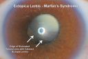
Ectopia Lentis and Cataract - Marfan's Syndrome1456 x angesehen40-year-old woman has had dislocated lenses since she was a child. The right eye is seeing well with an aphakic correction. The left eye unfortunately has declining vision for 4 months. OD 20/32, OS 20/125. Pinhole 20/50
|
|

Rubeosis Iridis1451 x angesehenPatient presents with Rubeosis Iridis in the right eye due to neovascular glaucoma. VA is 20/40 in the right eye. Will follow up in 3-months.
|
|

Uveitis - Iris Nodule - Koeppe Nodule1448 x angesehen29-year-old woman has had problems with uveitis in the right eye for about the last two years. Her symptoms are usually floaters. Occasionally the eye gets red, it has not been very uncomfortable, and it has always been in the right eye. Has had Iris Nodule like one in photo in left eye and Right eye. They clear with or without steroids after about a month.
|
|

Gonioscopy- Pigmentary Glaucoma Suspect1442 x angesehenPatient with no family history of glaucoma, comes in with elevated IOP. During Gonioscopy exam. brown pigment overlying the trabecular meshwork. Also, trans-illumination defects on the iris.
|
|

Juvenile Xanthogranuloma1424 x angesehenPresented with hyphaema after minor trauma
|
|

Gonioscopy; Scattered Peripheral Anterior Synechiae1414 x angesehenPatient comes in for evaluation for glaucoma. Patient also has a history of Uveitis. Last flare up was back in 1990. Patient's VA was 20/30, Right eye and 20/40-1, Left eye. Slit Lamp Gonioscopy reveals iris bow with scattered PAS around the angles of the anterior chamber. You can also see pigmentation in the trabecular meshwork. Patient will follow up in 3-months.
|
|

Gonioscopy; Scattered Peripheral Anterior Synechiae1409 x angesehenPatient comes in for evaluation for glaucoma. Patient also has a history of Uveitis. Last flare up was back in 1990. Patient's VA was 20/30, Right eye and 20/40-1, Left eye. Slit Lamp Gonioscopy reveals iris bow with scattered PAS around the angles of the anterior chamber. You can also see pigmentation in the trabecular meshwork. Patient will follow up in 3-months.
|
|

Gonioscopy; Scattered Peripheral Anterior Synechiae1406 x angesehenPatient comes in for evaluation for glaucoma. Patient also has a history of Uveitis. Last flare up was back in 1990. Patient's VA was 20/30, Right eye and 20/40-1, Left eye. Slit Lamp Gonioscopy reveals iris bow with scattered PAS around the angles of the anterior chamber. You can also see pigmentation in the trabecular meshwork. Patient will follow up in 3-months.
|
|

Cornea Ulcer/Abrasion1381 x angesehen
|
|

Endophthalmitis with Hypopion1379 x angesehen
|
|

Silicone Oil Bubbles in the Angle1367 x angesehenPatient is evaluated for glaucoma associated with ocular inflammation. VA is CF in the right eye and 20/50, left eye. Gonioscopy photos shows little silicone oil bubbles in the inferior angle of the right eye. Patient to consider removal of silicone oil.
|
|

Gonioscopy- Pigmentary Glaucoma Suspect1361 x angesehenPatient with no family history of glaucoma, comes in with elevated IOP. During Gonioscopy exam. brown pigment overlying the trabecular meshwork. Also, trans-illumination defects on the iris.
|
|

Displaced Scleral Buckle1359 x angesehenDisplaced Scleral Buckle in OD of a 75 yr old male
|
|

Foreign Body "Rust Ring" 1351 x angesehenPatient complained of redness and tearing with white discharge. Patient was working with a grinding wheel on a vehicle. Slit lamp exam shows a rust ring barley off center. FBS was removed with Algiers Brush.
|
|

"Jelly Bumps" Soft Contact Lens1350 x angesehenYoung female patient comes in wearing overnight contact lenses. Complains of irritation in both eyes. Denies sleeping in SCL. Slit lamp exam shows little jelly bumps underneath the contact lens in both eyes. Was issued a new pair and lectured on handling them.
|
|

"Jelly Bumps" Soft Contact Lens1342 x angesehenYoung female patient comes in wearing overnight contact lenses. Complains of irritation in both eyes. Denies sleeping in SCL. Slit lamp exam shows little jelly bumps underneath the contact lens in both eyes. Was issued a new pair and lectured on handling them.
|
|
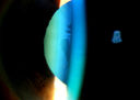
360-Degree's Pseudoexfoliation1339 x angesehenFollow-up on glaucoma. Patient has sever pseudoexfoliation in the right eye. VA is 20/25 with IOP at 27. Patient will return in 6-months for re-evaluation.
|
|

Endophthalmitis with Hypopion1335 x angesehen
|
|

Uveitis - Iris Nodule - Koeppe Nodule1327 x angesehen29-year-old woman has had problems with uveitis in the right eye for about the last two years. Her symptoms are usually floaters. Occasionally the eye gets red, it has not been very uncomfortable, and it has always been in the right eye. Has had Iris Nodule like one in photo in left eye and Right eye. They clear with or without steroids after about a month.
|
|

Gonioscopy; Scattered Peripheral Anterior Synechiae1327 x angesehenPatient comes in for evaluation for glaucoma. Patient also has a history of Uveitis. Last flare up was back in 1990. Patient's VA was 20/30, Right eye and 20/40-1, Left eye. Slit Lamp Gonioscopy reveals iris bow with scattered PAS around the angles of the anterior chamber. You can also see pigmentation in the trabecular meshwork. Patient will follow up in 3-months.
|
|

Dusted Pigment, Iris Nevus1322 x angesehen
|
|

Gonioscopy- Pigmentary Glaucoma Suspect1313 x angesehenPatient with no family history of glaucoma, comes in with elevated IOP. During Gonioscopy exam. brown pigment overlying the trabecular meshwork. Also, trans-illumination defects on the iris.
|
|

Corneal Abrasion with Foreign Body Present 1304 x angesehenER patient comes in with corneal abrasion in the left eye which was getting worst. VA 20/25. Slit lamp exam showed corneal abrasion superiorly at 2-o'clock. Flipped eyelid and foreign body appeared on the upper tarsal plate. Foreign body was removed.
|
|
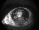
Iris Neovascularization1296 x angesehenDiabetic patient with advanced iris neovascularization
|
|

Uveitis - Iris Nodule - Koeppe Nodule1293 x angesehen29-year-old woman has had problems with uveitis in the right eye for about the last two years. Her symptoms are usually floaters. Occasionally the eye gets red, it has not been very uncomfortable, and it has always been in the right eye. Has had Iris Nodule like one in photo in left eye and Right eye. They clear with or without steroids after about a month.
|
|

Phacoantigenic Reaction1293 x angesehenPicture shows a Left eye enucleation with H&E staining.
Anterior capsule of lens is broken with evidence of granulomatous inflammation.
|
|

Ahmed Tube Placement1287 x angesehenPatient with severe glaucoma is seen post op for ahmed tube placement. VA is 20/70 and IOP is 8 in the left eye. Patient is stable.
|
|

Gonioscopy; Scattered Peripheral Anterior Synechiae1269 x angesehenPatient comes in for evaluation for glaucoma. Patient also has a history of Uveitis. Last flare up was back in 1990. Patient's VA was 20/30, Right eye and 20/40-1, Left eye. Slit Lamp Gonioscopy reveals iris bow with scattered PAS around the angles of the anterior chamber. You can also see pigmentation in the trabecular meshwork. Patient will follow up in 3-months.
|
|

Corneal Foreign Body 1268 x angesehenPatient complains of foreign body sensation in the right eye. Slit lamp photos shows a piece of glass metal embedded into the inferior part of the cornea at 7-o'clock in the right eye. Foreign body was removed.
|
|

Fuch's heterochromic iridocyclitis - uninvolved brown eye1267 x angesehen46-year-old man has had blurred vision in the left eye for the last two years. He had cataract surgery in that eye eight years ago. He has also had a lot of floaters in the left eye for several years. His left eye is blue and his right eye is brown. OD 20/16, OS 20/50.
|
|
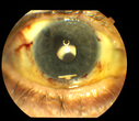
Ozurdex Implant in AC1267 x angesehen79yr old male presents Blurry vision with slight discomfort x 1 week in OD . Pt is S/P Ozurdex Implant x 2mos which is now surfaced in the Anterior Chamber.
|
|

Conjunctival Melanoma1267 x angesehenRight conjunctival melanoma extending into anterior orbit, right eye. Temporally and nasally with pigmented masses/nodules. VA was 20/30 without correction in the right eye. Follow up to proceed with proton beam therapy.
|
|

Giant Papillary Conjunctivitis, Left Upper Eyelid1255 x angesehenContact lens wearer, comes in for exam. Has rough feeling underneath both eyelids. Patient thought it was through SCL wear. Patient VA was 20/20. right eye, 20/30, left eye. Underneath the left upper eyelid, you can see papillary inflammation and redness.
|
|

Iris Nevus1245 x angesehenRoutine eye exam. Iris nevus located around 4-o'clock off the pupil.
|
|

Gonioscopy, Neovascularization in the Angle1245 x angesehenNeovascularization in the right eye, temporally. VA is 20/40, right eye with IOP at 25. Patient will proceed with Avastin injection to the right eye.
|
|

Accordioning Crystalline Lens with loss of Posterior Capsule1243 x angesehen71-year old male complains of blurred vision in the left eye. VA 20/40, right eye and 20/400, left eye without correction. Slit lamp exam shows Crystalline lens, both eyes. Right eye IOL is aligned and centered. Left eye shows an accordion of the crystalline lens. Retro Illumination shows the IOL bent inward in the left eye. There was a 6-diopter difference of astigmatism between the right and left eye. Patient will have surgery to correct the issue.
|
|

Gonioscopy, Pigment Dispersion Syndrome1241 x angesehenPatient comes in with elevated pressures. Goinoscopy shows heavy pigment dusted off the iris into the trabecular meshwork. Laser procedure was done to break up the pigment for more aqueous flow.
|
|
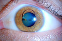
Silicone Oil in the Anterior Chamber1239 x angesehenPatient comes in for second opinion for retinal detachment in the left eye. VA is hand motion in the left eye. Poor view to look into the retina. Small silicone bubble at 12 o'clock superior in the anterior chamber. Surgery was not recommended due to the silicone oil in the left eye.
|
|

Gonioscopy- Pigmentary Glaucoma Suspect1229 x angesehenPatient with no family history of glaucoma, comes in with elevated IOP. During Gonioscopy exam. brown pigment overlying the trabecular meshwork. Also, trans-illumination defects on the iris.
|
|

Silicone Oil in the Anterior Chamber1228 x angesehenPatient comes in for second opinion for retinal detachment in the left eye. VA is hand motion in the left eye. Poor view to look into the retina. Small silicone bubble at 12 o'clock superior in the anterior chamber. Surgery was not recommended due to the silicone oil in the left eye.
|
|

Gonioscopy of Blood in the Angle1219 x angesehenYoung female who has a history of uveitis, glaucoma and juvenile rheumatoid arthritis. VA is hand motion in the left eye. Pigment changes with some blood in the anterior chamber seen here on Gonioscopy.
|
|
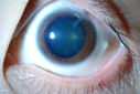
Flower Cataract1219 x angesehenPatient with ahmed tube and flower cataract
|
|

Prominent Corneal Nerves1218 x angesehenSlit lamp exam shows prominent corneal nerves present at 8-10-o'clock and 4-6-o'clock. Baseline work to rule out Fuch's dystrophy and recurrent corneal erosion.
|
|
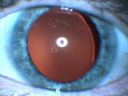
Ectopia Lentis - Marfan's Syndrome - Intermittent Pupillary Block Glaucoma Right Eye1213 x angesehen41-year-old woman decreased vision right eye and intermittent pupillary block glaucoma for 6 months from a dislocated natural lens in the eye, probably associated with Marfan syndrome.Vision OD is 20/160, PH 20/40; OS is 20/30, PH 20/25
|
|

Ghost Cell Glaucoma1210 x angesehenPost cataract surgery, patient presents with ghost cell in the anterior chamber after hemorrhage cleared. A small hyphema still is visible in the anterior chamber with a whitish ball appearance.
|
|

Gonioscopy; Scattered Peripheral Anterior Synechiae1202 x angesehenPatient comes in for evaluation for glaucoma. Patient also has a history of Uveitis. Last flare up was back in 1990. Patient's VA was 20/30, Right eye and 20/40-1, Left eye. Slit Lamp Gonioscopy reveals iris bow with scattered PAS around the angles of the anterior chamber. You can also see pigmentation in the trabecular meshwork. Patient will follow up in 3-months.
|
|

Cilia and Cement Intraocular Foreign Body1188 x angesehenConstruction worker jack-hammering 3 months prior to pictures. Asymptomatic. 20/20 OU. Corneal wound seen superiorly (not in pictures)
|
|

Dislocated IOL with Tube Shunt1184 x angesehenPatient comes in with a dislocated lens in the right eye. Decreased vision in the right eye. Slit lamp photos shows lens dislocated halfway inferior. Ahmed Tube shut is visible at 11-o'clock superiorly in the right eye.
|
|

Ghost Cell Glaucoma1176 x angesehenPost cataract surgery, patient presents with ghost cell in the anterior chamber after hemorrhage cleared. A small hyphema still is visible in the anterior chamber with a whitish ball appearance.
|
|

Ghost Cell Glaucoma1172 x angesehenPost cataract surgery, patient presents with ghost cell in the anterior chamber after hemorrhage cleared. A small hyphema still is visible in the anterior chamber with a whitish ball appearance.
|
|

Subluxed IOL with TID1160 x angesehenPatient comes in with blurred vision in the left eye. Slit lamp exam shows dislocated IOL, inferior, left eye. PXF material on the implant inferiorly.
|
|

Gonioscopy, Mass in the Angle of Anterior Chamber1154 x angesehenSlit Lamp and goinoscopy photos show a mass at 7-o'clock i the right eye. The mass extends beneath the iris behind the lens.
|
|
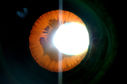
360-Degree's Pseudoexfoliation1153 x angesehenFollow-up on glaucoma. Patient has sever pseudoexfoliation in the right eye. VA is 20/25 with IOP at 27. Patient will return in 6-months for re-evaluation.
|
|
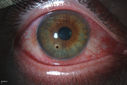
Corneal Foreign Body 1144 x angesehenPatient complains of foreign body sensation in the right eye. Slit lamp photos shows a piece of glass metal embedded into the inferior part of the cornea at 7-o'clock in the right eye. Foreign body was removed.
|
|

Gonioscopy; Scattered Peripheral Anterior Synechiae1138 x angesehenPatient comes in for evaluation for glaucoma. Patient also has a history of Uveitis. Last flare up was back in 1990. Patient's VA was 20/30, Right eye and 20/40-1, Left eye. Slit Lamp Gonioscopy reveals iris bow with scattered PAS around the angles of the anterior chamber. You can also see pigmentation in the trabecular meshwork. Patient will follow up in 3-months.
|
|

Free Floating Dislocated Lens in Vitreous1133 x angesehenPatient comes in aphakic with dislocated lens floating to the back of the eye when laying down. Lens is laying up against the endothelium of the cornea when patient is right side up..
|
|

Gonioscopy; Scattered Peripheral Anterior Synechiae1125 x angesehenPatient comes in for evaluation for glaucoma. Patient also has a history of Uveitis. Last flare up was back in 1990. Patient's VA was 20/30, Right eye and 20/40-1, Left eye. Slit Lamp Gonioscopy reveals iris bow with scattered PAS around the angles of the anterior chamber. You can also see pigmentation in the trabecular meshwork. Patient will follow up in 3-months.
|
|

Ghost Cell Glaucoma1123 x angesehenPost cataract surgery, patient presents with ghost cell in the anterior chamber after hemorrhage cleared. A small hyphema still is visible in the anterior chamber with a whitish ball appearance.
|
|

Gonioscopy, Pigment Dispersion Syndrome1123 x angesehenPatient comes in with elevated pressures. Goinoscopy shows heavy pigment dusted off the iris into the trabecular meshwork. Laser procedure was done to break up the pigment for more aqueous flow.
|
|

Fuch's heterochromic iridocyclitis - inflammed blue eye1118 x angesehen46-year-old man has had blurred vision in the left eye for the last two years. He had cataract surgery in that eye eight years ago. He has also had a lot of floaters in the left eye for several years. His left eye is blue and his right eye is brown. OD 20/16, OS 20/50.
|
|

Corneal Abrasion with Foreign Body Present 1106 x angesehenER patient comes in with corneal abrasion in the left eye which was getting worst. VA 20/25. Slit lamp exam showed corneal abrasion superiorly at 2-o'clock. Flipped eyelid and foreign body appeared on the upper tarsal plate. Foreign body was removed.
|
|

Pterygium and RK Scars1100 x angesehenPatient comes in for SCL fitting. VA is 20/30. right eye and 20/70, left eye. Slit lamp photos show 8-RK Scars in both eyes. Pterygium, nasally, left eye.
|
|

Ghost Cell Glaucoma1089 x angesehenPost cataract surgery, patient presents with ghost cell in the anterior chamber after hemorrhage cleared. A small hyphema still is visible in the anterior chamber with a whitish ball appearance.
|
|

Gonioscopy, Pigment Dispersion Syndrome1086 x angesehenPatient comes in with elevated pressures. Goinoscopy shows heavy pigment dusted off the iris into the trabecular meshwork. Laser procedure was done to break up the pigment for more aqueous flow.
|
|

Cornea Ulcer with Iris Prolaps1060 x angesehen
|
|

Gonioscopy; Scattered Peripheral Anterior Synechiae1055 x angesehenPatient comes in for evaluation for glaucoma. Patient also has a history of Uveitis. Last flare up was back in 1990. Patient's VA was 20/30, Right eye and 20/40-1, Left eye. Slit Lamp Gonioscopy reveals iris bow with scattered PAS around the angles of the anterior chamber. You can also see pigmentation in the trabecular meshwork. Patient will follow up in 3-months.
|
|

Peripheral Iridotomy1055 x angesehenSlit lamp photo of peripheral iridotomy for narrow angles. A hole was place at 10-o'clock superior temporally.
|
|
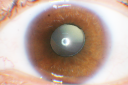
Ectopia Lentis and Cataract - Marfan's Syndrome1053 x angesehen40-year-old woman has had dislocated lenses since she was a child. The right eye is seeing well with an aphakic correction. The left eye unfortunately has declining vision for 4 months. OD 20/32, OS 20/125. Pinhole 20/50
|
|
| 381 Dateien auf 4 Seite(n) |
1 |
 |
|


