am meisten angesehen - Congenital Anomalies
|

Amelanotic - Light Colored CHRPE - Congenital Hypertrophy of the Pigment Epithelium5234 x angesehen56-year-old woman nov vision problems. OD is 20/20, OS is 20/16
|
|

Type 1 Retinal Arteriovenous Malformation4297 x angesehen42 year old female VA 20/32 OD, 20/20 OS MRI was negative.
|
|

Wyburn Mason Syndrome4106 x angesehen28 year old female, visual acuity OS 20/200
|
|

CAVERNOUS HEMONGIOMA2969 x angesehen28 year old male w/ 20/200 vision at time of exam. Patient c/o poor vision since childhood. No significant medical history or family medical history. A problem was only noted when patient enlisted in the Army.
|
|

Combined hamartoma of the retina and RPE2881 x angesehen16 year old female, diagnosed with a combined hamartoma of the retina and the retinal pigment epithelium.
|
|

Amelanotic - Light Colored CHRPE - Congenital Hypertrophy of the Pigment Epithelium2848 x angesehen56-year-old woman nov vision problems. OD is 20/20, OS is 20/16
|
|

OD Albinism with foveal hypoplasia2460 x angesehen28 year old female latina with vision of 20/400
|
|

CAVERNOUS HEMONGIOMA2321 x angesehen28 year old male. FA
|
|

Wyburn-Mason Syndrome 2141 x angesehen28 year old female, diagnosed with Wyburn-Mason Syndrome at age 7. At time of exam, vision in the left eye was 4/200.
|
|

Myelinated Nerve Fiber Layer Right Eye (white area)2136 x angesehen62-year-old man myelinated nerve fiber layer in the right eye. OD 20/25, OS 20/30.
|
|
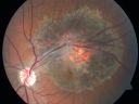
Retinal Pigment Epithelial Dysgenesis2083 x angesehen
|
|

CHRPE lesion in the left eye - Irregular pigmentation2050 x angesehen63 year old female with normal vision and CHRPE lesion in the right eye.
|
|

Sturge-Weber Encephalotigeminal Angiomatosis - Facial Hemangioma and Asymptomatic Ipsilateral Diffuse Choroidal Hemangioma2018 x angesehen61-year-old man with Sturge-Weber syndrome with a hemangioma on the left side of his face.
VISUAL ACUITY: Vision OD is 20/50, PH 20/30; OS 20/80, PH 20/30. IOP: OD 16, OS 19.
|
|

Myelinated Nerve Fiber Layer Right Eye (white area)1949 x angesehen62-year-old man myelinated nerve fiber layer in the right eye. OD 20/25, OS 20/30.
|
|

Coats' Disease in 14 Year Old Boy Treated with Cryotherapy and Avastin1854 x angesehen14 Year Old Boy with Coats Disease
|
|
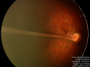
1769 x angesehenPHPV - right eye - optic nerve
|
|

Chorioretinal Scar1727 x angesehen21 year old male with a chorioretinal scar OD
|
|
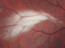
Myelinated Nerve Fiber Layer Right Eye (white area)1610 x angesehen62-year-old man myelinated nerve fiber layer in the right eye. OD 20/25, OS 20/30.
|
|

CHRPE lesion in the left eye - Irregular pigmentation1592 x angesehen63 year old female with normal vision and CHRPE lesion in the right eye.
|
|
Sturge-Weber Encephalotigeminal Angiomatosis - Facial Hemangioma and Asymptomatic Ipsilateral Diffuse Choroidal Hemangioma1526 x angesehen61-year-old man with Sturge-Weber syndrome with a hemangioma on the left side of his face.
VISUAL ACUITY: Vision OD is 20/50, PH 20/30; OS 20/80, PH 20/30. IOP: OD 16, OS 19.
|
|
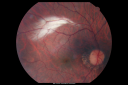
Myelinated Nerve Fiber Layer Right Eye (white area)1484 x angesehen62-year-old man myelinated nerve fiber layer in the right eye. OD 20/25, OS 20/30.
|
|

Hypertrophy of the Retinal Pigment Epithelium1481 x angesehenPatient comes in with vision changes in the right eye. VA is 20/30 in the right eye. Fundus and FAF photos show a large area of Hypertrophy in the right eye.
|
|

Myelinated Nerve Fiber Layer Right Eye (white area)1479 x angesehen62-year-old man myelinated nerve fiber layer in the right eye. OD 20/25, OS 20/30.
|
|

wyburn-mason sydrome1432 x angesehen28 year old female, visual acuity OS 20/200
|
|

Retinal Coloboma1425 x angesehen14-year old male presents with decreased vision in the left eye. Dx with iris and retinal coloboma in the left eye. Patient VA was 20/20, right eye, 20/100 left eye with pinhole improvement 20\50. Patient was fitted for SCL in the left eye.
|
|

Sturge-Weber Encephalotigeminal Angiomatosis - Facial Hemangioma and Ipsilateral Diffuse Choroidal Hemangioma1406 x angesehen61-year-old man with Sturge-Weber syndrome with a hemangioma on the left side of his face.
VISUAL ACUITY: Vision OD is 20/50, PH 20/30; OS 20/80, PH 20/30. IOP: OD 16, OS 19.
|
|

wyburn-mason syndrome fundus photos1397 x angesehen28 year old female, visual acuity 20/200
|
|

CHRPE lesion in the left eye - Irregular pigmentation1389 x angesehen63 year old female with normal vision and CHRPE lesion in the right eye.
|
|

CHRPE - Congenital Hypertrophy of the Pigment Epithelium - Peripheral1366 x angesehen55-year-old woman OD 20/30, OS 20/25
|
|

Coats' Disease with Cryotherapy 1359 x angesehenYoung male with Coat's Disease with Cryotherapy in the right eye. VA was 20/20, right eye. Left eye was completely normal. Patient will be followed up in 3-months.
|
|

Nearly complete coloboma1318 x angesehenNearly complete coloboma from necropsy of infant born with multiple congenital anomalies, incompatable with life, including holorprosencephaly. Infant expired on 3rd post-partum day
|
|

Myelinated Nerve Fiber Layer Right Eye (white area)1313 x angesehen62-year-old man myelinated nerve fiber layer in the right eye. OD 20/25, OS 20/30.
|
|

50 Year Old woman with Congenital Retinal Vascular Tortuosity - Normal Vision1312 x angesehen
|
|

Sturge-Weber Encephalotigeminal Angiomatosis - Facial Hemangioma and Asymptomatic Ipsilateral Diffuse Choroidal Hemangioma1293 x angesehen61-year-old man with Sturge-Weber syndrome with a hemangioma on the left side of his face.
VISUAL ACUITY: Vision OD is 20/50, PH 20/30; OS 20/80, PH 20/30. IOP: OD 16, OS 19.
|
|

CHRPE lesion in the left eye - Irregular pigmentation1242 x angesehen63 year old female with normal vision and CHRPE lesion in the right eye.
|
|

Sturge-Weber Encephalotigeminal Angiomatosis - Facial Hemangioma and Asymptomatic Ipsilateral Diffuse Choroidal Hemangioma1229 x angesehen61-year-old man with Sturge-Weber syndrome with a hemangioma on the left side of his face.
VISUAL ACUITY: Vision OD is 20/50, PH 20/30; OS 20/80, PH 20/30. IOP: OD 16, OS 19.
|
|

AVM1229 x angesehenFP of AVM in OS of a 48 yr old female.
|
|

Retinal Pigment Epithelial Dysgenesis1212 x angesehen
|
|

Myelinated Nerve Fiber Layer1200 x angesehen69-year-old woman OD is 20/30, OS is 20/40.
|
|

CHRPE - Congenital Hypertrophy of the Pigment Epithelium - Peripheral1165 x angesehen55-year-old woman OD 20/30, OS 20/25
|
|
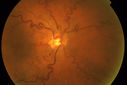
50 Year Old woman with Congenital Retinal Vascular Tortuosity - Normal Vision1149 x angesehen
|
|
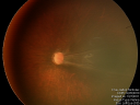
1131 x angesehenPHPV - Left eye - optic nerve
|
|

50 Year Old woman with Congenital Retinal Vascular Tortuosity - Normal Vision1117 x angesehen
|
|

Anomalous Branch Retinal Artery 1114 x angesehen59-year-old man OD 20/25, OS 20/25. TORTUOUS RETINAL ARTERY – LEF EYE
(POSSIBLE GROUP 2 WYBURN-MASON SYNDROME
POSSIBLE ACEPHALIC MIGRAINES )
|
|

Multifocal CHRPE also called "Bear Tracks"1105 x angesehen
|
|
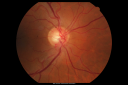
Anomalous Branch Retinal Artery 1092 x angesehen59-year-old man OD 20/25, OS 20/25. TORTUOUS RETINAL ARTERY – LEF EYE
(POSSIBLE GROUP 2 WYBURN-MASON SYNDROME
POSSIBLE ACEPHALIC MIGRAINES )
|
|
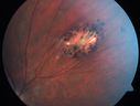
Congenital Hypertrophy of the Retinal Pigment Epithelium CHRPE has Satellite Lesions1077 x angesehen
|
|
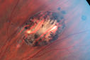
Congenital Hypertrophy of the Retinal Pigment Epithelium CHRPE has Satellite Lesions1071 x angesehen
|
|

Retinal Coloboma1070 x angesehenYoung 19-year old female with Charge Syndrome comes in for eye exam. VA is 20/400, right eye, 20/30, left eye. Fundus photography shows coloboma covering the macula and pushing the optic nerve in the right eye.
|
|

Foveal hypoplasia - aplasia1056 x angesehenSD OCT shows persistent plexiform layers in the center of the macula
EXTENDED HPI: He is having some trouble seeing his gadgets (like his watch). He was diagnosed with morning glory syndrome at about 3 yeras old. This last visit you saw fluid under the retina and asked him to come here. The vision is a little worse.
VA OD: Dcc20/40+2 NccJ2-2
VA OS: Dcc20/25-2 NccJ1-2
|
|

Nanopthalmous (Microphthalmous) Chorioretinal Folds and Choroidal Thickening Both Eyes B-Scan Ultrasound1047 x angesehen
|
|

Nanopthalmous (Microphthalmous) Chorioretinal Folds and Choroidal Thickening Both Eyes B-Scan Ultrasound1035 x angesehen
|
|

1027 x angesehenPHPV - right eye. Peripheral extension
|
|

CHRPE lesion in the left eye - Irregular pigmentation1025 x angesehen63 year old female with normal vision and CHRPE lesion in the right eye.
|
|

Sturge-Weber Encephalotigeminal Angiomatosis - Facial Hemangioma and Ipsilateral Diffuse Choroidal Hemangioma1020 x angesehen61-year-old man with Sturge-Weber syndrome with a hemangioma on the left side of his face.
VISUAL ACUITY: Vision OD is 20/50, PH 20/30; OS 20/80, PH 20/30. IOP: OD 16, OS 19.
|
|

50 Year Old woman with Congenital Retinal Vascular Tortuosity - Normal Vision1011 x angesehen
|
|

Sturge-Weber Encephalotigeminal Angiomatosis - Facial Hemangioma and Asymptomatic Ipsilateral Diffuse Choroidal Hemangioma994 x angesehen61-year-old man with Sturge-Weber syndrome with a hemangioma on the left side of his face.
VISUAL ACUITY: Vision OD is 20/50, PH 20/30; OS 20/80, PH 20/30. IOP: OD 16, OS 19.
|
|

Nanopthalmous (Microphthalmous) Chorioretinal Folds and Choroidal Thickening Both Eyes985 x angesehen67-year-old woman with microphthalmos/nanophthalmos Her vision has gradually been getting a little bit worse, especially in the right eye. Also the anterior chamber is getting crowded and there is concern about the need for cataract surgery. She has one sister and one cousin who also have nanophthalmos.
VISUAL ACUITY: OD 20/50, OS 20/80.
|
|

Sturge-Weber Encephalotigeminal Angiomatosis - Facial Hemangioma and Asymptomatic Ipsilateral Diffuse Choroidal Hemangioma980 x angesehen61-year-old man with Sturge-Weber syndrome with a hemangioma on the left side of his face.
VISUAL ACUITY: Vision OD is 20/50, PH 20/30; OS 20/80, PH 20/30. IOP: OD 16, OS 19.
|
|

Retinal Pigment Epithelial Dysgenesis971 x angesehen
|
|

Anomalous Branch Retinal Artery 953 x angesehen59-year-old man OD 20/25, OS 20/25. TORTUOUS RETINAL ARTERY – LEF EYE
(POSSIBLE GROUP 2 WYBURN-MASON SYNDROME
POSSIBLE ACEPHALIC MIGRAINES )
|
|

Retinal Pigment Epithelial Dysgenesis947 x angesehen
|
|

Sturge-Weber Encephalotigeminal Angiomatosis - Facial Hemangioma and Asymptomatic Ipsilateral Diffuse Choroidal Hemangioma943 x angesehen61-year-old man with Sturge-Weber syndrome with a hemangioma on the left side of his face.
VISUAL ACUITY: Vision OD is 20/50, PH 20/30; OS 20/80, PH 20/30. IOP: OD 16, OS 19.
|
|
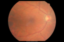
Sturge-Weber Encephalotigeminal Angiomatosis - Facial Hemangioma and Asymptomatic Ipsilateral Diffuse Choroidal Hemangioma937 x angesehen61-year-old man with Sturge-Weber syndrome with a hemangioma on the left side of his face.
VISUAL ACUITY: Vision OD is 20/50, PH 20/30; OS 20/80, PH 20/30. IOP: OD 16, OS 19.
|
|

Choroidal Hemangioma - Diffuse - Enhanced Depth Imaging Spectral Domain OCT Line Scan - Abnormal Eye931 x angesehenChoroidal Thickness is 697 Microns from Diffuse Choroidal Hemangioma
|
|
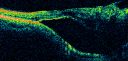
Retinal Pigment Epithelial Dysgenesis926 x angesehen
|
|

Myelinated Nerve Fiber Layer - Optic Nerve908 x angesehenRight eye subtle and inferior - left eye obvious
|
|

906 x angesehenPHPV - Left eye macular area. The patient was diagnosed as having regressed ROP even with a history of full term birth with no antecedent perinatal history of disease
|
|

Retinal Pigment Epithelial Dysgenesis898 x angesehen
|
|

Coats' Disease in 14 Year Old Boy Treated with Cryotherapy and Avastin895 x angesehen14 Year Old Boy with Coats Disease
|
|

Retinal Pigment Epithelial Dysgenesis893 x angesehen
|
|
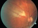
Myelinated Nerve Fiber Layer891 x angesehen69-year-old woman OD is 20/30, OS is 20/40.
|
|

Myelinated Nerve Fiber Layer889 x angesehen69-year-old woman OD is 20/30, OS is 20/40.
|
|

Coats' Disease in 14 Year Old Boy Treated with Cryotherapy and Avastin872 x angesehen14 Year Old Boy with Coats Disease
|
|

Sturge-Weber Encephalotigeminal Angiomatosis - Facial Hemangioma and Asymptomatic Ipsilateral Diffuse Choroidal Hemangioma857 x angesehen61-year-old man with Sturge-Weber syndrome with a hemangioma on the left side of his face.
VISUAL ACUITY: Vision OD is 20/50, PH 20/30; OS 20/80, PH 20/30. IOP: OD 16, OS 19.
|
|
Retinal Pigment Epithelial Dysgenesis856 x angesehen
|
|

Anomalous Branch Retinal Artery 856 x angesehen59-year-old man OD 20/25, OS 20/25. TORTUOUS RETINAL ARTERY – LEF EYE
(POSSIBLE GROUP 2 WYBURN-MASON SYNDROME
POSSIBLE ACEPHALIC MIGRAINES )
|
|
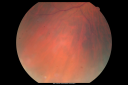
Sturge-Weber Encephalotigeminal Angiomatosis - Facial Hemangioma and Asymptomatic Ipsilateral Diffuse Choroidal Hemangioma854 x angesehen61-year-old man with Sturge-Weber syndrome with a hemangioma on the left side of his face.
VISUAL ACUITY: Vision OD is 20/50, PH 20/30; OS 20/80, PH 20/30. IOP: OD 16, OS 19.
|
|
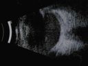
Nanopthalmous (Microphthalmous) Chorioretinal Folds and Choroidal Thickening Both Eyes B-Scan Ultrasound853 x angesehen
|
|

Retinal Pigment Epithelial Dysgenesis834 x angesehen
|
|

Retinal Pigment Epithelial Dysgenesis828 x angesehen
|
|

Retinal Pigment Epithelial Dysgenesis827 x angesehen
|
|
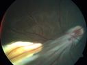
Familial Exudative Vitreoretinopathy - FEVR - Stage 4b OD822 x angesehen10 year old child with poor vision OD from birth. The left eye had vascular remodelling in the temporal periphery with preretinal abnormalitlies seen on OCT. The patient never returned for a fluorescein angiogram. Left eye is either stage 1 or stage 2. no family history
|
|

Choroidal Hemangioma - Diffuse - Enhanced Depth Imaging Spectral Domain OCT Line Scan - Normal Eye817 x angesehenNormal Choroidal Thickness in Right Eye
|
|

Retinal Pigment Epithelial Dysgenesis815 x angesehen
|
|

Nanopthalmous (Microphthalmous) Chorioretinal Folds and Choroidal Thickening Both Eyes B-Scan Ultrasound815 x angesehen
|
|
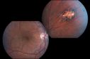
Congenital Hypertrophy of the Retinal Pigment Epithelium CHRPE has Satellite Lesions813 x angesehen
|
|

Nanopthalmous (Microphthalmous) Chorioretinal Folds and Choroidal Thickening Both Eyes B-Scan Ultrasound812 x angesehen
|
|

Coats' Disease in 14 Year Old Boy Treated with Cryotherapy and Avastin812 x angesehen14 Year Old Boy with Coats Disease
|
|
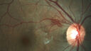
Type 1 Retinal Arteriovenous Malformation812 x angesehen42 year old female VA 20/32 OD, 20/20 OS MRI was negative.
|
|

Retinal Coloboma799 x angesehenYoung 19-year old female with Charge Syndrome comes in for eye exam. VA is 20/400, right eye, 20/30, left eye. Fundus photography shows coloboma covering the macula and pushing the optic nerve in the right eye.
|
|

Retinal Pigment Epithelial Dysgenesis797 x angesehen
|
|

Anomalous Branch Retinal Artery 785 x angesehen59-year-old man OD 20/25, OS 20/25. TORTUOUS RETINAL ARTERY – LEF EYE
(POSSIBLE GROUP 2 WYBURN-MASON SYNDROME
POSSIBLE ACEPHALIC MIGRAINES )
|
|

Coloboma - optic nerve, choroid, and retina - left eye781 x angesehen80 year old man with glaucoma in the right eye. The left eye has a coloboma of the optic nerve, choroid and retina.
VA 20/25 OD, 20/32 OS
|
|

Retinal Pigment Epithelial Dysgenesis775 x angesehen
|
|
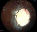
775 x angesehenYoung 19-year old female with Charge Syndrome comes in for eye exam. VA is 20/400, right eye, 20/30, left eye. Fundus photography shows coloboma covering the macula and pushing the optic nerve in the right eye.
|
|

Foveal hypoplasia - aplasia773 x angesehenSD OCT shows persistent plexiform layers in the center of the macula
EXTENDED HPI: He is having some trouble seeing his gadgets (like his watch). He was diagnosed with morning glory syndrome at about 3 yeras old. This last visit you saw fluid under the retina and asked him to come here. The vision is a little worse.
VA OD: Dcc20/40+2 NccJ2-2
VA OS: Dcc20/25-2 NccJ1-2
|
|

Congenital hypertrophy of the retinal pigment epithelium CHRPE773 x angesehenFAF, Optos Photo and FA
|
|

Coats' Disease - 14 year old - Asymptomatic767 x angesehenPresented with macular exudates. Temporal macroaneurysms responded to laser. Ultra-wide field FA shows nonperfusion
|
|

Myelinated Nerve Fiber Layer765 x angesehen69-year-old woman OD is 20/30, OS is 20/40.
|
|
| 578 Dateien auf 6 Seite(n) |
1 |
 |
|
|
|


