Les plus populaires - Choroidal Melanoma and Choroidal Nevus
|

CHRPE - Gardner Syndrome - FAMILIAL COLORECTAL POLYPOSIS - 11 year old femalevu 4045 fois11-year-old child with CHRPE OS has six undescended adult teeth and also she has had a cyst removed from her arm pit and she has three more in her arm pit and one on her face and she is seeing a dermatologist for that in the near future. The mother reports no health problems, however, the patient’s grandmother does have a history of intestinal polyps, as does the patient’s grandmother’s brothers son.
|
|

CHRPE - Gardner Syndrome - FAMILIAL COLORECTAL POLYPOSIS - 11 year old femalevu 2051 fois11-year-old child with CHRPE OS has six undescended adult teeth and also she has had a cyst removed from her arm pit and she has three more in her arm pit and one on her face and she is seeing a dermatologist for that in the near future. The mother reports no health problems, however, the patient’s grandmother does have a history of intestinal polyps, as does the patient’s grandmother’s brothers son.
|
|

Amelanotic Choroidal Nevus vu 2004 fois45-year-old man OD 20/20, OS 20/20. 5x5 mm flat, yellow choroidal lesion just inferonasal to the optic nerve, two disc-diameters from the nerve with scalloped edges. It looks like a confluent drusen, but it is not elevated.
|
|

Multiple - Congenital Hypertrophy of the Pigment Epithelium - CHRPE - Gardner Syndromevu 1916 fois63 Year Old woman with a history of colon cancer at age 32. She has also had thyroid cancer. Two of her 6 siblings died of cancer, one of sarcoma and one of lung cancer. Vision 20/30, 20/25. Fundus shows CHRPE in both eyes.
|
|

CHRPE - Gardner Syndrome - FAMILIAL COLORECTAL POLYPOSIS - 11 year old femalevu 1665 fois11-year-old child with CHRPE OS has six undescended adult teeth and also she has had a cyst removed from her arm pit and she has three more in her arm pit and one on her face and she is seeing a dermatologist for that in the near future. The mother reports no health problems, however, the patient’s grandmother does have a history of intestinal polyps, as does the patient’s grandmother’s brothers son.
|
|

Amelanotic Choroidal Nevus vu 1658 fois45-year-old man OD 20/20, OS 20/20. 5x5 mm flat, yellow choroidal lesion just inferonasal to the optic nerve, two disc-diameters from the nerve with scalloped edges. It looks like a confluent drusen, but it is not elevated.
|
|
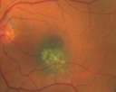
Macular Choroidal Nevusvu 1641 fois78-year-old woman has choroidal nevus in the macula of the left eye. You asked her to come in here for an evaluation and nevus was noted in the past about 7 years ago. She has not noticed any change in vision in that eye.
VISUAL ACUITY: OU 20/30.
|
|

Choroidal Nevus with Overlying Drusenvu 1636 fois48-year-old woman Vision OD is 20/16, OS is 20/20.
|
|

CHRPE - Gardner Syndrome - FAMILIAL COLORECTAL POLYPOSIS - 11 year old femalevu 1619 fois11-year-old child with CHRPE OS has six undescended adult teeth and also she has had a cyst removed from her arm pit and she has three more in her arm pit and one on her face and she is seeing a dermatologist for that in the near future. The mother reports no health problems, however, the patient’s grandmother does have a history of intestinal polyps, as does the patient’s grandmother’s brothers son.
|
|

Grouped pigmentation - Bear Tracksvu 1605 fois71 year old caucasian woman with incidental finding of extensive grouped RPE hypertrophy. Routine colonoscopy performed in the preceding year was normal.
|
|

Choroidal Nevus with Overlying Drusenvu 1522 fois48-year-old woman Vision OD is 20/16, OS is 20/20.
|
|

Melanomavu 1514 fois40 Year old male
|
|

Choroidal Melanomavu 1489 fois
|
|

Melanomavu 1468 foisYoung male with large Melanoma
|
|

Amelanotic Nevus Initial Visitvu 1456 fois
|
|

Pigmented Choroidal Mass Right Eye - Melanoma vs. Nevus vs. Unilateral Melanocytic Proliferation - Initial Visit B Scanvu 1382 fois71-year-old man is vision has been bothering him for about a month. He does not notice one eye particularly worse than the other. OD is 20/40, OS is 20/25 - Initial Visit
|
|
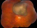
Choroidal Melanomavu 1377 fois
|
|

Choroidal Melanoma - Exudative Retinal Detachment 82 Year Old Man B Scan - Transversevu 1299 fois82-year-old man who had 3 falls 2 weeks ago. After the falls he started checking his vision and noticed there was a veil over the left eye, which he had seen for about two weeks in the superior visual field. OD 20/25, OS 20/40
(patient was medically never well enough for brachytherapy and died 6 months later from heart disease)
|
|

Choroidal Melanoma with Retinal Detachmentvu 1268 fois57 year old female who underwent proton beam therapy for Choroidal Melanoma. VA was 20/400 at post op visit.
|
|

Chorodial Melanomavu 1247 fois80 year old male came in for retinal evaluation and presented with a melanoma.
|
|

Multiple - Congenital Hypertrophy of the Pigment Epithelium - CHRPE - Gardner Syndromevu 1244 fois63 Year Old woman with a history of colon cancer at age 32. She has also had thyroid cancer. Two of her 6 siblings died of cancer, one of sarcoma and one of lung cancer. Vision 20/30, 20/25. Fundus shows CHRPE in both eyes.
|
|

Peripapillary Choroidal Nevusvu 1236 fois37-year-old woman was seen in the office on July 27, 2011. She has photographs from 2009 and 2010 showing a peripapillary choroidal nevus in the right eye. VISUAL ACUITY: OD 20/20
|
|

Multiple - Congenital Hypertrophy of the Pigment Epithelium - CHRPE - Gardner Syndromevu 1231 fois63 Year Old woman with a history of colon cancer at age 32. She has also had thyroid cancer. Two of her 6 siblings died of cancer, one of sarcoma and one of lung cancer. Vision 20/30, 20/25. Fundus shows CHRPE in both eyes.
|
|
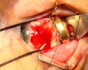
Radioactive Plaque Placement for Choroidal Melanoma Plaque Placed but not Sutured to Eye Yetvu 1210 fois
|
|

Radioactive Plaque Placement for Choroidal Melanoma Iodine Radioactive Seeds in Plaquevu 1179 fois
|
|

Choroidal Melanoma with Radiation Retinopathyvu 1179 foisPatient comes with follow up on Choroidal Melanoma, Right Eye that was treated back in June of 2009 with Radioactive Implant. Vein Occlusion is also present with VA - Hand Motion. Hemorrhages visible with hard exudates from the Radiation Retinopathy.
|
|
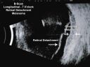
Choroidal Melanoma - Exudative Retinal Detachment 82 Year Old Man B Scan Ultrasound Longitudinalvu 1157 fois82-year-old man who had 3 falls 2 weeks ago. After the falls he started checking his vision and noticed there was a veil over the left eye, which he had seen for about two weeks in the superior visual field. OD 20/25, OS 20/40
(patient was medically never well enough for brachytherapy and died 6 months later from heart disease)
|
|

Radioactive Plaque Placement for Choroidal Melanoma Eye is Anesthetized Prior to Surgeryvu 1118 fois
|
|

Choroidal Nevus with Drusenvu 1104 foisLow risk choroidal nevus with many overlying drusen
|
|

CHOROIDAL Melanomavu 1099 fois
|
|

Amelanotic Choroidal Nevus vu 1071 fois45-year-old man OD 20/20, OS 20/20. 5x5 mm flat, yellow choroidal lesion just inferonasal to the optic nerve, two disc-diameters from the nerve with scalloped edges. It looks like a confluent drusen, but it is not elevated.
|
|

Melanoma - Large Ciliary Body Photo shows tumor visible through the pupilvu 1066 fois
|
|

Radioactive Plaque Placement for Choroidal Melanoma Radioactive Plaque Placement for Choroidal Melanoma vu 1058 fois
|
|

Choroidal Tumor consistent with Uveal Melanomavu 1058 foisPatient comes in for second opinion for 2-melanomas in the right eye. Patient's VA is 20/20, both eyes. Fundus exam reveals melanoma superior to the macula which measured at 0.7-mm thick. Second melanoma is nasally which measured at 4.1-mm thick and has low internal reflectivity. Patient will be followed up with MRI and ocular protocol.
|
|

Radioactive Plaque Placement for Choroidal Melanoma Plaque Sutured to Globevu 1054 fois
|
|

Radioactive Plaque Placement for Choroidal Melanoma Eye After Surgery with Conjunctiva Closedvu 1041 fois
|
|

Peripapillary Choroidal Nevusvu 1040 fois37-year-old woman was seen in the office on July 27, 2011. She has photographs from 2009 and 2010 showing a peripapillary choroidal nevus in the right eye. VISUAL ACUITY: OD 20/20
|
|

Choroidal Melanoma with Sub Retinal Fluidvu 1038 foisFundus photography shows choroidal melanoma superiorily between 11-12-o'clock in the left eye. VA is 20/150 in the left eye.
|
|

Choroidal Melanomavu 1037 foisPatient comes in with spot in vision in the right eye. Fundus photography shows medium size melanoma adjacent to the optic nerve and measures 13mm across horizontally and 6mm diameter in thickness. Patient will undergo proton beam therapy.
|
|
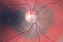
Peripapillary Choroidal Nevusvu 1035 fois37-year-old woman was seen in the office on July 27, 2011. She has photographs from 2009 and 2010 showing a peripapillary choroidal nevus in the right eye. VISUAL ACUITY: OD 20/20
|
|

Melanoma of Optic Nervevu 1027 foisPatient comes in for evaluation on Optic Nerve Melanoma in the right eye. Patient's VA is 20/40 in the right eye. Fundus photograph reveals melanoma invading optic nerve and ultrasound reveals behind the optic nerve as well.
|
|
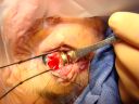
Radioactive Plaque Placement for Choroidal Melanoma Plaque Placed but not Sutured to Eye Yevu 1024 fois
|
|

Choroidal Melanoma with Retinal Detachmentvu 1012 fois57 year old female who underwent proton beam therapy for Choroidal Melanoma. VA was 20/400 at post op visit.
|
|
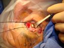
Radioactive Plaque Placement for Choroidal Melanoma Plaque Placed but not Sutured to Eye Yevu 1000 fois
|
|

Choroidal Nevus with Overlying Drusenvu 996 fois48-year-old woman Vision OD is 20/16, OS is 20/20.
|
|

Choroidal Nevusvu 996 foisPatient comes in with pigmented spot in the right eye. VA is 20/25. Fundus photography shows elevated choroidal nevus inferiorily in the right eye. Will be reevaluated in 3-months...
|
|

Amelanotic Choroidal Nevus vu 985 fois45-year-old man OD 20/20, OS 20/20. 5x5 mm flat, yellow choroidal lesion just inferonasal to the optic nerve, two disc-diameters from the nerve with scalloped edges. It looks like a confluent drusen, but it is not elevated.
|
|

Amelanotic Choroidal Nevus vu 982 fois45-year-old man OD 20/20, OS 20/20. 5x5 mm flat, yellow choroidal lesion just inferonasal to the optic nerve, two disc-diameters from the nerve with scalloped edges. It looks like a confluent drusen, but it is not elevated.
|
|

Choroidal Melanomavu 974 foisFemale patient comes in with a visual defect in the right eye. VA is 20/40 in the right eye. Fundus photography shows choroidal melanoma with retinal detachment temporally at 3-o'clock in the right eye.
|
|

Pigmented Choroidal Mass Right Eye - Melanoma vs. Nevus vs. Unilateral Melanocytic Proliferation - Initial Visit and 4 Month Follow-Up - Color Photovu 972 foisPigmented lesion is visible in the superior half of the macular on the follow-up visit.
|
|

Radioactive Plaque Placement for Choroidal Melanoma Transillumination of Globe to Localize Melanoma Arrow Points to Tumor Shadowvu 964 fois
|
|
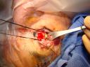
Radioactive Plaque Placement for Choroidal Melanoma Plaque Sutured to Globevu 951 fois
|
|

Radioactive Plaque Placement for Choroidal Melanoma Transillumination of Globe to Localize Melanoma vu 942 fois
|
|

Amelanotic Nevus - 1 year Follow-up Visit - No Changevu 928 fois
|
|

Radioactive Plaque Placement for Choroidal Melanoma Gold Plaque about the size of a Quartervu 927 fois
|
|
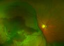
Large Choroidal Melanoma with Retinal Detachmentvu 923 foisPatient notices a decline in her vision for the past 3-weeks in her right eye. VA was light perception in the right eye. Fundus photo shows a very large melanoma temporal to the optic nerve and retinal detachment inferiorly. Patient will under go proton beam therapy.
|
|
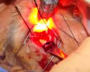
Radioactive Plaque Placement for Choroidal Melanoma Transillumination of Globe to Localize Melanoma vu 921 fois
|
|

Radioactive Plaque Placement for Choroidal Melanoma Transillumination of Globe to Localize Melanoma vu 915 fois
|
|
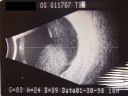
Melanoma - Large Ciliary Body B-Scan ultrasound shows tumore filling much of the eyevu 908 fois
|
|

Treated Choroidal Melanomavu 903 foisFollow up patient for melanoma in the right eye that was treated with proton beam therapy. VA is 20/50 in the right eye. Fundus photo shows scattered hemorrhages inferiorly with tumor temporal inferior at 8-o'clock. Patient will be followed up in 6-months
|
|
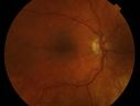
Multiple - Congenital Hypertrophy of the Pigment Epithelium - CHRPE - Gardner Syndromevu 897 fois63 Year Old woman with a history of colon cancer at age 32. She has also had thyroid cancer. Two of her 6 siblings died of cancer, one of sarcoma and one of lung cancer. Vision 20/30, 20/25. Fundus shows CHRPE in both eyes.
|
|

Uveal Choroidal Melanomavu 897 foisPatient comes in for evaluation on a Choroidal Melanoma in the right eye. VA was 20/25 in both eyes. The melanoma is in the temporal aspect of the right eye. It measured at 0.7mm elevated after doing a BSCAN Ultrasound.
|
|
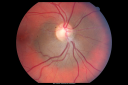
Peripapillary Choroidal Nevusvu 896 fois37-year-old woman was seen in the office on July 27, 2011. She has photographs from 2009 and 2010 showing a peripapillary choroidal nevus in the right eye. VISUAL ACUITY: OD 20/20
|
|

Melanoma - Large Ciliary Body Photo shows tumor visible throught the pupilvu 894 fois
|
|

Radioactive Plaque Placement for Choroidal Melanoma Lead Patch after Surgeryvu 894 fois
|
|
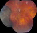
6mm x 11mm Melanomavu 880 fois76 year old male present with a 6mm x 1mm melanoma. Large melanoma very nasal OS. Asteroid Hyalosis present OS only. VA 20/40 OD 20/40 OS.
|
|

Choroidal Melanoma - Exudative Retinal Detachment 82 Year Old Man B Scan Ultrasound Transvercevu 873 fois82-year-old man who had 3 falls 2 weeks ago. After the falls he started checking his vision and noticed there was a veil over the left eye, which he had seen for about two weeks in the superior visual field. OD 20/25, OS 20/40
(patient was medically never well enough for brachytherapy and died 6 months later from heart disease)
|
|

Choroidal Nevus with Drusenvu 869 foisLow risk choroidal nevus with many overlying drusen
|
|

Treated Melanomavu 865 foisTreated melanoma in the right eye. VA is light perception in the right eye.
|
|

Choroidal Nevus in Macula vu 864 fois
|
|

Pigmented Choroidal Mass Right Eye - Melanoma vs. Nevus vs. Unilateral Melanocytic Proliferation - Initial Visit B Scanvu 856 fois71-year-old man is vision has been bothering him for about a month. He does not notice one eye particularly worse than the other. OD is 20/40, OS is 20/25 - Initial Visit
|
|

Radioactive Plaque Placement for Choroidal Melanoma Transillumination of Globe to Localize Melanoma Arrow Points to Tumor Shadowvu 855 fois
|
|
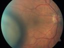
Choroidal Melanoma (Medium) Exudative Retinal Detachment vu 844 fois87-year-old man noticed a new onset floater a few months ago. OD is 20/40, OS is 20/30
|
|

Choroidal Melanomavu 844 foisPost op Radioactive Implant with hard exudates surrounding the Melanoma with hemorrhage present.
|
|
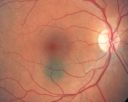
Choroidal Nevus in Macula vu 834 fois
|
|

Ciliary Body Melanomavu 830 fois71-year old male who was recently diagnosed with a large ciliary body melanoma that is pushing into the anterior chamber of the left eye. Patient is going to proceed with proton therapy.
|
|

Choroidal Melanoma - Exudative Retinal Detachment 82 Year Old Man B Scan Ultrasound Longitudinalvu 829 fois82-year-old man who had 3 falls 2 weeks ago. After the falls he started checking his vision and noticed there was a veil over the left eye, which he had seen for about two weeks in the superior visual field. OD 20/25, OS 20/40
(patient was medically never well enough for brachytherapy and died 6 months later from heart disease)
|
|

Metastasis Choroidal Melanoma vu 789 fois71-year old female who has undergone chemotherapy treatment for breast cancer. Choroidal Melanoma was from metastasis from her breast cancer. VA is 20/20 as the tumor is in the superior aspect above the optic nerve.
|
|

Melanosis Oculi and Choroidal Melanomavu 779 fois88 year old female with 2.5 mm elevated choroidal melanoma and melanosis oculi. She also has geographic atrophy from AMD. The right eye is the better eye with 20/40 vision. After observation with growth, brachytherapy was done.
|
|

Melanoma - Large Ciliary Body A scan shows medium to low internal reflectivityvu 778 fois
|
|

vu 773 fois
|
|

Choroidal Nevus with Drusenvu 762 foisLow risk choroidal nevus with many overlying drusen
|
|

Choroidal Malignant Melanoma - early phase FAvu 753 foisearly phase of FA at 23 seconds (Choroidal malignant melanoma status post plaque radiotherapy OD in 1986, patient also has a submacular hemorrhage OD, and radiation retinopathy OD.)
|
|

Pigmented Choroidal Mass Right Eye - Melanoma vs. Nevus vs. Unilateral Melanocytic Proliferation - 4 Month Follow-Up Imagesvu 750 fois71-year-old man his vision 4 month follow-up. Vision is 20/60 and patient sees flashing lights. Lesion has grown slightly in the superior half of the macula.
|
|

Choroidal Nevus with Drusenvu 741 foisLow risk choroidal nevus with many overlying drusen
|
|

Choroidal Melanoma Mediumvu 730 fois33-year-old man with a history of decreased in the right eye. He had blurring in the eye about a year ago. He reports that it improved spontaneously until it was “nearly resolvedâ€. He said he had some persistent distortion, but that over the past couple of weeks he has noticed this dark spot recurring in the right eye, the left eye is unaffected. He does not have any flashes, floaters or change in his peripheral visual acuity. 20/80 OD, 20/16 OS. No improvement with pinhole. IOP: 12 OD, 11 OS.
POSTERIOR SEGMENT EXAMINATION: Shows a clear view with no PVD in either eye. C/D ratio is 0.2 OU. Large retinal vessels are unremarkable. The macula of the right eye has a brown choroidal lesion with pigment overlying the surface and orange spots along its lateral surface. The left macula is normal.
B-SCAN: B-scan ecography of the ultrasound of the right eye shows a mass about 5 mm high with a moderate to low internal reflectivity on A-scan echogram. There is serous fluid inferiorly on the echogram
|
|

Choroidal Melanomavu 728 foisNotice the orange strands presently on the tumor.
|
|

Choroidal Melanoma (Medium) Exudative Retinal Detachment vu 710 fois87-year-old man noticed a new onset floater a few months ago. OD is 20/40, OS is 20/30
|
|

Choroidal Melanoma (Medium) Exudative Retinal Detachment vu 707 fois87-year-old man noticed a new onset floater a few months ago. OD is 20/40, OS is 20/30
|
|

Radiation Retinopathy and Macular Pucker 4 years following Brachytherapy (Plaque) for Nasal Melanomavu 706 fois67-year-old woman has a macular pucker in the left eye. She had radiation for a melanoma in that eye 4 years aog. She had cataract surgery 2 years ago and a posterior sub-Tenon Kenalog injection in 7 months ago. Her vision in the left eye is hazy and she has trouble seeing out of it. VA OS 20/70.
|
|
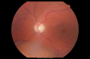
Choroidal Melanoma - Exudative Retinal Detachment 82 Year Old Man vu 701 fois82-year-old man who had 3 falls 2 weeks ago. After the falls he started checking his vision and noticed there was a veil over the left eye, which he had seen for about two weeks in the superior visual field. OD 20/25, OS 20/40
(patient was medically never well enough for brachytherapy and died 6 months later from heart disease)
|
|

Pigmented Choroidal Mass Right Eye - Melanoma vs. Nevus vs. Unilateral Melanocytic Proliferation - 4 Month Follow-Up Imagesvu 699 fois71-year-old man his vision 4 month follow-up. Vision is 20/60 and patient sees flashing lights. Lesion has grown slightly in the superior half of the macula.
|
|

Choroidal Melanoma Mediumvu 697 fois33-year-old man with a history of decreased in the right eye. He had blurring in the eye about a year ago. He reports that it improved spontaneously until it was “nearly resolvedâ€. He said he had some persistent distortion, but that over the past couple of weeks he has noticed this dark spot recurring in the right eye, the left eye is unaffected. He does not have any flashes, floaters or change in his peripheral visual acuity. 20/80 OD, 20/16 OS. No improvement with pinhole. IOP: 12 OD, 11 OS.
POSTERIOR SEGMENT EXAMINATION: Shows a clear view with no PVD in either eye. C/D ratio is 0.2 OU. Large retinal vessels are unremarkable. The macula of the right eye has a brown choroidal lesion with pigment overlying the surface and orange spots along its lateral surface. The left macula is normal.
B-SCAN: B-scan ecography of the ultrasound of the right eye shows a mass about 5 mm high with a moderate to low internal reflectivity on A-scan echogram. There is serous fluid inferiorly on the echogram
|
|

Ciliary Body Melanomavu 690 fois71-year old male who was recently diagnosed with a large ciliary body melanoma that is pushing into the anterior chamber of the left eye. Patient is going to proceed with proton therapy.
|
|

Pigmented Choroidal Mass Right Eye - Melanoma vs. Nevus vs. Unilateral Melanocytic Proliferation - 4 Month Follow-Up Imagesvu 686 fois71-year-old man his vision 4 month follow-up. Vision is 20/60 and patient sees flashing lights. Lesion has grown slightly in the superior half of the macula.
|
|

Pigmented Choroidal Mass Right Eye - Melanoma vs. Nevus vs. Unilateral Melanocytic Proliferation - Initial Visitvu 683 fois71-year-old man is vision has been bothering him for about a month. He does not notice one eye particularly worse than the other. OD is 20/40, OS is 20/25 - Initial Visit
|
|

Choroidal Melanomavu 677 foisChoroidal Melanoma with Retinal Detachment present inferior.
|
|
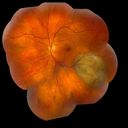
Choroidal Melanomavu 676 fois
|
|

Pigmented Choroidal Mass Right Eye - Melanoma vs. Nevus vs. Unilateral Melanocytic Proliferation - 4 Month Follow-Up Imagesvu 675 fois71-year-old man his vision 4 month follow-up. Vision is 20/60 and patient sees flashing lights. Lesion has grown slightly in the superior half of the macula.
|
|

Choroidal Melanoma - Exudative Retinal Detachment 82 Year Old Man vu 674 fois82-year-old man who had 3 falls 2 weeks ago. After the falls he started checking his vision and noticed there was a veil over the left eye, which he had seen for about two weeks in the superior visual field. OD 20/25, OS 20/40
(patient was medically never well enough for brachytherapy and died 6 months later from heart disease)
|
|
| 543 photos sur 6 page(s) |
1 |
 |
|
|
|


