Most viewed - Choroidal Melanoma and Choroidal Nevus
|

Amelanotic Choroidal Nevus - Multimodal Imaging421 views61 year old man with amelanotic choroidal nevus. Vision Normal - Stable over 4 years.
|
|

Amelanotic Choroidal Nevus - Multimodal Imaging421 views61 year old man with amelanotic choroidal nevus. Vision Normal - Stable over 4 years.
|
|

Amelanotic Choroidal Nevus - Multimodal Imaging420 views61 year old man with amelanotic choroidal nevus. Vision Normal - Stable over 4 years.
|
|

Melanosis Oculi and Choroidal Melanoma418 views88 year old female with 2.5 mm elevated choroidal melanoma and melanosis oculi. She also has geographic atrophy from AMD. The right eye is the better eye with 20/40 vision. After observation with growth, brachytherapy was done.
|
|

Amelanotic Choroidal Nevus - Multimodal Imaging418 views61 year old man with amelanotic choroidal nevus. Vision Normal - Stable over 4 years.
|
|
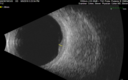
Amelanotic Choroidal Nevus - Multimodal Imaging415 views61 year old man with amelanotic choroidal nevus. Vision Normal - Stable over 4 years.
|
|
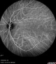
Amelanotic Choroidal Nevus - Multimodal Imaging414 views61 year old man with amelanotic choroidal nevus. Vision Normal - Stable over 4 years.
|
|

Amelanotic Choroidal Nevus - Multimodal Imaging411 views61 year old man with amelanotic choroidal nevus. Vision Normal - Stable over 4 years.
|
|

Amelanotic Choroidal Nevus - Multimodal Imaging411 views61 year old man with amelanotic choroidal nevus. Vision Normal - Stable over 4 years.
|
|

Atypical High Risk Choroidal Nevus vs Macular Melanoma410 views70 year old female with vision loss in the left eye for a few weeks. She was referred to a tumor center and being managed as an "atypical high risk choroidal nevus". The size is 6 mm x 6.5 mm x 1.6 mm (elevated).
Vision 20/40 - having serial avastin injections.
|
|

Choroidal Nevus with Drusen410 views82 year old female with flat nevus for 3 years.
|
|
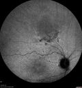
Amelanotic Choroidal Nevus - Multimodal Imaging410 views61 year old man with amelanotic choroidal nevus. Vision Normal - Stable over 4 years.
|
|

Amelanotic Choroidal Nevus409 views60 year old man. Nevus is stable over 6 years. Images are Photo, FAF, OCT, FA, ICG, and B-Scan
|
|

Amelanotic Choroidal Nevus - Multimodal Imaging406 views61 year old man with amelanotic choroidal nevus. Vision Normal - Stable over 4 years.
|
|

Amelanotic Choroidal Nevus - Multimodal Imaging404 views61 year old man with amelanotic choroidal nevus. Vision Normal - Stable over 4 years.
|
|

Melanosis Oculi and Choroidal Melanoma403 views88 year old female with 2.5 mm elevated choroidal melanoma and melanosis oculi. She also has geographic atrophy from AMD. The right eye is the better eye with 20/40 vision. After observation with growth, brachytherapy was done.
|
|

Choroidal Nevus with Drusen402 views82 year old female with flat nevus for 3 years.
|
|

Atypical High Risk Choroidal Nevus vs Macular Melanoma401 views70 year old female with vision loss in the left eye for a few weeks. She was referred to a tumor center and being managed as an "atypical high risk choroidal nevus". The size is 6 mm x 6.5 mm x 1.6 mm (elevated).
Vision 20/40 - having serial avastin injections.
|
|

Amelanotic Choroidal Nevus - Multimodal Imaging399 views61 year old man with amelanotic choroidal nevus. Vision Normal - Stable over 4 years.
|
|

Atypical High Risk Choroidal Nevus vs Macular Melanoma398 views70 year old female with vision loss in the left eye for a few weeks. She was referred to a tumor center and being managed as an "atypical high risk choroidal nevus". The size is 6 mm x 6.5 mm x 1.6 mm (elevated).
Vision 20/40 - having serial avastin injections.
|
|
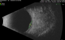
Atypical High Risk Choroidal Nevus vs Macular Melanoma398 views70 year old female with vision loss in the left eye for a few weeks. She was referred to a tumor center and being managed as an "atypical high risk choroidal nevus". The size is 6 mm x 6.5 mm x 1.6 mm (elevated).
Vision 20/40 - having serial avastin injections.
|
|
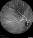
Amelanotic Choroidal Nevus - Multimodal Imaging397 views61 year old man with amelanotic choroidal nevus. Vision Normal - Stable over 4 years.
|
|

Radiation Retinopathy and Macular Pucker 4 years following Brachytherapy (Plaque) for Nasal Melanoma394 views67-year-old woman has a macular pucker in the left eye. She had radiation for a melanoma in that eye 4 years aog. She had cataract surgery 2 years ago and a posterior sub-Tenon Kenalog injection in 7 months ago. Her vision in the left eye is hazy and she has trouble seeing out of it. VA OS 20/70.
|
|
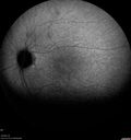
Amelanotic Choroidal Nevus394 views60 year old man. Nevus is stable over 6 years. Images are Photo, FAF, OCT, FA, ICG, and B-Scan
|
|

Amelanotic Choroidal Nevus - Multimodal Imaging391 views61 year old man with amelanotic choroidal nevus. Vision Normal - Stable over 4 years.
|
|

Amelanotic Choroidal Nevus - Multimodal Imaging390 views61 year old man with amelanotic choroidal nevus. Vision Normal - Stable over 4 years.
|
|

Melanocytoma - Lightly Pigmented388 views42 year old female with very mild visual field and vision loss in the left eye: 20/20 OU
|
|

Treated Choroidal Melanoma385 viewsPatient that underwent proton therapy for choroidal melanoma. Fundus photo shows treated melanoma temporal inferior with scattered hemorrhages inferior of the retina.
|
|

Amelanotic Choroidal Nevus - Multimodal Imaging384 views61 year old man with amelanotic choroidal nevus. Vision Normal - Stable over 4 years.
|
|

Amelanotic Choroidal Nevus383 views60 year old man. Nevus is stable over 6 years. Images are Photo, FAF, OCT, FA, ICG, and B-Scan
|
|
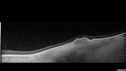
Choroidal Nevus with Drusen380 views82 year old female with flat nevus for 3 years.
|
|

Atypical High Risk Choroidal Nevus vs Macular Melanoma379 views70 year old female with vision loss in the left eye for a few weeks. She was referred to a tumor center and being managed as an "atypical high risk choroidal nevus". The size is 6 mm x 6.5 mm x 1.6 mm (elevated).
Vision 20/40 - having serial avastin injections.
|
|

Amelanotic Choroidal Nevus - Multimodal Imaging376 views61 year old man with amelanotic choroidal nevus. Vision Normal - Stable over 4 years.
|
|

Amelanotic Choroidal Nevus376 views60 year old man. Nevus is stable over 6 years. Images are Photo, FAF, OCT, FA, ICG, and B-Scan
|
|

Amelanotic Choroidal Nevus374 views60 year old man. Nevus is stable over 6 years. Images are Photo, FAF, OCT, FA, ICG, and B-Scan
|
|
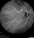
Amelanotic Choroidal Nevus374 views60 year old man. Nevus is stable over 6 years. Images are Photo, FAF, OCT, FA, ICG, and B-Scan
|
|

Amelanotic Choroidal Nevus365 views60 year old man. Nevus is stable over 6 years. Images are Photo, FAF, OCT, FA, ICG, and B-Scan
|
|

Amelanotic Choroidal Nevus365 views60 year old man. Nevus is stable over 6 years. Images are Photo, FAF, OCT, FA, ICG, and B-Scan
|
|

Amelanotic Choroidal Nevus365 views60 year old man. Nevus is stable over 6 years. Images are Photo, FAF, OCT, FA, ICG, and B-Scan
|
|

Amelanotic Choroidal Nevus361 views60 year old man. Nevus is stable over 6 years. Images are Photo, FAF, OCT, FA, ICG, and B-Scan
|
|

Amelanotic Choroidal Nevus357 views60 year old man. Nevus is stable over 6 years. Images are Photo, FAF, OCT, FA, ICG, and B-Scan
|
|

Amelanotic Choroidal Nevus353 views60 year old man. Nevus is stable over 6 years. Images are Photo, FAF, OCT, FA, ICG, and B-Scan
|
|

Amelanotic Choroidal Nevus350 views60 year old man. Nevus is stable over 6 years. Images are Photo, FAF, OCT, FA, ICG, and B-Scan
|
|

Amelanotic Choroidal Nevus350 views60 year old man. Nevus is stable over 6 years. Images are Photo, FAF, OCT, FA, ICG, and B-Scan
|
|

Amelanotic Choroidal Nevus349 views60 year old man. Nevus is stable over 6 years. Images are Photo, FAF, OCT, FA, ICG, and B-Scan
|
|

Amelanotic Choroidal Nevus343 views60 year old man. Nevus is stable over 6 years. Images are Photo, FAF, OCT, FA, ICG, and B-Scan
|
|

Amelanotic Choroidal Nevus342 views60 year old man. Nevus is stable over 6 years. Images are Photo, FAF, OCT, FA, ICG, and B-Scan
|
|

Amelanotic Choroidal Nevus341 views60 year old man. Nevus is stable over 6 years. Images are Photo, FAF, OCT, FA, ICG, and B-Scan
|
|

Amelanotic Choroidal Nevus341 views60 year old man. Nevus is stable over 6 years. Images are Photo, FAF, OCT, FA, ICG, and B-Scan
|
|

Amelanotic Choroidal Nevus334 views60 year old man. Nevus is stable over 6 years. Images are Photo, FAF, OCT, FA, ICG, and B-Scan
|
|

Amelanotic Choroidal Nevus333 views60 year old man. Nevus is stable over 6 years. Images are Photo, FAF, OCT, FA, ICG, and B-Scan
|
|

Amelanotic Choroidal Nevus327 views60 year old man. Nevus is stable over 6 years. Images are Photo, FAF, OCT, FA, ICG, and B-Scan
|
|

Choroidal nevus310 viewsStable for 8 years.
|
|

Amelanotic Choroidal Nevus308 views60 year old man. Nevus is stable over 6 years. Images are Photo, FAF, OCT, FA, ICG, and B-Scan
|
|

Choroidal nevus302 viewsStable for 8 years.
|
|

Large chronic choroidal nevus299 viewsAutofluorescence image shows guttering at the bottom of the lesion suggesting chronicity. This was documented 23 years prior to these photos
|
|
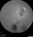
Choroidal nevus288 viewsStable for 8 years.
|
|

Large chronic choroidal nevus287 viewsAutofluorescence image shows guttering at the bottom of the lesion suggesting chronicity. This was documented 23 years prior to these photos
|
|
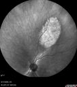
Choroidal nevus277 viewsStable for 8 years.
|
|
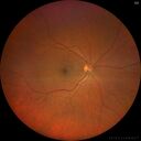
Choroidal nevus275 viewsStable for 8 years.
|
|

Large chronic choroidal nevus275 viewsAutofluorescence image shows guttering at the bottom of the lesion suggesting chronicity. This was documented 23 years prior to these photos
|
|

Large chronic choroidal nevus275 viewsAutofluorescence image shows guttering at the bottom of the lesion suggesting chronicity. This was documented 23 years prior to these photos
|
|

Large chronic choroidal nevus275 viewsAutofluorescence image shows guttering at the bottom of the lesion suggesting chronicity. This was documented 23 years prior to these photos
|
|

Choroidal nevus274 viewsStable for 8 years.
|
|

Large chronic choroidal nevus274 viewsAutofluorescence image shows guttering at the bottom of the lesion suggesting chronicity. This was documented 23 years prior to these photos
|
|

Large chronic choroidal nevus271 viewsAutofluorescence image shows guttering at the bottom of the lesion suggesting chronicity. This was documented 23 years prior to these photos
|
|

Choroidal nevus269 viewsStable for 8 years.
|
|

Large chronic choroidal nevus266 viewsAutofluorescence image shows guttering at the bottom of the lesion suggesting chronicity. This was documented 23 years prior to these photos
|
|

Large chronic choroidal nevus262 viewsAutofluorescence image shows guttering at the bottom of the lesion suggesting chronicity. This was documented 23 years prior to these photos
|
|

Large chronic choroidal nevus245 viewsAutofluorescence image shows guttering at the bottom of the lesion suggesting chronicity. This was documented 23 years prior to these photos
|
|

Large chronic choroidal nevus239 viewsAutofluorescence image shows guttering at the bottom of the lesion suggesting chronicity. This was documented 23 years prior to these photos
|
|

Class 2 choroidal melanoma239 views48 year old man with no visual complaints referred by optometrist.
Brachytherapy was done and biopsy showed a class 2 melanoma. 2 years later the patient developed liver metastases.
|
|

Class 2 choroidal melanoma218 views48 year old man with no visual complaints referred by optometrist.
Brachytherapy was done and biopsy showed a class 2 melanoma. 2 years later the patient developed liver metastases.
|
|

Class 2 choroidal melanoma209 views48 year old man with no visual complaints referred by optometrist.
Brachytherapy was done and biopsy showed a class 2 melanoma. 2 years later the patient developed liver metastases.
|
|

Class 2 choroidal melanoma205 views48 year old man with no visual complaints referred by optometrist.
Brachytherapy was done and biopsy showed a class 2 melanoma. 2 years later the patient developed liver metastases.
|
|

Class 2 choroidal melanoma196 views48 year old man with no visual complaints referred by optometrist.
Brachytherapy was done and biopsy showed a class 2 melanoma. 2 years later the patient developed liver metastases.
|
|

Class 2 choroidal melanoma194 views48 year old man with no visual complaints referred by optometrist.
Brachytherapy was done and biopsy showed a class 2 melanoma. 2 years later the patient developed liver metastases.
|
|

Class 2 choroidal melanoma193 views48 year old man with no visual complaints referred by optometrist.
Brachytherapy was done and biopsy showed a class 2 melanoma. 2 years later the patient developed liver metastases.
|
|

Class 2 choroidal melanoma190 views48 year old man with no visual complaints referred by optometrist.
Brachytherapy was done and biopsy showed a class 2 melanoma. 2 years later the patient developed liver metastases.
|
|

Class 2 choroidal melanoma185 views48 year old man with no visual complaints referred by optometrist.
Brachytherapy was done and biopsy showed a class 2 melanoma. 2 years later the patient developed liver metastases.
|
|

Class 2 choroidal melanoma183 views48 year old man with no visual complaints referred by optometrist.
Brachytherapy was done and biopsy showed a class 2 melanoma. 2 years later the patient developed liver metastases.
|
|

Class 2 choroidal melanoma182 views48 year old man with no visual complaints referred by optometrist.
Brachytherapy was done and biopsy showed a class 2 melanoma. 2 years later the patient developed liver metastases.
|
|

Class 2 choroidal melanoma179 views48 year old man with no visual complaints referred by optometrist.
Brachytherapy was done and biopsy showed a class 2 melanoma. 2 years later the patient developed liver metastases.
|
|

Class 2 choroidal melanoma163 views48 year old man with no visual complaints referred by optometrist.
Brachytherapy was done and biopsy showed a class 2 melanoma. 2 years later the patient developed liver metastases.
|
|
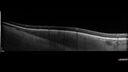
Choroidal Nevus with Halo159 views
|
|

Small choroidal melanoma150 views28 year old man Serial images suggested that the lesion is growing
OD: sc20/16
OS: sc20/40+2
IOP: TP: OD:19 OS:16
|
|

Serous Macular Detachment from Small Choroidal Melanoma148 views72 year old female misdiagnosed as CSR who had small choroidal melanoms.
|
|
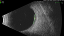
Serous Macular Detachment from Small Choroidal Melanoma143 views72 year old female misdiagnosed as CSR who had small choroidal melanoms.
|
|
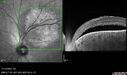
Small choroidal melanoma139 views28 year old man Serial images suggested that the lesion is growing
OD: sc20/16
OS: sc20/40+2
IOP: TP: OD:19 OS:16
|
|
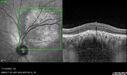
Small choroidal melanoma139 views28 year old man Serial images suggested that the lesion is growing
OD: sc20/16
OS: sc20/40+2
IOP: TP: OD:19 OS:16
|
|

Serous Macular Detachment from Small Choroidal Melanoma133 views72 year old female misdiagnosed as CSR who had small choroidal melanoms.
|
|

Serous Macular Detachment from Small Choroidal Melanoma131 views72 year old female misdiagnosed as CSR who had small choroidal melanoms.
|
|

Small choroidal melanoma122 views28 year old man Serial images suggested that the lesion is growing
OD: sc20/16
OS: sc20/40+2
IOP: TP: OD:19 OS:16
|
|

Small choroidal melanoma121 views28 year old man Serial images suggested that the lesion is growing
OD: sc20/16
OS: sc20/40+2
IOP: TP: OD:19 OS:16
|
|
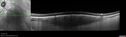
Choroidal Nevus with Halo115 views
|
|

Serous Macular Detachment from Small Choroidal Melanoma115 views72 year old female misdiagnosed as CSR who had small choroidal melanoms.
|
|

Serous Macular Detachment from Small Choroidal Melanoma115 views72 year old female misdiagnosed as CSR who had small choroidal melanoms.
|
|

Serous Macular Detachment from Small Choroidal Melanoma114 views72 year old female misdiagnosed as CSR who had small choroidal melanoms.
|
|

Small choroidal melanoma113 views28 year old man Serial images suggested that the lesion is growing
OD: sc20/16
OS: sc20/40+2
IOP: TP: OD:19 OS:16
|
|
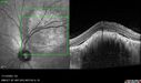
Small choroidal melanoma113 views28 year old man Serial images suggested that the lesion is growing
OD: sc20/16
OS: sc20/40+2
IOP: TP: OD:19 OS:16
|
|
|
|


