Most viewed - Tumors of the Retina and Retinal Pigment Epithelium
|

Retinal Capillary Hemangioma - Endophytic and Exophytic568 views46 year old female with normal vision - her father died of pancreatic cancer
|
|

Primary Ocular Lymphoma - 051812 567 views
|
|

Ocular Lymphoma - non-Hodgkins - B-Cell567 viewsMultifocal sub-RPE lesions in the left eye of a patient with history of Non-hodgkins B-cell Lymphoma last active 2 years ago
|
|

Primary Ocular Lymphoma - 051812 564 views
|
|

Ocular Lymphoma - non-Hodgkins - B-Cell - Right eye has equatorial drusen559 viewsMultifocal sub-RPE lesions in the left eye of a patient with history of Non-hodgkins B-cell Lymphoma last active 2 years ago
|
|
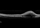
Retinal Capillary Hemangioma - Endophytic and Exophytic559 views46 year old female with normal vision - her father died of pancreatic cancer
|
|

Primary Ocular Lymphoma - 020513 - FUndus Autofluorescence554 views
|
|

Ocular Lymphoma - non-Hodgkins - B-Cell553 viewsMultifocal sub-RPE lesions in the left eye of a patient with history of Non-hodgkins B-cell Lymphoma last active 2 years ago
|
|

Retinal Capillary Hemangioma - Endophytic and Exophytic553 views46 year old female with normal vision - her father died of pancreatic cancer
|
|

Primary Ocular Lymphoma - White Mass Left Eye - 112712 552 views
|
|
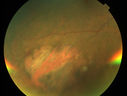
Primary Ocular Lymphoma - 051812 551 views
|
|
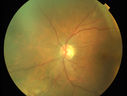
Primary Ocular Lymphoma - 051812 550 views
|
|
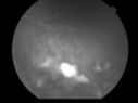
Uveitis and Retinal Whitening - 04-27-12 - Prior to PPV OD - Possible Primary Ocular Lymphoma 548 views
|
|
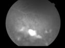
Uveitis and Retinal Whitening - 04-27-12 - Prior to PPV OD - Possible Primary Ocular Lymphoma 547 views
|
|
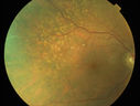
Primary Ocular Lymphoma - 051812 547 views
|
|

Retinal Capillary Hemangioma - Endophytic and Exophytic547 views46 year old female with normal vision - her father died of pancreatic cancer
|
|
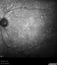
Primary Ocular Lymphoma - 020513 546 views
|
|

Retinal Capillary Hemangioma - Endophytic and Exophytic544 views46 year old female with normal vision - her father died of pancreatic cancer
|
|

Primary Ocular Lymphoma - 051812 543 views
|
|

Primary Ocular Lymphoma - White Mass Left Eye - 112712 540 views
|
|

Retinal Capillary Hemangioma due to Von Hipple-Lindau Syndrome539 viewsA 47-year old female comes in for a second opinion for retinal capillary Hemangioma in the left eye. VA was HM (hand motion) in the left eye. She has had multiple procedures done for the left eye including, Retinal Detachment surgery, YAG Laser PI, YAG Laser Capsulotomy, and Cataract surgery.
|
|
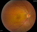
Cavernous Choroidal Hemangioma539 viewsPatient presents with Cavernous Choroidal Hemangioma in the right eye. VA is 20/50 after manifest refraction. Patient also notices decreased vision with shadow close to her central vision in the right eye. Patient will undergo photodynamic therapy in the right eye.
|
|

Retinal Capillary Hemangioma - Endophytic and Exophytic538 views46 year old female with normal vision - her father died of pancreatic cancer
|
|
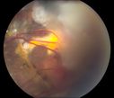
Retinal Capillary Hemangioma due to Von Hipple-Lindau Syndrome536 viewsA 47-year old female comes in for a second opinion for retinal capillary Hemangioma in the left eye. VA was HM (hand motion) in the left eye. She has had multiple procedures done for the left eye including, Retinal Detachment surgery, YAG Laser PI, YAG Laser Capsulotomy, and Cataract surgery.
|
|

Ocular Lymphoma - non-Hodgkins - B-Cell - OCT shows multifocal sub-RPE masses 536 viewsMultifocal sub-RPE lesions in the left eye of a patient with history of Non-hodgkins B-cell Lymphoma last active 2 years ago
|
|

Primary Ocular Lymphoma - 051812 532 views
|
|
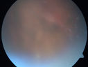
Primary Ocular Lymphoma - White Mass Left Eye - 112712 526 views
|
|

Primary Ocular Lymphoma - 051812 524 views
|
|

Ocular Lymphoma - non-Hodgkins - B-Cell - Right eye has equatorial drusen524 viewsMultifocal sub-RPE lesions in the left eye of a patient with history of Non-hodgkins B-cell Lymphoma last active 2 years ago
|
|

Retinal Capillary Hemangioma - Endophytic and Exophytic521 views46 year old female with normal vision - her father died of pancreatic cancer
|
|

Ocular Lymphoma - non-Hodgkins - B-Cell519 viewsMultifocal sub-RPE lesions in the left eye of a patient with history of Non-hodgkins B-cell Lymphoma last active 2 years ago
|
|

Primary Ocular Lymphoma - 051812 518 views
|
|

Retinal Capillary Hemangioma - Endophytic and Exophytic518 views46 year old female with normal vision - her father died of pancreatic cancer
|
|

Retinal Capillary Hemangioma due to Von Hipple-Lindau Syndrome516 viewsA 47-year old female comes in for a second opinion for retinal capillary Hemangioma in the left eye. VA was HM (hand motion) in the left eye. She has had multiple procedures done for the left eye including, Retinal Detachment surgery, YAG Laser PI, YAG Laser Capsulotomy, and Cataract surgery.
|
|

Cavernous Choroidal Hemangioma516 viewsPatient presents with Cavernous Choroidal Hemangioma in the right eye. VA is 20/50 after manifest refraction. Patient also notices decreased vision with shadow close to her central vision in the right eye. Patient will undergo photodynamic therapy in the right eye.
|
|

Retinal Capillary Hemangioma - Endophytic and Exophytic516 views46 year old female with normal vision - her father died of pancreatic cancer
|
|

Ocular Lymphoma - non-Hodgkins - B-Cell514 viewsMultifocal sub-RPE lesions in the left eye of a patient with history of Non-hodgkins B-cell Lymphoma last active 2 years ago
|
|

Retinal Capillary Hemangioma - Endophytic and Exophytic511 views46 year old female with normal vision - her father died of pancreatic cancer
|
|
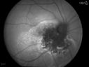
Cavernous Choroidal Hemangioma510 viewsPatient presents with Cavernous Choroidal Hemangioma in the right eye. VA is 20/50 after manifest refraction. Patient also notices decreased vision with shadow close to her central vision in the right eye. Patient will undergo photodynamic therapy in the right eye.
|
|

Cavernous Choroidal Hemangioma509 viewsPatient presents with Cavernous Choroidal Hemangioma in the right eye. VA is 20/50 after manifest refraction. Patient also notices decreased vision with shadow close to her central vision in the right eye. Patient will undergo photodynamic therapy in the right eye.
|
|

Ocular Lymphoma - non-Hodgkins - B-Cell509 viewsMultifocal sub-RPE lesions in the left eye of a patient with history of Non-hodgkins B-cell Lymphoma last active 2 years ago
|
|
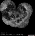
Ocular Lymphoma - non-Hodgkins - B-Cell508 viewsMultifocal sub-RPE lesions in the left eye of a patient with history of Non-hodgkins B-cell Lymphoma last active 2 years ago
|
|

Ocular Lymphoma - non-Hodgkins - B-Cell507 viewsMultifocal sub-RPE lesions in the left eye of a patient with history of Non-hodgkins B-cell Lymphoma last active 2 years ago
|
|

Retinal Capillary Hemangioma - Endophytic and Exophytic501 views46 year old female with normal vision - her father died of pancreatic cancer
|
|

Vasoproliferative Tumor 7 Years after Retinal Detachment Repair497 views65 year old female presents 7 years following retinal detachment repair with peripheral white mass with hemorrhage.
|
|

Retinal Capillary Hemangioma due to Von Hipple-Lindau Syndrome496 viewsA 47-year old female comes in for a second opinion for retinal capillary Hemangioma in the left eye. VA was HM (hand motion) in the left eye. She has had multiple procedures done for the left eye including, Retinal Detachment surgery, YAG Laser PI, YAG Laser Capsulotomy, and Cataract surgery.
|
|
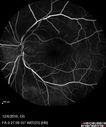
Ocular Lymphoma - non-Hodgkins - B-Cell493 viewsMultifocal sub-RPE lesions in the left eye of a patient with history of Non-hodgkins B-cell Lymphoma last active 2 years ago
|
|

Vasoproliferative Tumor 7 Years after Retinal Detachment Repair493 views65 year old female presents 7 years following retinal detachment repair with peripheral white mass with hemorrhage.
|
|

Ocular Lymphoma - non-Hodgkins - B-Cell491 viewsMultifocal sub-RPE lesions in the left eye of a patient with history of Non-hodgkins B-cell Lymphoma last active 2 years ago
|
|
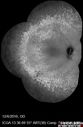
Ocular Lymphoma - non-Hodgkins - B-Cell490 viewsMultifocal sub-RPE lesions in the left eye of a patient with history of Non-hodgkins B-cell Lymphoma last active 2 years ago
|
|

Ocular Lymphoma - non-Hodgkins - B-Cell481 viewsMultifocal sub-RPE lesions in the left eye of a patient with history of Non-hodgkins B-cell Lymphoma last active 2 years ago
|
|

Retinal Capillary Hemangioma due to Von Hipple-Lindau Syndrome471 viewsA 47-year old female comes in for a second opinion for retinal capillary Hemangioma in the left eye. VA was HM (hand motion) in the left eye. She has had multiple procedures done for the left eye including, Retinal Detachment surgery, YAG Laser PI, YAG Laser Capsulotomy, and Cataract surgery.
|
|

Retinal Capillary Hemangioma - Endophytic and Exophytic469 views46 year old female with normal vision - her father died of pancreatic cancer
|
|
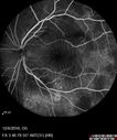
Ocular Lymphoma - non-Hodgkins - B-Cell467 viewsMultifocal sub-RPE lesions in the left eye of a patient with history of Non-hodgkins B-cell Lymphoma last active 2 years ago
|
|

Ocular Lymphoma - non-Hodgkins - B-Cell460 viewsMultifocal sub-RPE lesions in the left eye of a patient with history of Non-hodgkins B-cell Lymphoma last active 2 years ago
|
|

Ocular Lymphoma - non-Hodgkins - B-Cell - OCT shows multifocal sub-RPE masses 459 viewsMultifocal sub-RPE lesions in the left eye of a patient with history of Non-hodgkins B-cell Lymphoma last active 2 years ago
|
|
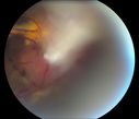
Retinal Capillary Hemangioma due to Von Hipple-Lindau Syndrome450 viewsA 47-year old female comes in for a second opinion for retinal capillary Hemangioma in the left eye. VA was HM (hand motion) in the left eye. She has had multiple procedures done for the left eye including, Retinal Detachment surgery, YAG Laser PI, YAG Laser Capsulotomy, and Cataract surgery.
|
|

Retinal Capillary Hemangioma - Endophytic and Exophytic448 views46 year old female with normal vision - her father died of pancreatic cancer
|
|

Ocular Lymphoma - non-Hodgkins - B-Cell442 viewsMultifocal sub-RPE lesions in the left eye of a patient with history of Non-hodgkins B-cell Lymphoma last active 2 years ago
|
|

Ocular Lymphoma - non-Hodgkins - B-Cell - Right eye has equatorial drusen438 viewsMultifocal sub-RPE lesions in the left eye of a patient with history of Non-hodgkins B-cell Lymphoma last active 2 years ago
|
|
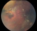
Retinal Capillary Hemangioma due to Von Hipple-Lindau Syndrome434 viewsA 47-year old female comes in for a second opinion for retinal capillary Hemangioma in the left eye. VA was HM (hand motion) in the left eye. She has had multiple procedures done for the left eye including, Retinal Detachment surgery, YAG Laser PI, YAG Laser Capsulotomy, and Cataract surgery.
|
|

Ocular Lymphoma - non-Hodgkins - B-Cell - Right eye has equatorial drusen427 viewsMultifocal sub-RPE lesions in the left eye of a patient with history of Non-hodgkins B-cell Lymphoma last active 2 years ago
|
|

Ocular Lymphoma - non-Hodgkins - B-Cell424 viewsMultifocal sub-RPE lesions in the left eye of a patient with history of Non-hodgkins B-cell Lymphoma last active 2 years ago
|
|

Retinal Capillary Hemangioma due to Von Hipple-Lindau Syndrome423 viewsA 47-year old female comes in for a second opinion for retinal capillary Hemangioma in the left eye. VA was HM (hand motion) in the left eye. She has had multiple procedures done for the left eye including, Retinal Detachment surgery, YAG Laser PI, YAG Laser Capsulotomy, and Cataract surgery.
|
|

Ocular Lymphoma - non-Hodgkins - B-Cell - Right eye has equatorial drusen419 viewsMultifocal sub-RPE lesions in the left eye of a patient with history of Non-hodgkins B-cell Lymphoma last active 2 years ago
|
|

Ocular Lymphoma - Initially presented like auto-immune retinopathy385 views43 year old man: initial presentation - The vision in the left eye is blurry and opaque for about 2 weeks. In the central of the vision, when he covers the right eye, in the center of the left eye there is a film with a light in the middle. This has never happened before. He has not been sick for the last few months. A few weeks ago he saw waves on the floor when looking down but that has gone away. Since the vision loss started the vision has not changed much. The central vision is not getting better. Â
PMHx – Borderline HTN
VA OD: Dcc20/20
VA OS: Dcc20/40-1 IOP: TP: OD:15 OS:19
1+ NS
No uveitis
Labs normal. One positive antiretinal antibody - Arrestin
VA improved over 2 months to normal. Then the patient had 2 more episodes of self-limited vision loss (no treatment). Then 2 years later he presented with a diagnosis of CNS lymphoma and later developed subretinal deposits (last images)
|
|

Ocular Lymphoma - Flare up despite chemotherapy and radiation therapy - This was treated with intravitreal methotrexate385 viewsVision in the left eye has declined to LP in this image following ocular recurrence of lymphoma
|
|

Ocular Lymphoma - Initially presented like auto-immune retinopathy375 views43 year old man: initial presentation - The vision in the left eye is blurry and opaque for about 2 weeks. In the central of the vision, when he covers the right eye, in the center of the left eye there is a film with a light in the middle. This has never happened before. He has not been sick for the last few months. A few weeks ago he saw waves on the floor when looking down but that has gone away. Since the vision loss started the vision has not changed much. The central vision is not getting better. Â
PMHx – Borderline HTN
VA OD: Dcc20/20
VA OS: Dcc20/40-1 IOP: TP: OD:15 OS:19
1+ NS
No uveitis
Labs normal. One positive antiretinal antibody - Arrestin
VA improved over 2 months to normal. Then the patient had 2 more episodes of self-limited vision loss (no treatment). Then 2 years later he presented with a diagnosis of CNS lymphoma and later developed subretinal deposits (last images)
|
|
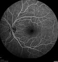
Ocular Lymphoma - Initially presented like auto-immune retinopathy372 views43 year old man: initial presentation - The vision in the left eye is blurry and opaque for about 2 weeks. In the central of the vision, when he covers the right eye, in the center of the left eye there is a film with a light in the middle. This has never happened before. He has not been sick for the last few months. A few weeks ago he saw waves on the floor when looking down but that has gone away. Since the vision loss started the vision has not changed much. The central vision is not getting better. Â
PMHx – Borderline HTN
VA OD: Dcc20/20
VA OS: Dcc20/40-1 IOP: TP: OD:15 OS:19
1+ NS
No uveitis
Labs normal. One positive antiretinal antibody - Arrestin
VA improved over 2 months to normal. Then the patient had 2 more episodes of self-limited vision loss (no treatment). Then 2 years later he presented with a diagnosis of CNS lymphoma and later developed subretinal deposits (last images)
|
|

Ocular Lymphoma - Initially presented like auto-immune retinopathy367 views43 year old man: initial presentation - The vision in the left eye is blurry and opaque for about 2 weeks. In the central of the vision, when he covers the right eye, in the center of the left eye there is a film with a light in the middle. This has never happened before. He has not been sick for the last few months. A few weeks ago he saw waves on the floor when looking down but that has gone away. Since the vision loss started the vision has not changed much. The central vision is not getting better. Â
PMHx – Borderline HTN
VA OD: Dcc20/20
VA OS: Dcc20/40-1 IOP: TP: OD:15 OS:19
1+ NS
No uveitis
Labs normal. One positive antiretinal antibody - Arrestin
VA improved over 2 months to normal. Then the patient had 2 more episodes of self-limited vision loss (no treatment). Then 2 years later he presented with a diagnosis of CNS lymphoma and later developed subretinal deposits (last images)
|
|

Ocular Lymphoma - Initially presented like auto-immune retinopathy363 views43 year old man: initial presentation - The vision in the left eye is blurry and opaque for about 2 weeks. In the central of the vision, when he covers the right eye, in the center of the left eye there is a film with a light in the middle. This has never happened before. He has not been sick for the last few months. A few weeks ago he saw waves on the floor when looking down but that has gone away. Since the vision loss started the vision has not changed much. The central vision is not getting better. Â
PMHx – Borderline HTN
VA OD: Dcc20/20
VA OS: Dcc20/40-1 IOP: TP: OD:15 OS:19
1+ NS
No uveitis
Labs normal. One positive antiretinal antibody - Arrestin
VA improved over 2 months to normal. Then the patient had 2 more episodes of self-limited vision loss (no treatment). Then 2 years later he presented with a diagnosis of CNS lymphoma and later developed subretinal deposits (last images)
|
|
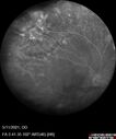
Retinal Angioma358 views53 year old female with no-symptoms and no significant medical history (or family history)
|
|

Retinal Angioma355 views53 year old female with no-symptoms and no significant medical history (or family history)
|
|

Ocular Lymphoma - Initially presented like auto-immune retinopathy355 views43 year old man: initial presentation - The vision in the left eye is blurry and opaque for about 2 weeks. In the central of the vision, when he covers the right eye, in the center of the left eye there is a film with a light in the middle. This has never happened before. He has not been sick for the last few months. A few weeks ago he saw waves on the floor when looking down but that has gone away. Since the vision loss started the vision has not changed much. The central vision is not getting better. Â
PMHx – Borderline HTN
VA OD: Dcc20/20
VA OS: Dcc20/40-1 IOP: TP: OD:15 OS:19
1+ NS
No uveitis
Labs normal. One positive antiretinal antibody - Arrestin
VA improved over 2 months to normal. Then the patient had 2 more episodes of self-limited vision loss (no treatment). Then 2 years later he presented with a diagnosis of CNS lymphoma and later developed subretinal deposits (last images)
|
|

Ocular Lymphoma - Initially presented like auto-immune retinopathy354 views43 year old man: initial presentation - The vision in the left eye is blurry and opaque for about 2 weeks. In the central of the vision, when he covers the right eye, in the center of the left eye there is a film with a light in the middle. This has never happened before. He has not been sick for the last few months. A few weeks ago he saw waves on the floor when looking down but that has gone away. Since the vision loss started the vision has not changed much. The central vision is not getting better. Â
PMHx – Borderline HTN
VA OD: Dcc20/20
VA OS: Dcc20/40-1 IOP: TP: OD:15 OS:19
1+ NS
No uveitis
Labs normal. One positive antiretinal antibody - Arrestin
VA improved over 2 months to normal. Then the patient had 2 more episodes of self-limited vision loss (no treatment). Then 2 years later he presented with a diagnosis of CNS lymphoma and later developed subretinal deposits (last images)
|
|

Ocular Lymphoma - Initially presented like auto-immune retinopathy353 views43 year old man: initial presentation - The vision in the left eye is blurry and opaque for about 2 weeks. In the central of the vision, when he covers the right eye, in the center of the left eye there is a film with a light in the middle. This has never happened before. He has not been sick for the last few months. A few weeks ago he saw waves on the floor when looking down but that has gone away. Since the vision loss started the vision has not changed much. The central vision is not getting better. Â
PMHx – Borderline HTN
VA OD: Dcc20/20
VA OS: Dcc20/40-1 IOP: TP: OD:15 OS:19
1+ NS
No uveitis
Labs normal. One positive antiretinal antibody - Arrestin
VA improved over 2 months to normal. Then the patient had 2 more episodes of self-limited vision loss (no treatment). Then 2 years later he presented with a diagnosis of CNS lymphoma and later developed subretinal deposits (last images)
|
|

Ocular Lymphoma - Initially presented like auto-immune retinopathy351 views43 year old man: initial presentation - The vision in the left eye is blurry and opaque for about 2 weeks. In the central of the vision, when he covers the right eye, in the center of the left eye there is a film with a light in the middle. This has never happened before. He has not been sick for the last few months. A few weeks ago he saw waves on the floor when looking down but that has gone away. Since the vision loss started the vision has not changed much. The central vision is not getting better. Â
PMHx – Borderline HTN
VA OD: Dcc20/20
VA OS: Dcc20/40-1 IOP: TP: OD:15 OS:19
1+ NS
No uveitis
Labs normal. One positive antiretinal antibody - Arrestin
VA improved over 2 months to normal. Then the patient had 2 more episodes of self-limited vision loss (no treatment). Then 2 years later he presented with a diagnosis of CNS lymphoma and later developed subretinal deposits (last images)
|
|

Retinal Angioma349 views53 year old female with no-symptoms and no significant medical history (or family history)
|
|
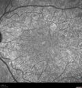
Ocular Lymphoma - Initially presented like auto-immune retinopathy348 views43 year old man: initial presentation - The vision in the left eye is blurry and opaque for about 2 weeks. In the central of the vision, when he covers the right eye, in the center of the left eye there is a film with a light in the middle. This has never happened before. He has not been sick for the last few months. A few weeks ago he saw waves on the floor when looking down but that has gone away. Since the vision loss started the vision has not changed much. The central vision is not getting better. Â
PMHx – Borderline HTN
VA OD: Dcc20/20
VA OS: Dcc20/40-1 IOP: TP: OD:15 OS:19
1+ NS
No uveitis
Labs normal. One positive antiretinal antibody - Arrestin
VA improved over 2 months to normal. Then the patient had 2 more episodes of self-limited vision loss (no treatment). Then 2 years later he presented with a diagnosis of CNS lymphoma and later developed subretinal deposits (last images)
|
|

Retinal Angioma347 views53 year old female with no-symptoms and no significant medical history (or family history)
|
|
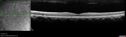
Ocular Lymphoma - Initially presented like auto-immune retinopathy347 views43 year old man: initial presentation - The vision in the left eye is blurry and opaque for about 2 weeks. In the central of the vision, when he covers the right eye, in the center of the left eye there is a film with a light in the middle. This has never happened before. He has not been sick for the last few months. A few weeks ago he saw waves on the floor when looking down but that has gone away. Since the vision loss started the vision has not changed much. The central vision is not getting better. Â
PMHx – Borderline HTN
VA OD: Dcc20/20
VA OS: Dcc20/40-1 IOP: TP: OD:15 OS:19
1+ NS
No uveitis
Labs normal. One positive antiretinal antibody - Arrestin
VA improved over 2 months to normal. Then the patient had 2 more episodes of self-limited vision loss (no treatment). Then 2 years later he presented with a diagnosis of CNS lymphoma and later developed subretinal deposits (last images)
|
|

Ocular Lymphoma - Initially presented like auto-immune retinopathy346 views43 year old man: initial presentation - The vision in the left eye is blurry and opaque for about 2 weeks. In the central of the vision, when he covers the right eye, in the center of the left eye there is a film with a light in the middle. This has never happened before. He has not been sick for the last few months. A few weeks ago he saw waves on the floor when looking down but that has gone away. Since the vision loss started the vision has not changed much. The central vision is not getting better. Â
PMHx – Borderline HTN
VA OD: Dcc20/20
VA OS: Dcc20/40-1 IOP: TP: OD:15 OS:19
1+ NS
No uveitis
Labs normal. One positive antiretinal antibody - Arrestin
VA improved over 2 months to normal. Then the patient had 2 more episodes of self-limited vision loss (no treatment). Then 2 years later he presented with a diagnosis of CNS lymphoma and later developed subretinal deposits (last images)
|
|
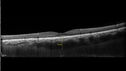
Ocular Lymphoma - Initially presented like auto-immune retinopathy344 views43 year old man: initial presentation - The vision in the left eye is blurry and opaque for about 2 weeks. In the central of the vision, when he covers the right eye, in the center of the left eye there is a film with a light in the middle. This has never happened before. He has not been sick for the last few months. A few weeks ago he saw waves on the floor when looking down but that has gone away. Since the vision loss started the vision has not changed much. The central vision is not getting better. Â
PMHx – Borderline HTN
VA OD: Dcc20/20
VA OS: Dcc20/40-1 IOP: TP: OD:15 OS:19
1+ NS
No uveitis
Labs normal. One positive antiretinal antibody - Arrestin
VA improved over 2 months to normal. Then the patient had 2 more episodes of self-limited vision loss (no treatment). Then 2 years later he presented with a diagnosis of CNS lymphoma and later developed subretinal deposits (last images)
|
|

Ocular Lymphoma - Initially presented like auto-immune retinopathy344 views43 year old man: initial presentation - The vision in the left eye is blurry and opaque for about 2 weeks. In the central of the vision, when he covers the right eye, in the center of the left eye there is a film with a light in the middle. This has never happened before. He has not been sick for the last few months. A few weeks ago he saw waves on the floor when looking down but that has gone away. Since the vision loss started the vision has not changed much. The central vision is not getting better. Â
PMHx – Borderline HTN
VA OD: Dcc20/20
VA OS: Dcc20/40-1 IOP: TP: OD:15 OS:19
1+ NS
No uveitis
Labs normal. One positive antiretinal antibody - Arrestin
VA improved over 2 months to normal. Then the patient had 2 more episodes of self-limited vision loss (no treatment). Then 2 years later he presented with a diagnosis of CNS lymphoma and later developed subretinal deposits (last images)
|
|

Ocular Lymphoma - Initially presented like auto-immune retinopathy340 views43 year old man: initial presentation - The vision in the left eye is blurry and opaque for about 2 weeks. In the central of the vision, when he covers the right eye, in the center of the left eye there is a film with a light in the middle. This has never happened before. He has not been sick for the last few months. A few weeks ago he saw waves on the floor when looking down but that has gone away. Since the vision loss started the vision has not changed much. The central vision is not getting better. Â
PMHx – Borderline HTN
VA OD: Dcc20/20
VA OS: Dcc20/40-1 IOP: TP: OD:15 OS:19
1+ NS
No uveitis
Labs normal. One positive antiretinal antibody - Arrestin
VA improved over 2 months to normal. Then the patient had 2 more episodes of self-limited vision loss (no treatment). Then 2 years later he presented with a diagnosis of CNS lymphoma and later developed subretinal deposits (last images)
|
|

Ocular Lymphoma - Initially presented like auto-immune retinopathy339 views43 year old man: initial presentation - The vision in the left eye is blurry and opaque for about 2 weeks. In the central of the vision, when he covers the right eye, in the center of the left eye there is a film with a light in the middle. This has never happened before. He has not been sick for the last few months. A few weeks ago he saw waves on the floor when looking down but that has gone away. Since the vision loss started the vision has not changed much. The central vision is not getting better. Â
PMHx – Borderline HTN
VA OD: Dcc20/20
VA OS: Dcc20/40-1 IOP: TP: OD:15 OS:19
1+ NS
No uveitis
Labs normal. One positive antiretinal antibody - Arrestin
VA improved over 2 months to normal. Then the patient had 2 more episodes of self-limited vision loss (no treatment). Then 2 years later he presented with a diagnosis of CNS lymphoma and later developed subretinal deposits (last images)
|
|

Retinal Cavernous Hemangioma339 views27 year old female with normal vision - MRI was normal
|
|

Ocular Lymphoma - Initially presented like auto-immune retinopathy337 views43 year old man: initial presentation - The vision in the left eye is blurry and opaque for about 2 weeks. In the central of the vision, when he covers the right eye, in the center of the left eye there is a film with a light in the middle. This has never happened before. He has not been sick for the last few months. A few weeks ago he saw waves on the floor when looking down but that has gone away. Since the vision loss started the vision has not changed much. The central vision is not getting better. Â
PMHx – Borderline HTN
VA OD: Dcc20/20
VA OS: Dcc20/40-1 IOP: TP: OD:15 OS:19
1+ NS
No uveitis
Labs normal. One positive antiretinal antibody - Arrestin
VA improved over 2 months to normal. Then the patient had 2 more episodes of self-limited vision loss (no treatment). Then 2 years later he presented with a diagnosis of CNS lymphoma and later developed subretinal deposits (last images)
|
|

Retinal Cavernous Hemangioma337 views27 year old female with normal vision - MRI was normal
|
|

Ocular Lymphoma - Initially presented like auto-immune retinopathy336 views43 year old man: initial presentation - The vision in the left eye is blurry and opaque for about 2 weeks. In the central of the vision, when he covers the right eye, in the center of the left eye there is a film with a light in the middle. This has never happened before. He has not been sick for the last few months. A few weeks ago he saw waves on the floor when looking down but that has gone away. Since the vision loss started the vision has not changed much. The central vision is not getting better. Â
PMHx – Borderline HTN
VA OD: Dcc20/20
VA OS: Dcc20/40-1 IOP: TP: OD:15 OS:19
1+ NS
No uveitis
Labs normal. One positive antiretinal antibody - Arrestin
VA improved over 2 months to normal. Then the patient had 2 more episodes of self-limited vision loss (no treatment). Then 2 years later he presented with a diagnosis of CNS lymphoma and later developed subretinal deposits (last images)
|
|

Ocular Lymphoma - Initially presented like auto-immune retinopathy335 views43 year old man: initial presentation - The vision in the left eye is blurry and opaque for about 2 weeks. In the central of the vision, when he covers the right eye, in the center of the left eye there is a film with a light in the middle. This has never happened before. He has not been sick for the last few months. A few weeks ago he saw waves on the floor when looking down but that has gone away. Since the vision loss started the vision has not changed much. The central vision is not getting better. Â
PMHx – Borderline HTN
VA OD: Dcc20/20
VA OS: Dcc20/40-1 IOP: TP: OD:15 OS:19
1+ NS
No uveitis
Labs normal. One positive antiretinal antibody - Arrestin
VA improved over 2 months to normal. Then the patient had 2 more episodes of self-limited vision loss (no treatment). Then 2 years later he presented with a diagnosis of CNS lymphoma and later developed subretinal deposits (last images)
|
|

Ocular Lymphoma - Initially presented like auto-immune retinopathy335 views43 year old man: initial presentation - The vision in the left eye is blurry and opaque for about 2 weeks. In the central of the vision, when he covers the right eye, in the center of the left eye there is a film with a light in the middle. This has never happened before. He has not been sick for the last few months. A few weeks ago he saw waves on the floor when looking down but that has gone away. Since the vision loss started the vision has not changed much. The central vision is not getting better. Â
PMHx – Borderline HTN
VA OD: Dcc20/20
VA OS: Dcc20/40-1 IOP: TP: OD:15 OS:19
1+ NS
No uveitis
Labs normal. One positive antiretinal antibody - Arrestin
VA improved over 2 months to normal. Then the patient had 2 more episodes of self-limited vision loss (no treatment). Then 2 years later he presented with a diagnosis of CNS lymphoma and later developed subretinal deposits (last images)
|
|

Retinal Angioma334 views53 year old female with no-symptoms and no significant medical history (or family history)
|
|

Ocular Lymphoma - Initially presented like auto-immune retinopathy334 views43 year old man: initial presentation - The vision in the left eye is blurry and opaque for about 2 weeks. In the central of the vision, when he covers the right eye, in the center of the left eye there is a film with a light in the middle. This has never happened before. He has not been sick for the last few months. A few weeks ago he saw waves on the floor when looking down but that has gone away. Since the vision loss started the vision has not changed much. The central vision is not getting better. Â
PMHx – Borderline HTN
VA OD: Dcc20/20
VA OS: Dcc20/40-1 IOP: TP: OD:15 OS:19
1+ NS
No uveitis
Labs normal. One positive antiretinal antibody - Arrestin
VA improved over 2 months to normal. Then the patient had 2 more episodes of self-limited vision loss (no treatment). Then 2 years later he presented with a diagnosis of CNS lymphoma and later developed subretinal deposits (last images)
|
|

Ocular Lymphoma - Initially presented like auto-immune retinopathy333 views43 year old man: initial presentation - The vision in the left eye is blurry and opaque for about 2 weeks. In the central of the vision, when he covers the right eye, in the center of the left eye there is a film with a light in the middle. This has never happened before. He has not been sick for the last few months. A few weeks ago he saw waves on the floor when looking down but that has gone away. Since the vision loss started the vision has not changed much. The central vision is not getting better. Â
PMHx – Borderline HTN
VA OD: Dcc20/20
VA OS: Dcc20/40-1 IOP: TP: OD:15 OS:19
1+ NS
No uveitis
Labs normal. One positive antiretinal antibody - Arrestin
VA improved over 2 months to normal. Then the patient had 2 more episodes of self-limited vision loss (no treatment). Then 2 years later he presented with a diagnosis of CNS lymphoma and later developed subretinal deposits (last images)
|
|
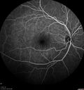
Ocular Lymphoma - Initially presented like auto-immune retinopathy332 views43 year old man: initial presentation - The vision in the left eye is blurry and opaque for about 2 weeks. In the central of the vision, when he covers the right eye, in the center of the left eye there is a film with a light in the middle. This has never happened before. He has not been sick for the last few months. A few weeks ago he saw waves on the floor when looking down but that has gone away. Since the vision loss started the vision has not changed much. The central vision is not getting better. Â
PMHx – Borderline HTN
VA OD: Dcc20/20
VA OS: Dcc20/40-1 IOP: TP: OD:15 OS:19
1+ NS
No uveitis
Labs normal. One positive antiretinal antibody - Arrestin
VA improved over 2 months to normal. Then the patient had 2 more episodes of self-limited vision loss (no treatment). Then 2 years later he presented with a diagnosis of CNS lymphoma and later developed subretinal deposits (last images)
|
|

Ocular Lymphoma - Initially presented like auto-immune retinopathy331 views43 year old man: initial presentation - The vision in the left eye is blurry and opaque for about 2 weeks. In the central of the vision, when he covers the right eye, in the center of the left eye there is a film with a light in the middle. This has never happened before. He has not been sick for the last few months. A few weeks ago he saw waves on the floor when looking down but that has gone away. Since the vision loss started the vision has not changed much. The central vision is not getting better. Â
PMHx – Borderline HTN
VA OD: Dcc20/20
VA OS: Dcc20/40-1 IOP: TP: OD:15 OS:19
1+ NS
No uveitis
Labs normal. One positive antiretinal antibody - Arrestin
VA improved over 2 months to normal. Then the patient had 2 more episodes of self-limited vision loss (no treatment). Then 2 years later he presented with a diagnosis of CNS lymphoma and later developed subretinal deposits (last images)
|
|

Ocular Lymphoma - Initially presented like auto-immune retinopathy331 views43 year old man: initial presentation - The vision in the left eye is blurry and opaque for about 2 weeks. In the central of the vision, when he covers the right eye, in the center of the left eye there is a film with a light in the middle. This has never happened before. He has not been sick for the last few months. A few weeks ago he saw waves on the floor when looking down but that has gone away. Since the vision loss started the vision has not changed much. The central vision is not getting better. Â
PMHx – Borderline HTN
VA OD: Dcc20/20
VA OS: Dcc20/40-1 IOP: TP: OD:15 OS:19
1+ NS
No uveitis
Labs normal. One positive antiretinal antibody - Arrestin
VA improved over 2 months to normal. Then the patient had 2 more episodes of self-limited vision loss (no treatment). Then 2 years later he presented with a diagnosis of CNS lymphoma and later developed subretinal deposits (last images)
|
|
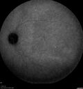
Ocular Lymphoma - Initially presented like auto-immune retinopathy331 views43 year old man: initial presentation - The vision in the left eye is blurry and opaque for about 2 weeks. In the central of the vision, when he covers the right eye, in the center of the left eye there is a film with a light in the middle. This has never happened before. He has not been sick for the last few months. A few weeks ago he saw waves on the floor when looking down but that has gone away. Since the vision loss started the vision has not changed much. The central vision is not getting better. Â
PMHx – Borderline HTN
VA OD: Dcc20/20
VA OS: Dcc20/40-1 IOP: TP: OD:15 OS:19
1+ NS
No uveitis
Labs normal. One positive antiretinal antibody - Arrestin
VA improved over 2 months to normal. Then the patient had 2 more episodes of self-limited vision loss (no treatment). Then 2 years later he presented with a diagnosis of CNS lymphoma and later developed subretinal deposits (last images)
|
|

Ocular Lymphoma - Initially presented like auto-immune retinopathy327 views43 year old man: initial presentation - The vision in the left eye is blurry and opaque for about 2 weeks. In the central of the vision, when he covers the right eye, in the center of the left eye there is a film with a light in the middle. This has never happened before. He has not been sick for the last few months. A few weeks ago he saw waves on the floor when looking down but that has gone away. Since the vision loss started the vision has not changed much. The central vision is not getting better. Â
PMHx – Borderline HTN
VA OD: Dcc20/20
VA OS: Dcc20/40-1 IOP: TP: OD:15 OS:19
1+ NS
No uveitis
Labs normal. One positive antiretinal antibody - Arrestin
VA improved over 2 months to normal. Then the patient had 2 more episodes of self-limited vision loss (no treatment). Then 2 years later he presented with a diagnosis of CNS lymphoma and later developed subretinal deposits (last images)
|
|
|
|


