Les plus populaires - Peripheral Retinal Degeneration
|

Peripheral reticular pigment degeneration - pavingstone degeneration - age-related macular degenerationvu 1946 fois60 year old healthy man with intermediate AMD and peripheral pigment degeneration and pavinstone degeneration. Vision is 20/20 OU and the patient is asymptomatic. No significant history of AMD and he is a non-smoker.
|
|

Reticular peripheral pigment degenerationvu 1683 fois
|
|

Reticular pigment degenerationvu 1573 fois78 year old man with wet AMD- Peripheral retina show reticular pigment degeneration
|
|

Eccentric Disciform Scar - Breakthrough Vitreous Hemorrhage - InferoTemporal Subretinal Fibrosis - Ectopic Choroidal Neovascular Membranesvu 1253 fois88-year-old man has noticed for the last few months the vision in the right eye is foggy, like there is a film in the eye or a haze, His vision is 20/16 in each eye.
Right eye - inferotemporal periphery there is a hemorrhage at the equator at about 7:00 o’clock, which is about 4-disc diameters across and adjacent to that there is scarring.
Left eye - large disciform scar inferotemporally
|
|
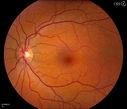
Pseudoxanthoma Elasticum with Angioid Streaksvu 1173 foisYoung female patient who comes in for routine eye exam, VA 20/20 in both eyes. Fundus photos shows a Peu'd orange appearance in the periphery of the retina and angioid streaks around the optic nerve.
|
|

Peripheral Exudative Hemorrhagic Chorioretinopathyvu 1103 fois70 year old man with recurrent peripheral choroidal hemorrhages - no treatment - b-scan shows hemorrhage shrinking over a month
|
|

Eccentric Disciform Scar - Breakthrough Vitreous Hemorrhage - InferoTemporal Subretinal Fibrosis - Ectopic Choroidal Neovascular Membranesvu 1088 fois88-year-old man has noticed for the last few months the vision in the right eye is foggy, like there is a film in the eye or a haze, His vision is 20/16 in each eye.
Right eye - inferotemporal periphery there is a hemorrhage at the equator at about 7:00 o’clock, which is about 4-disc diameters across and adjacent to that there is scarring.
Left eye - large disciform scar inferotemporally
|
|

Pigmented Lattice Degenerationvu 1053 foisPigmented Lattice - Incidental Finding
|
|

Peripheral Exudative Hemorrhagic Chorioretinopathyvu 1050 fois70 year old man with recurrent peripheral choroidal hemorrhages - no treatment - b-scan shows hemorrhage shrinking over a month
|
|

Lattice Degenerationvu 941 fois
|
|

ECTOPIC DISCIFORM SCAR/PERIPHERAL CHOROIDAL NEOVASCULAR MEMBRANE – EXUDATE –INFLAMMATION IN REACTION TO THE PERIPHERAL VASCULAR LESION vu 886 fois53-year-old woman has noticed some blurred vision in the right eye for a few years. In retrospect, she thinks it is possible since she was a teenager the right eye was always the weaker eye and that the left eye has always been the better eye. VISUAL ACUITY: Vision OD is 20/400
|
|

Peripheral Exudative Hemorrhagic Chorioretinopathyvu 882 fois70 year old man with recurrent peripheral choroidal hemorrhages - no treatment - b-scan shows hemorrhage shrinking over a month
|
|

Eccentric Disciform Scar - Breakthrough Vitreous Hemorrhage - InferoTemporal Subretinal Fibrosis - Ectopic Choroidal Neovascular Membranesvu 881 fois88-year-old man has noticed for the last few months the vision in the right eye is foggy, like there is a film in the eye or a haze, His vision is 20/16 in each eye.
Right eye - inferotemporal periphery there is a hemorrhage at the equator at about 7:00 o’clock, which is about 4-disc diameters across and adjacent to that there is scarring.
Left eye - large disciform scar inferotemporally
|
|

ECTOPIC DISCIFORM SCAR/PERIPHERAL CHOROIDAL NEOVASCULAR MEMBRANE – EXUDATE –INFLAMMATION IN REACTION TO THE PERIPHERAL VASCULAR LESION vu 857 fois53-year-old woman has noticed some blurred vision in the right eye for a few years. In retrospect, she thinks it is possible since she was a teenager the right eye was always the weaker eye and that the left eye has always been the better eye. VISUAL ACUITY: Vision OD is 20/400
|
|
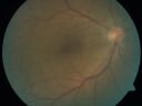
ECTOPIC DISCIFORM SCAR/PERIPHERAL CHOROIDAL NEOVASCULAR MEMBRANE – EXUDATE –INFLAMMATION IN REACTION TO THE PERIPHERAL VASCULAR LESION vu 841 fois53-year-old woman has noticed some blurred vision in the right eye for a few years. In retrospect, she thinks it is possible since she was a teenager the right eye was always the weaker eye and that the left eye has always been the better eye. VISUAL ACUITY: Vision OD is 20/400
|
|

Peripheral Retinal Degenerationvu 838 foisPatient comes in with double vision. VA was 20/20 in both eyes. Fundus exam shows Retinal Degenerative changes in both eyes. Offer to correct double vision with temporary Fresnel prism
|
|

Equatorial Drusenvu 837 fois61 year old female with normal visoin
|
|

ECTOPIC DISCIFORM SCAR/PERIPHERAL CHOROIDAL NEOVASCULAR MEMBRANE – EXUDATE –INFLAMMATION IN REACTION TO THE PERIPHERAL VASCULAR LESION vu 828 fois53-year-old woman has noticed some blurred vision in the right eye for a few years. In retrospect, she thinks it is possible since she was a teenager the right eye was always the weaker eye and that the left eye has always been the better eye. VISUAL ACUITY: Vision OD is 20/400
|
|

Peripheral Reticular Pigment Degenerationvu 826 fois81 year old man with a choroidal nevus in the right eye. VA 20/20 OU. There is extensive peripheral pigment degeneration
|
|

Eccentric Disciform Scar - Breakthrough Vitreous Hemorrhage - InferoTemporal Subretinal Fibrosis - Ectopic Choroidal Neovascular Membranesvu 808 fois88-year-old man has noticed for the last few months the vision in the right eye is foggy, like there is a film in the eye or a haze, His vision is 20/16 in each eye.
Right eye - inferotemporal periphery there is a hemorrhage at the equator at about 7:00 o’clock, which is about 4-disc diameters across and adjacent to that there is scarring.
Left eye - large disciform scar inferotemporally
|
|
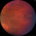
Peripheral reticular pigment degeneration - pavingstone degeneration - age-related macular degenerationvu 801 fois60 year old healthy man with intermediate AMD and peripheral pigment degeneration and pavinstone degeneration. Vision is 20/20 OU and the patient is asymptomatic. No significant history of AMD and he is a non-smoker.
|
|
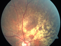
Equatorial Drusenvu 797 fois61 year old female with normal visoin
|
|

Eccentric Disciform Scar - Breakthrough Vitreous Hemorrhage - InferoTemporal Subretinal Fibrosis - Ectopic Choroidal Neovascular Membranesvu 790 fois88-year-old man has noticed for the last few months the vision in the right eye is foggy, like there is a film in the eye or a haze, His vision is 20/16 in each eye.
Right eye - inferotemporal periphery there is a hemorrhage at the equator at about 7:00 o’clock, which is about 4-disc diameters across and adjacent to that there is scarring.
Left eye - large disciform scar inferotemporally
|
|

Eccentric Disciform Scar - Breakthrough Vitreous Hemorrhage - InferoTemporal Subretinal Fibrosis - Ectopic Choroidal Neovascular Membranesvu 771 fois88-year-old man has noticed for the last few months the vision in the right eye is foggy, like there is a film in the eye or a haze, His vision is 20/16 in each eye.
Right eye - inferotemporal periphery there is a hemorrhage at the equator at about 7:00 o’clock, which is about 4-disc diameters across and adjacent to that there is scarring.
Left eye - large disciform scar inferotemporally
|
|

Peripheral Exudative Hemorrhagic Chorioretinopathyvu 765 fois70 year old man with recurrent peripheral choroidal hemorrhages - no treatment - b-scan shows hemorrhage shrinking over a month
|
|

Eccentric Disciform Scar - Breakthrough Vitreous Hemorrhage - InferoTemporal Subretinal Fibrosis - Ectopic Choroidal Neovascular Membranesvu 760 fois88-year-old man has noticed for the last few months the vision in the right eye is foggy, like there is a film in the eye or a haze, His vision is 20/16 in each eye.
Right eye - inferotemporal periphery there is a hemorrhage at the equator at about 7:00 o’clock, which is about 4-disc diameters across and adjacent to that there is scarring.
Left eye - large disciform scar inferotemporally
|
|

Peripheral Exudative Hemorrhagic Chorioretinopathyvu 745 fois70 year old man with recurrent peripheral choroidal hemorrhages - no treatment - b-scan shows hemorrhage shrinking over a month
|
|
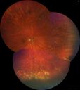
Peripheral reticular pigment degeneration - pavingstone degeneration - age-related macular degenerationvu 734 fois60 year old healthy man with intermediate AMD and peripheral pigment degeneration and pavinstone degeneration. Vision is 20/20 OU and the patient is asymptomatic. No significant history of AMD and he is a non-smoker.
|
|

Peripheral exudative hemorrhagic chorioretinopathy - PECHR - Subfoveal fluidvu 731 fois85 year old female with
Fluctuating vision in the right eye.
VA 20/200 OD, 20/32 OS
|
|

Eccentric Disciform Scar - Breakthrough Vitreous Hemorrhage - InferoTemporal Subretinal Fibrosis - Ectopic Choroidal Neovascular Membranesvu 730 fois88-year-old man has noticed for the last few months the vision in the right eye is foggy, like there is a film in the eye or a haze, His vision is 20/16 in each eye.
Right eye - inferotemporal periphery there is a hemorrhage at the equator at about 7:00 o’clock, which is about 4-disc diameters across and adjacent to that there is scarring.
Left eye - large disciform scar inferotemporally
|
|

Peripheral Exudative Hemorrhagic Chorioretinopathyvu 716 fois70 year old man with recurrent peripheral choroidal hemorrhages - no treatment - b-scan shows hemorrhage shrinking over a month
|
|

Eccentric Disciform Scar - Breakthrough Vitreous Hemorrhage - InferoTemporal Subretinal Fibrosis - Ectopic Choroidal Neovascular Membranesvu 712 fois88-year-old man has noticed for the last few months the vision in the right eye is foggy, like there is a film in the eye or a haze, His vision is 20/16 in each eye.
Right eye - inferotemporal periphery there is a hemorrhage at the equator at about 7:00 o’clock, which is about 4-disc diameters across and adjacent to that there is scarring.
Left eye - large disciform scar inferotemporally
|
|
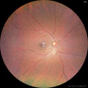
Pigmented Cystic Retinal Tuftvu 710 foisAsymptomatic 20 year old male
|
|

Eccentric Disciform Scar - Breakthrough Vitreous Hemorrhage - InferoTemporal Subretinal Fibrosis - Ectopic Choroidal Neovascular Membranesvu 707 fois88-year-old man has noticed for the last few months the vision in the right eye is foggy, like there is a film in the eye or a haze, His vision is 20/16 in each eye.
Right eye - inferotemporal periphery there is a hemorrhage at the equator at about 7:00 o’clock, which is about 4-disc diameters across and adjacent to that there is scarring.
Left eye - large disciform scar inferotemporally
|
|

ECTOPIC DISCIFORM SCAR/PERIPHERAL CHOROIDAL NEOVASCULAR MEMBRANE – EXUDATE –INFLAMMATION IN REACTION TO THE PERIPHERAL VASCULAR LESION vu 706 fois53-year-old woman has noticed some blurred vision in the right eye for a few years. In retrospect, she thinks it is possible since she was a teenager the right eye was always the weaker eye and that the left eye has always been the better eye. VISUAL ACUITY: Vision OD is 20/400
|
|

Peripheral Exudative Hemorrhagic Chorioretinopathyvu 699 fois70 year old man with recurrent peripheral choroidal hemorrhages - no treatment - b-scan shows hemorrhage shrinking over a month
|
|

Peripheral exudative hemorrhagic chorioretinopathy - PECHR - Subfoveal fluidvu 695 fois85 year old female with
Fluctuating vision in the right eye.
VA 20/200 OD, 20/32 OS
|
|

Peripheral Exudative Hemorrhagic Chorioretinopathyvu 693 fois93 year old female with shadow in side vision in the left eye. Referred for retinal detachment - Exudative lesion subsided in about 6 months without treatment.
Vision 20/63 OD and 20/100 OS (from dry AMD)
|
|
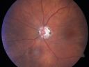
Eccentric Disciform Scar - Breakthrough Vitreous Hemorrhage - InferoTemporal Subretinal Fibrosis - Ectopic Choroidal Neovascular Membranesvu 687 fois88-year-old man has noticed for the last few months the vision in the right eye is foggy, like there is a film in the eye or a haze, His vision is 20/16 in each eye.
Right eye - inferotemporal periphery there is a hemorrhage at the equator at about 7:00 o’clock, which is about 4-disc diameters across and adjacent to that there is scarring.
Left eye - large disciform scar inferotemporally
|
|

ECTOPIC DISCIFORM SCAR/PERIPHERAL CHOROIDAL NEOVASCULAR MEMBRANE – EXUDATE –INFLAMMATION IN REACTION TO THE PERIPHERAL VASCULAR LESION vu 684 fois53-year-old woman has noticed some blurred vision in the right eye for a few years. In retrospect, she thinks it is possible since she was a teenager the right eye was always the weaker eye and that the left eye has always been the better eye. VISUAL ACUITY: Vision OD is 20/400
|
|
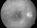
ECTOPIC DISCIFORM SCAR/PERIPHERAL CHOROIDAL NEOVASCULAR MEMBRANE – EXUDATE –INFLAMMATION IN REACTION TO THE PERIPHERAL VASCULAR LESION vu 678 fois53-year-old woman has noticed some blurred vision in the right eye for a few years. In retrospect, she thinks it is possible since she was a teenager the right eye was always the weaker eye and that the left eye has always been the better eye. VISUAL ACUITY: Vision OD is 20/400
|
|

ECTOPIC DISCIFORM SCAR/PERIPHERAL CHOROIDAL NEOVASCULAR MEMBRANE – EXUDATE –INFLAMMATION IN REACTION TO THE PERIPHERAL VASCULAR LESION vu 674 fois53-year-old woman has noticed some blurred vision in the right eye for a few years. In retrospect, she thinks it is possible since she was a teenager the right eye was always the weaker eye and that the left eye has always been the better eye. VISUAL ACUITY: Vision OD is 20/400
|
|

Peripheral Exudative Hemorrhagic Chorioretinopathyvu 674 fois70 year old man with recurrent peripheral choroidal hemorrhages - no treatment - b-scan shows hemorrhage shrinking over a month
|
|

Equatorial Drusenvu 667 fois61 year old female with normal visoin
|
|

ECTOPIC DISCIFORM SCAR/PERIPHERAL CHOROIDAL NEOVASCULAR MEMBRANE – EXUDATE –INFLAMMATION IN REACTION TO THE PERIPHERAL VASCULAR LESION vu 666 fois53-year-old woman has noticed some blurred vision in the right eye for a few years. In retrospect, she thinks it is possible since she was a teenager the right eye was always the weaker eye and that the left eye has always been the better eye. VISUAL ACUITY: Vision OD is 20/400
|
|

Pigmented Cystic Retinal Tuftvu 666 foisAsymptomatic 20 year old male
|
|

Eccentric Disciform Scar - Breakthrough Vitreous Hemorrhage - InferoTemporal Subretinal Fibrosis - Ectopic Choroidal Neovascular Membranesvu 663 fois88-year-old man has noticed for the last few months the vision in the right eye is foggy, like there is a film in the eye or a haze, His vision is 20/16 in each eye.
Right eye - inferotemporal periphery there is a hemorrhage at the equator at about 7:00 o’clock, which is about 4-disc diameters across and adjacent to that there is scarring.
Left eye - large disciform scar inferotemporally
|
|

Peripheral Retinal Degenerationvu 659 foisPatient comes in with double vision. VA was 20/20 in both eyes. Fundus exam shows Retinal Degenerative changes in both eyes. Offer to correct double vision with temporary Fresnel prism
|
|

Bullous Retinoschisis - Confirmed with SD OCTvu 657 foisAtypical location for retinoschisis - SD-OCT is used to confirm that this is retinoschisis and not a retinal detachment
|
|

Peripheral Retinal Degenerationvu 654 foisPatient comes in with double vision. VA was 20/20 in both eyes. Fundus exam shows Retinal Degenerative changes in both eyes. Offer to correct double vision with temporary Fresnel prism
|
|

Bullous Retinoschisis - Confirmed with SD OCTvu 654 foisAtypical location for retinoschisis - SD-OCT is used to confirm that this is retinoschisis and not a retinal detachment
|
|

Bullous Retinoschisis - Confirmed with SD OCTvu 654 foisAtypical location for retinoschisis - SD-OCT is used to confirm that this is retinoschisis and not a retinal detachment
|
|

Pseudoxanthoma Elasticum with Angioid Streaksvu 653 foisYoung female patient who comes in for routine eye exam, VA 20/20 in both eyes. Fundus photos shows a Peu'd orange appearance in the periphery of the retina and angioid streaks around the optic nerve.
|
|
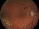
Peripheral exudative hemorrhagic chorioretinopathy - PECHR - Subfoveal fluidvu 651 fois85 year old female with
Fluctuating vision in the right eye.
VA 20/200 OD, 20/32 OS
|
|

Peripheral Retinal Degenerationvu 650 foisPatient comes in with double vision. VA was 20/20 in both eyes. Fundus exam shows Retinal Degenerative changes in both eyes. Offer to correct double vision with temporary Fresnel prism
|
|
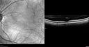
Bullous Retinoschisis - Confirmed with SD OCTvu 649 foisAtypical location for retinoschisis - SD-OCT is used to confirm that this is retinoschisis and not a retinal detachment
|
|
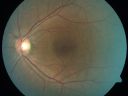
ECTOPIC DISCIFORM SCAR/PERIPHERAL CHOROIDAL NEOVASCULAR MEMBRANE – EXUDATE –INFLAMMATION IN REACTION TO THE PERIPHERAL VASCULAR LESION vu 639 fois53-year-old woman has noticed some blurred vision in the right eye for a few years. In retrospect, she thinks it is possible since she was a teenager the right eye was always the weaker eye and that the left eye has always been the better eye. VISUAL ACUITY: Vision OD is 20/400
|
|

Eccentric Disciform Scar - Breakthrough Vitreous Hemorrhage - InferoTemporal Subretinal Fibrosis - Ectopic Choroidal Neovascular Membranesvu 630 fois88-year-old man has noticed for the last few months the vision in the right eye is foggy, like there is a film in the eye or a haze, His vision is 20/16 in each eye.
Right eye - inferotemporal periphery there is a hemorrhage at the equator at about 7:00 o’clock, which is about 4-disc diameters across and adjacent to that there is scarring.
Left eye - large disciform scar inferotemporally
|
|

Eccentric Disciform Scar - Breakthrough Vitreous Hemorrhage - InferoTemporal Subretinal Fibrosis - Ectopic Choroidal Neovascular Membranesvu 630 fois88-year-old man has noticed for the last few months the vision in the right eye is foggy, like there is a film in the eye or a haze, His vision is 20/16 in each eye.
Right eye - inferotemporal periphery there is a hemorrhage at the equator at about 7:00 o’clock, which is about 4-disc diameters across and adjacent to that there is scarring.
Left eye - large disciform scar inferotemporally
|
|

Lattice Degenerationvu 628 foisAsymptomatic
|
|
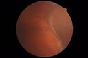
Peripheral Exudative Hemorrhagic Chorioretinopathyvu 625 fois93 year old female with shadow in side vision in the left eye. Referred for retinal detachment - Exudative lesion subsided in about 6 months without treatment.
Vision 20/63 OD and 20/100 OS (from dry AMD)
|
|

ECTOPIC DISCIFORM SCAR/PERIPHERAL CHOROIDAL NEOVASCULAR MEMBRANE – EXUDATE –INFLAMMATION IN REACTION TO THE PERIPHERAL VASCULAR LESION vu 623 fois53-year-old woman has noticed some blurred vision in the right eye for a few years. In retrospect, she thinks it is possible since she was a teenager the right eye was always the weaker eye and that the left eye has always been the better eye. VISUAL ACUITY: Vision OD is 20/400
|
|

Bullous Retinoschisis - Confirmed with SD OCTvu 618 foisAtypical location for retinoschisis - SD-OCT is used to confirm that this is retinoschisis and not a retinal detachment
|
|

ECTOPIC DISCIFORM SCAR/PERIPHERAL CHOROIDAL NEOVASCULAR MEMBRANE – EXUDATE –INFLAMMATION IN REACTION TO THE PERIPHERAL VASCULAR LESION vu 615 fois53-year-old woman has noticed some blurred vision in the right eye for a few years. In retrospect, she thinks it is possible since she was a teenager the right eye was always the weaker eye and that the left eye has always been the better eye. VISUAL ACUITY: Vision OD is 20/400
|
|

Peripheral exudative hemorrhagic chorioretinopathy - PECHR - Subfoveal fluidvu 613 fois85 year old female with
Fluctuating vision in the right eye.
VA 20/200 OD, 20/32 OS
|
|
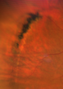
Pigmented Lattice Degenerationvu 606 foisPigmented Lattice - Incidental Finding
|
|

Peripheral Retinal Degenerationvu 597 foisPatient comes in with double vision. VA was 20/20 in both eyes. Fundus exam shows Retinal Degenerative changes in both eyes. Offer to correct double vision with temporary Fresnel prism
|
|

ECTOPIC DISCIFORM SCAR/PERIPHERAL CHOROIDAL NEOVASCULAR MEMBRANE – EXUDATE –INFLAMMATION IN REACTION TO THE PERIPHERAL VASCULAR LESION vu 593 fois53-year-old woman has noticed some blurred vision in the right eye for a few years. In retrospect, she thinks it is possible since she was a teenager the right eye was always the weaker eye and that the left eye has always been the better eye. VISUAL ACUITY: Vision OD is 20/400
|
|

Peripheral exudative hemorrhagic chorioretinopathy - PECHR - Subfoveal fluidvu 591 fois85 year old female with
Fluctuating vision in the right eye.
VA 20/200 OD, 20/32 OS
|
|

Pseudoxanthoma Elasticum with Angioid Streaksvu 590 foisYoung female patient who comes in for routine eye exam, VA 20/20 in both eyes. Fundus photos shows a Peu'd orange appearance in the periphery of the retina and angioid streaks around the optic nerve.
|
|
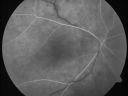
ECTOPIC DISCIFORM SCAR/PERIPHERAL CHOROIDAL NEOVASCULAR MEMBRANE – EXUDATE –INFLAMMATION IN REACTION TO THE PERIPHERAL VASCULAR LESION vu 586 fois53-year-old woman has noticed some blurred vision in the right eye for a few years. In retrospect, she thinks it is possible since she was a teenager the right eye was always the weaker eye and that the left eye has always been the better eye. VISUAL ACUITY: Vision OD is 20/400
|
|

Bullous Retinoschisis - Confirmed with SD OCTvu 586 foisAtypical location for retinoschisis - SD-OCT is used to confirm that this is retinoschisis and not a retinal detachment
|
|

Peripheral exudative hemorrhagic chorioretinopathy - PECHR - Subfoveal fluidvu 585 fois85 year old female with
Fluctuating vision in the right eye.
VA 20/200 OD, 20/32 OS
|
|

Peripheral Retinal Degenerationvu 583 foisPatient comes in with double vision. VA was 20/20 in both eyes. Fundus exam shows Retinal Degenerative changes in both eyes. Offer to correct double vision with temporary Fresnel prism
|
|

Peripheral exudative hemorrhagic chorioretinopathy - PECHR - Subfoveal fluidvu 583 fois85 year old female with
Fluctuating vision in the right eye.
VA 20/200 OD, 20/32 OS
|
|

Eccentric Disciform Scar - Breakthrough Vitreous Hemorrhage - InferoTemporal Subretinal Fibrosis - Ectopic Choroidal Neovascular Membranesvu 579 fois88-year-old man has noticed for the last few months the vision in the right eye is foggy, like there is a film in the eye or a haze, His vision is 20/16 in each eye.
Right eye - inferotemporal periphery there is a hemorrhage at the equator at about 7:00 o’clock, which is about 4-disc diameters across and adjacent to that there is scarring.
Left eye - large disciform scar inferotemporally
|
|

Peripheral exudative hemorrhagic chorioretinopathy - PECHR - Subfoveal fluidvu 578 fois85 year old female with
Fluctuating vision in the right eye.
VA 20/200 OD, 20/32 OS
|
|

ECTOPIC DISCIFORM SCAR/PERIPHERAL CHOROIDAL NEOVASCULAR MEMBRANE – EXUDATE –INFLAMMATION IN REACTION TO THE PERIPHERAL VASCULAR LESION vu 574 fois53-year-old woman has noticed some blurred vision in the right eye for a few years. In retrospect, she thinks it is possible since she was a teenager the right eye was always the weaker eye and that the left eye has always been the better eye. VISUAL ACUITY: Vision OD is 20/400
|
|

Equatorial Drusenvu 572 fois61 year old female with normal visoin
|
|
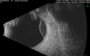
Peripheral Exudative Hemorrhagic Chorioretinopathyvu 569 fois93 year old female with shadow in side vision in the left eye. Referred for retinal detachment - Exudative lesion subsided in about 6 months without treatment.
Vision 20/63 OD and 20/100 OS (from dry AMD)
|
|

Pseudoxanthoma Elasticum with Angioid Streaksvu 566 foisYoung female patient who comes in for routine eye exam, VA 20/20 in both eyes. Fundus photos shows a Peu'd orange appearance in the periphery of the retina and angioid streaks around the optic nerve.
|
|

Equatorial Drusenvu 563 fois61 year old female with normal visoin
|
|

Eccentric Disciform Scar - Breakthrough Vitreous Hemorrhage - InferoTemporal Subretinal Fibrosis - Ectopic Choroidal Neovascular Membranesvu 561 fois88-year-old man has noticed for the last few months the vision in the right eye is foggy, like there is a film in the eye or a haze, His vision is 20/16 in each eye.
Right eye - inferotemporal periphery there is a hemorrhage at the equator at about 7:00 o’clock, which is about 4-disc diameters across and adjacent to that there is scarring.
Left eye - large disciform scar inferotemporally
|
|

Peripheral reticular pigment degeneration - pavingstone degeneration - age-related macular degenerationvu 560 fois60 year old healthy man with intermediate AMD and peripheral pigment degeneration and pavinstone degeneration. Vision is 20/20 OU and the patient is asymptomatic. No significant history of AMD and he is a non-smoker.
|
|

Equatorial Drusenvu 558 fois61 year old female with normal visoin
|
|

Peripheral Retinal Degenerationvu 556 foisPatient comes in with double vision. VA was 20/20 in both eyes. Fundus exam shows Retinal Degenerative changes in both eyes. Offer to correct double vision with temporary Fresnel prism
|
|

Equatorial Drusenvu 556 fois61 year old female with normal visoin
|
|
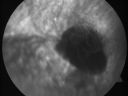
Eccentric Disciform Scar - Breakthrough Vitreous Hemorrhage - InferoTemporal Subretinal Fibrosis - Ectopic Choroidal Neovascular Membranesvu 554 fois88-year-old man has noticed for the last few months the vision in the right eye is foggy, like there is a film in the eye or a haze, His vision is 20/16 in each eye.
Right eye - inferotemporal periphery there is a hemorrhage at the equator at about 7:00 o’clock, which is about 4-disc diameters across and adjacent to that there is scarring.
Left eye - large disciform scar inferotemporally
|
|

Pseudoxanthoma Elasticum with Angioid Streaksvu 550 foisYoung female patient who comes in for routine eye exam, VA 20/20 in both eyes. Fundus photos shows a Peu'd orange appearance in the periphery of the retina and angioid streaks around the optic nerve.
|
|

Peripheral exudative hemorrhagic chorioretinopathy - PECHR - Subfoveal fluidvu 548 fois85 year old female with
Fluctuating vision in the right eye.
VA 20/200 OD, 20/32 OS
|
|

Peripheral exudative hemorrhagic chorioretinopathy - PECHR - Subfoveal fluidvu 547 fois85 year old female with
Fluctuating vision in the right eye.
VA 20/200 OD, 20/32 OS
|
|

Peripheral Exudative Hemorrhagic Chorioretinopathyvu 546 fois93 year old female with shadow in side vision in the left eye. Referred for retinal detachment - Exudative lesion subsided in about 6 months without treatment.
Vision 20/63 OD and 20/100 OS (from dry AMD)
|
|

Lattice Degeneration with Tears - Pre and Post laservu 546 fois60 year old female with new floaters VA is good
|
|

Pseudoxanthoma Elasticum with Angioid Streaksvu 544 foisYoung female patient who comes in for routine eye exam, VA 20/20 in both eyes. Fundus photos shows a Peu'd orange appearance in the periphery of the retina and angioid streaks around the optic nerve.
|
|

Eccentric Disciform Scar - Breakthrough Vitreous Hemorrhage - InferoTemporal Subretinal Fibrosis - Ectopic Choroidal Neovascular Membranesvu 541 fois88-year-old man has noticed for the last few months the vision in the right eye is foggy, like there is a film in the eye or a haze, His vision is 20/16 in each eye.
Right eye - inferotemporal periphery there is a hemorrhage at the equator at about 7:00 o’clock, which is about 4-disc diameters across and adjacent to that there is scarring.
Left eye - large disciform scar inferotemporally
|
|

Bullous Retinoschisis - Confirmed with SD OCTvu 541 foisAtypical location for retinoschisis - SD-OCT is used to confirm that this is retinoschisis and not a retinal detachment
|
|

Equatorial Drusenvu 526 fois61 year old female with normal visoin
|
|

Peripheral Retinal Degenerationvu 525 foisPatient comes in with double vision. VA was 20/20 in both eyes. Fundus exam shows Retinal Degenerative changes in both eyes. Offer to correct double vision with temporary Fresnel prism
|
|

Peripheral reticular pigment degeneration - pavingstone degeneration - age-related macular degenerationvu 519 fois60 year old healthy man with intermediate AMD and peripheral pigment degeneration and pavinstone degeneration. Vision is 20/20 OU and the patient is asymptomatic. No significant history of AMD and he is a non-smoker.
|
|

Bullous Retinoschisis - Confirmed with SD OCTvu 516 foisAtypical location for retinoschisis - SD-OCT is used to confirm that this is retinoschisis and not a retinal detachment
|
|
| 217 photos sur 3 page(s) |
1 |
|


