am meisten angesehen - Non-infectious Posterior Uveitis
|
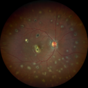
Multifocal Choroiditis and Subretinal Fibrosis with new Subretinal Neovascular Membrane768 x angesehen26 year old African American Girl Saw me at age 10 (16 years ago) with acute multifocal choroiditis and panuveitis which evolved into subretinal fibrosis. She was initially treated with 3 years of methotrexate. Vision ended up 20/20 in both eyes. Her eyes have been inactive since and she was lost to follow-up.
A week ago she noticed sudden decreased vision in the right eye. Within the past few weeks there are more floaters in the right eye.
VA OD: sc6’/200 PHNI
VA OS: sc20/16
|
|

Multiple Evanescent White Dot Syndrome - Acute Idiopathic Blind Spot Enlargement767 x angesehen33-year-old woman one week ago she awoke and saw a red spots flashing out of her right eye. She notices them to the right side, which is the temporal visual field out of the right eye, then they changed to bursts of white lights without colors and then she notices straight ahead a couple spots. She has headaches in the back of her head. OD 20/20, OS 20/20
|
|

Acute Annular Outer Retinopathy - VA loss 2 days Right Eye Only - 25 Year Old Woman766 x angesehenInitial visit (June 11, 2011): 2 days of vision loss right eye. Wedge suddenly of VA loss OD, too dardkto see through. No flashes or floaters. VA 20/20 OU.
|
|

Acute Posterior Multifocal Placoid Pigment Epitheliopathy - APMPPE - Indocyanine Green Angiogram766 x angesehen 25-year-old man with one and half weeks ago he woke up with night chills. Also around that time his joints started hurting him. He also has headaches, although the headache at this point is not severe. His vision has been changing for about the last week and a half. He sees some spots in his vision and they are blurred spots. They are not in the central vision.
VISUAL ACUITY: OD 20/32, OS 20/32
|
|

Acute Macular Neuroretinopathy - (AMN) - Spot in Vision OS - 40 Year old Female766 x angesehenSD OCT shows disruption of the outer retina nasal to the fovea
|
|

Serpiginouse Choroiditis (Chorioretinitis) - Acute Right Eye - Old Left Eye VA 20/25 OD , 20/50 OS761 x angesehen63-year-old woman has serpiginous choroiditis (date - March 2011). The right eye has not been previously involved, and then she noticed new onset floaters in the right eye for the last two weeks. Her vision in the right eye is hazy because of that.
VISUAL ACUITY: Vision OD is 20/25, OS is 20/50
|
|

Punctate Inner Choroidopathy - PIC and Choroidal Neovascular Membrane - Right Eye - Color Fundus Photograph760 x angesehen34-year-old woman with a change in vision in the right eye about eight days ago. Her vision was hazy and she saw waviness in the vision. She does have a history of aches and pains and she was diagnosed with Sjogren’s and 2 months ago she had some fevers and some sinus drainage and that went on for about two months. She is a moderate myope. OD 20/25, OS 20/20.
|
|
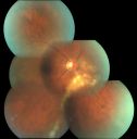
Serpiginouse Choroiditis (Chorioretinitis) - Acute Right Eye - Old Left Eye VA 20/25 OD , 20/50 OS - 1 Week After Intravitreal Kenalog OD758 x angesehen63-year-old woman has serpiginous choroiditis 1 week after intravitreal kenalog treatment
|
|

Multifocal Choroiditis - Linear Scars - Schlaegel lines758 x angesehen66 year old woman with macular pucker right eye. Images show typical linear peripheral scars for MFC or POHS - this is unilateral.
|
|

Acute Macular Neuroretinopathy - (AMN) - Spot in Vision OS - 40 Year old Female757 x angesehenICG angiogram is normal
|
|
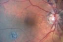
Multifocal Choroiditis Old Scars - Also Peripapillary Scar 82 Year Old Woman756 x angesehen82-year-old woman with multifocal chorioretinal scars, more in the right than the left eye. OD is 20/40, OS is 20/20.
|
|

Acute Macular Neuroretinopathy - (AMN) - Spot in Vision OS - 40 Year old Female756 x angesehen4 months later spot is nearly gone and vision is back to normal (almost) - 20/20
|
|

Acute Annular Outer Retinopathy for 3 Months - 25 YO Female Fundus Photograph755 x angesehen25-year-old female who, about three months ago, put on her sunglasses and noticed a smudge in the vision and at that point she noticed the right eye was hazy. She had a cold a week or two before the vision loss, but she didn’t have any fever. She has no focal neurological problems in her past history. VA 20/20 OU
|
|

Punctate Inner Choroidopathy - PIC and Choroidal Neovascular Membrane - Right Eye - SD OCT754 x angesehen34-year-old woman with a change in vision in the right eye about eight days ago. Her vision was hazy and she saw waviness in the vision. She does have a history of aches and pains and she was diagnosed with Sjogren’s and 2 months ago she had some fevers and some sinus drainage and that went on for about two months. She is a moderate myope. OD 20/25, OS 20/20.
|
|

Atypical Birdshot Chorioretinitis and Retinal Arterial Macroaneurysm Right Eye - HLA-B29 Positive750 x angesehen55-year-old woman has multifocal choroiditis in both eyes because she was HLA A-29 positive. She has presumptive birdshot chorioretinopathy. She has not noticed any vision changes. OD 20/40, OS 20/20.
|
|

Acute Annular Outer Retinopathy - VA loss 2 days Right Eye Only - 25 Year Old Woman749 x angesehenInitial visit (June 11, 2011): 2 days of vision loss right eye. Wedge suddenly of VA loss OD, too dardkto see through. No flashes or floaters. VA 20/20 OU.
|
|

Psuedo-retinitis Pigmentosa - Bone Spicules One Eye - Probably Acute Zonal Occult Outer Retinopathy (AZOOR)748 x angesehen65-year-old woman has pseudoretinitis pigmentosa in the right eye only, most likely from acute zonal occult outer retinopathy. OD 20/25, OS 20/25
|
|

Multifocal Choroiditis and Subretinal Fibrosis - 32 yo Female New Lesion OS745 x angesehen32-year-old woman vision loss in the right eye associated with macular scarring and multifocal choroiditis in 1999 with new vision loss in left eye: OD 20/400, OS 20/50.
|
|
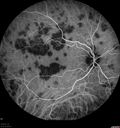
Acute Posterior Multifocal Placoid Pigment Epitheliopathy - APMPPE - Indocyanine Green Angiogram745 x angesehen 25-year-old man with one and half weeks ago he woke up with night chills. Also around that time his joints started hurting him. He also has headaches, although the headache at this point is not severe. His vision has been changing for about the last week and a half. He sees some spots in his vision and they are blurred spots. They are not in the central vision.
VISUAL ACUITY: OD 20/32, OS 20/32
|
|

Acute Posterior Multifocal Placoid Pigment Epitheliopathy - APMPPE - Fundus Photo - Color743 x angesehen 25-year-old man with one and half weeks ago he woke up with night chills. Also around that time his joints started hurting him. He also has headaches, although the headache at this point is not severe. His vision has been changing for about the last week and a half. He sees some spots in his vision and they are blurred spots. They are not in the central vision.
VISUAL ACUITY: OD 20/32, OS 20/32
|
|
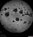
Acute Posterior Multifocal Placoid Pigment Epitheliopathy - APMPPE - Indocyanine Green Angiogram742 x angesehen 25-year-old man with one and half weeks ago he woke up with night chills. Also around that time his joints started hurting him. He also has headaches, although the headache at this point is not severe. His vision has been changing for about the last week and a half. He sees some spots in his vision and they are blurred spots. They are not in the central vision.
VISUAL ACUITY: OD 20/32, OS 20/32
|
|

Acute Macular Neuroretinopathy - (AMN) - Spot in Vision OS - 40 Year old Female742 x angesehen
|
|

Multifocal Choroiditis and Subretinal Fibrosis - 32 yo Female FA New Lesion OS741 x angesehen32-year-old woman vision loss in the right eye associated with macular scarring and multifocal choroiditis in 1999 with new vision loss in left eye: OD 20/400, OS 20/50.
|
|

Acute Posterior Multifocal Placoid Pigment Epitheliopathy - APMPPE - Fluorescein Angiogram741 x angesehen 25-year-old man with one and half weeks ago he woke up with night chills. Also around that time his joints started hurting him. He also has headaches, although the headache at this point is not severe. His vision has been changing for about the last week and a half. He sees some spots in his vision and they are blurred spots. They are not in the central vision.
VISUAL ACUITY: OD 20/32, OS 20/32
|
|
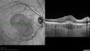
Punctate Inner Choroidopathy - PIC and Choroidal Neovascular Membrane - Right Eye - SD OCT740 x angesehen34-year-old woman with a change in vision in the right eye about eight days ago. Her vision was hazy and she saw waviness in the vision. She does have a history of aches and pains and she was diagnosed with Sjogren’s and 2 months ago she had some fevers and some sinus drainage and that went on for about two months. She is a moderate myope. OD 20/25, OS 20/20.
|
|

Acute Macular Neuroretinopathy - (AMN) - Spot in Vision OS - 40 Year old Female740 x angesehen
|
|
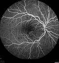
Punctate Inner Choroidopathy - PIC and Choroidal Neovascular Membrane - Right Eye - Fluorescein Angiogram738 x angesehen34-year-old woman with a change in vision in the right eye about eight days ago. Her vision was hazy and she saw waviness in the vision. She does have a history of aches and pains and she was diagnosed with Sjogren’s and 2 months ago she had some fevers and some sinus drainage and that went on for about two months. She is a moderate myope. OD 20/25, OS 20/20.
|
|
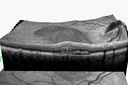
Punctate Inner Choroidopathy - PIC and Choroidal Neovascular Membrane - Right Eye - SD OCT VIDEO TAKES TIME TO LOAD738 x angesehen34-year-old woman with a change in vision in the right eye about eight days ago. Her vision was hazy and she saw waviness in the vision. She does have a history of aches and pains and she was diagnosed with Sjogren’s and 2 months ago she had some fevers and some sinus drainage and that went on for about two months. She is a moderate myope. OD 20/25, OS 20/20.
|
|

Uveitis and Cystoid Macular Edema - Probably Sarcoidosis738 x angesehenVA 20/63 OD 20/50 OS - 3 months vision loss - second episode by history of uveitis
|
|

Acute Macular Neuroretinopathy - 40 year old female738 x angesehen
|
|

Acute Posterior Multifocal Placoid Pigment Epitheliopathy - 4 years after onset737 x angesehen4 years after presentation - vision 20/80 OD 20/16 OS and there is subretinal fibrosis
|
|

Acute Zonal Occult Outer Retinopathy736 x angesehen45-year-old man noticed a few years ago peripheral vision blurriness in the left eye and also some central vision loss. Previous to that, as far as he knows, the two eyes were okay.
VISUAL ACUITY: OD: 20/20; OS: 20/40.
|
|

Multifocal Choroidits Vitiligo ( Voght- Kayanagi-Harada Syndrome )736 x angesehen36-year old patient with decreased vision in both eyes with fatigue for over a year. VA 20/25-OD, 20/60 -OS. Patient was treated with Avastin and oral prednisone.
|
|
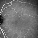
Acute Macular Neuroretinopathy - (AMN) - Spot in Vision OS - 40 Year old Female736 x angesehenICG angiogram is normal
|
|

Acute Macular Neuroretinopathy - (AMN) - Spot in Vision OS - 40 Year old Female734 x angesehenICG angiogram is normal
|
|

Punctate Inner Choroidopathy - Recurrent CNVM 4 years after initial treatment Left Eye - Fluorescein Angiogram733 x angesehenJune 7, 2012. This pleasant 35-year-old woman noticed decreasing vision in the left eye recently. She returns now noticing the vision in the left eye is declining. OD 20/50, OS 20/80. OS: Vertical C/D ratio is 0.3. There is a scar just inferonasal to the fovea, which has grown some towards the fovea with a little blood just under the fovea.
|
|

Multiple Evanescent White Dot Syndrome - Acute Idiopathic Blind Spot Enlargement732 x angesehen33-year-old woman one week ago she awoke and saw a red spots flashing out of her right eye. She notices them to the right side, which is the temporal visual field out of the right eye, then they changed to bursts of white lights without colors and then she notices straight ahead a couple spots. She has headaches in the back of her head. OD 20/20, OS 20/20
|
|

Acute Annular Outer Retinopathy - VA loss 2 days Right Eye Only - 25 Year Old Woman732 x angesehenInitial visit (June 11, 2011): 2 days of vision loss right eye. Wedge suddenly of VA loss OD, too dardkto see through. No flashes or floaters. VA 20/20 OU.
|
|

Punctate Inner Choroidopathy - PIC and Choroidal Neovascular Membrane - Right Eye - Fluorescein Angiogram731 x angesehen34-year-old woman with a change in vision in the right eye about eight days ago. Her vision was hazy and she saw waviness in the vision. She does have a history of aches and pains and she was diagnosed with Sjogren’s and 2 months ago she had some fevers and some sinus drainage and that went on for about two months. She is a moderate myope. OD 20/25, OS 20/20.
|
|

Normal Fellow Eye730 x angesehen65-year-old woman has pseudoretinitis pigmentosa in the right eye only, most likely from acute zonal occult outer retinopathy. OD 20/25, OS 20/25
|
|

Punctate Inner Choroidopathy - PIC and Choroidal Neovascular Membrane - Right Eye - SD OCT730 x angesehen34-year-old woman with a change in vision in the right eye about eight days ago. Her vision was hazy and she saw waviness in the vision. She does have a history of aches and pains and she was diagnosed with Sjogren’s and 2 months ago she had some fevers and some sinus drainage and that went on for about two months. She is a moderate myope. OD 20/25, OS 20/20.
|
|

Acute Zonal Occult Outer Retinopathy with Severe Peripheral Vision Loss Left Eye Post Treatment for CME728 x angesehen 58-year-old man developed a peripheral vision problem about 25 years ago. OD 20/30, OS 20/40.
|
|

Acute Annular Outer Retinopathy 728 x angesehenInitial visit (June 11, 2011): 2 days of vision loss right eye. Wedge suddenly of VA loss OD, too dardkto see through. No flashes or floaters. VA 20/20 OU.
|
|
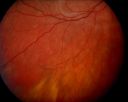
Acute Annular Outer Retinopathy - VA loss 2 days Right Eye Only - 25 Year Old Woman - AZOOR - White Line Around Fresh Lesions728 x angesehenInitial visit (June 11, 2011): 2 days of vision loss right eye. Wedge suddenly of VA loss OD, too dardkto see through. No flashes or floaters. VA 20/20 OU.
|
|

Acute Annular Outer Retinopathy for 3 Months - 25 YO Female Fluorescein Angiogram728 x angesehen25-year-old female who, about three months ago, put on her sunglasses and noticed a smudge in the vision and at that point she noticed the right eye was hazy. She had a cold a week or two before the vision loss, but she didn’t have any fever. She has no focal neurological problems in her past history. VA 20/20 OU
|
|

727 x angesehen
|
|

Acute Posterior Multifocal Placoid Pigment Epitheliopathy - APMPPE - Indocyanine Green Angiogram727 x angesehen 25-year-old man with one and half weeks ago he woke up with night chills. Also around that time his joints started hurting him. He also has headaches, although the headache at this point is not severe. His vision has been changing for about the last week and a half. He sees some spots in his vision and they are blurred spots. They are not in the central vision.
VISUAL ACUITY: OD 20/32, OS 20/32
|
|

Acute Macular Neuroretinopathy - (AMN) - Spot in Vision OS - 40 Year old Female727 x angesehenPhotos shows gray area nasal to the fovea
|
|

Punctate Inner Choroidopathy - PIC and Choroidal Neovascular Membrane - Right Eye - Color Fundus Photograph724 x angesehen34-year-old woman with a change in vision in the right eye about eight days ago. Her vision was hazy and she saw waviness in the vision. She does have a history of aches and pains and she was diagnosed with Sjogren’s and 2 months ago she had some fevers and some sinus drainage and that went on for about two months. She is a moderate myope. OD 20/25, OS 20/20.
|
|

Acute Macular Neuroretinopathy - (AMN) - Spot in Vision OS - 40 Year old Female723 x angesehen
|
|

Acute Posterior Multifocal Placoid Pigment Epitheliopathy - APMPPE - Fluorescein Angiogram722 x angesehen 25-year-old man with one and half weeks ago he woke up with night chills. Also around that time his joints started hurting him. He also has headaches, although the headache at this point is not severe. His vision has been changing for about the last week and a half. He sees some spots in his vision and they are blurred spots. They are not in the central vision.
VISUAL ACUITY: OD 20/32, OS 20/32
|
|
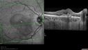
Punctate Inner Choroidopathy - PIC and Choroidal Neovascular Membrane - Right Eye - SD OCT721 x angesehen34-year-old woman with a change in vision in the right eye about eight days ago. Her vision was hazy and she saw waviness in the vision. She does have a history of aches and pains and she was diagnosed with Sjogren’s and 2 months ago she had some fevers and some sinus drainage and that went on for about two months. She is a moderate myope. OD 20/25, OS 20/20.
|
|

Multiple Evanescent White Dot Syndrome - Acute Idiopathic Blind Spot Enlargement717 x angesehen33-year-old woman one week ago she awoke and saw a red spots flashing out of her right eye. She notices them to the right side, which is the temporal visual field out of the right eye, then they changed to bursts of white lights without colors and then she notices straight ahead a couple spots. She has headaches in the back of her head. OD 20/20, OS 20/20
|
|

Punctate Inner Choroidopathy - Recurrent CNVM 4 years after initial treatment Left Eye 716 x angesehenJune 7, 2012. This pleasant 35-year-old woman noticed decreasing vision in the left eye recently. She returns now noticing the vision in the left eye is declining. OD 20/50, OS 20/80. OS: Vertical C/D ratio is 0.3. There is a scar just inferonasal to the fovea, which has grown some towards the fovea with a little blood just under the fovea.
|
|

Acute Macular Neuroretinopathy - (AMN) - Spot in Vision OS - 40 Year old Female716 x angesehen
|
|

Acute Zonal Occult Outer Retinopathy with Severe Peripheral Vision Loss Left Eye Post Treatment for CME715 x angesehen 58-year-old man developed a peripheral vision problem about 25 years ago. OD 20/30, OS 20/40.
|
|
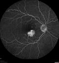
Punctate Inner Choroidopathy - PIC and Choroidal Neovascular Membrane - Right Eye - Fluorescein Angiogram715 x angesehen34-year-old woman with a change in vision in the right eye about eight days ago. Her vision was hazy and she saw waviness in the vision. She does have a history of aches and pains and she was diagnosed with Sjogren’s and 2 months ago she had some fevers and some sinus drainage and that went on for about two months. She is a moderate myope. OD 20/25, OS 20/20.
|
|
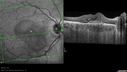
Punctate Inner Choroidopathy - PIC and Choroidal Neovascular Membrane - Right Eye - SD OCT715 x angesehen34-year-old woman with a change in vision in the right eye about eight days ago. Her vision was hazy and she saw waviness in the vision. She does have a history of aches and pains and she was diagnosed with Sjogren’s and 2 months ago she had some fevers and some sinus drainage and that went on for about two months. She is a moderate myope. OD 20/25, OS 20/20.
|
|

Acute Macular Neuroretinopathy - (AMN) - Spot in Vision OS - 40 Year old Female714 x angesehenICG angiogram is normal
|
|

Acute Zonal Occult Outer Retinopathy with Severe Peripheral Vision Loss Left Eye 712 x angesehen 58-year-old man developed a peripheral vision problem about 25 years ago. OD 20/30, OS 20/40.
|
|

Multiple Evanescent White Dot Syndrome - Atypical Yellow Foveal Spot Right Eye711 x angesehen12-year-old had decreased vision in the left eye from reported amblyopia since around the year 2000. His right eye was doing fine until two days ago, when he had sudden severe vision loss. He sees a blurred spot in the middle of the vision in the right eye wherever he looks.
OD 20/60 with eccentric movement, OS 20/200. Pinhole 20/70. IOP: OD 14, OS 10.
|
|

Serpiginouse Choroiditis (Chorioretinitis) - Acute Right Eye - Old Left Eye VA 20/25 OD , 20/50 OS - 1 Week After Intravitreal Kenalog OD711 x angesehen63-year-old woman has serpiginous choroiditis 1 week after intravitreal kenalog treatment
|
|

Psuedo-retinitis Pigmentosa - Bone Spicules One Eye - Probably Acute Zonal Occult Outer Retinopathy (AZOOR)711 x angesehen65-year-old woman has pseudoretinitis pigmentosa in the right eye only, most likely from acute zonal occult outer retinopathy. OD 20/25, OS 20/25
|
|
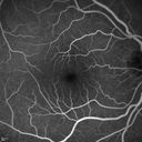
Acute Macular Neuroretinopathy - (AMN) - Spot in Vision OS - 40 Year old Female711 x angesehenFluorescein Angiogram is normal
|
|

Punctate Inner Choroidopathy - Recurrent CNVM 4 years after initial treatment Left Eye 708 x angesehenJune 7, 2012. This pleasant 35-year-old woman noticed decreasing vision in the left eye recently. She returns now noticing the vision in the left eye is declining. OD 20/50, OS 20/80. OS: Vertical C/D ratio is 0.3. There is a scar just inferonasal to the fovea, which has grown some towards the fovea with a little blood just under the fovea.
|
|
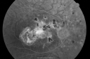
Multifocal Choroiditis and Subretinal Fibrosis - 32 yo Female Old Scar OD707 x angesehen32-year-old woman vision loss in the right eye associated with macular scarring and multifocal choroiditis in 1999 with new vision loss in left eye: OD 20/400, OS 20/50.
|
|

Acute Macular Neuroretinopathy - (AMN) - Spot in Vision OS - 40 Year old Female707 x angesehen
|
|

Acute Macular Neuroretinopathy - (AMN) - Spot in Vision OS - 40 Year old Female706 x angesehenICG angiogram is normal
|
|
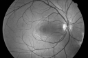
Multiple Evanescent White Dot Syndrome - Acute Idiopathic Blind Spot Enlargement705 x angesehen33-year-old woman one week ago she awoke and saw a red spots flashing out of her right eye. She notices them to the right side, which is the temporal visual field out of the right eye, then they changed to bursts of white lights without colors and then she notices straight ahead a couple spots. She has headaches in the back of her head. OD 20/20, OS 20/20
|
|

Multifocal Choroiditis - Linear Scars - Schlaegel lines705 x angesehen66 year old woman with macular pucker right eye. Images show typical linear peripheral scars for MFC or POHS - this is unilateral.
|
|

Acute Zonal Occult Outer Retinopathy with Severe Peripheral Vision Loss Left Eye 704 x angesehen 58-year-old man developed a peripheral vision problem about 25 years ago. OD 20/30, OS 20/40.
|
|

Acute Zonal Occult Outer Retinopathy with Severe Peripheral Vision Loss Left Eye 702 x angesehen 58-year-old man developed a peripheral vision problem about 25 years ago. OD 20/30, OS 20/40.
|
|

Birdshot Chorioretinitis - Retinal Vasculitis - HLA A29 Positive 702 x angesehen64-year-old woman three or four year’s fluctuations in her vision. She mostly notices them in bright light her vision glares out and she has difficulty seeing. PMHx: malaria in 1948 that was treated. She was told at one point she could possibly have had Familial Mediterranean fever.
She does intermittently get rashes around her arms. She has arthritis in her knees and ankles.
VISUAL ACUITY: OD 20/40, OS 20/50 - HLA - A29 Positive
|
|

Acute Zonal Occult Outer Retinopathy701 x angesehen45-year-old man noticed a few years ago peripheral vision blurriness in the left eye and also some central vision loss. Previous to that, as far as he knows, the two eyes were okay.
VISUAL ACUITY: OD: 20/20; OS: 20/40.
|
|

Multiple Evanescent White Dot Syndrome - Acute Idiopathic Blind Spot Enlargement701 x angesehen33-year-old woman one week ago she awoke and saw a red spots flashing out of her right eye. She notices them to the right side, which is the temporal visual field out of the right eye, then they changed to bursts of white lights without colors and then she notices straight ahead a couple spots. She has headaches in the back of her head. OD 20/20, OS 20/20
|
|

Punctate Inner Choroidopathy - PIC and Choroidal Neovascular Membrane - Right Eye - Fluorescein Angiogram701 x angesehen34-year-old woman with a change in vision in the right eye about eight days ago. Her vision was hazy and she saw waviness in the vision. She does have a history of aches and pains and she was diagnosed with Sjogren’s and 2 months ago she had some fevers and some sinus drainage and that went on for about two months. She is a moderate myope. OD 20/25, OS 20/20.
|
|

Acute Posterior Multifocal Placoid Pigment Epitheliopathy - APMPPE - Indocyanine Green Angiogram699 x angesehen 25-year-old man with one and half weeks ago he woke up with night chills. Also around that time his joints started hurting him. He also has headaches, although the headache at this point is not severe. His vision has been changing for about the last week and a half. He sees some spots in his vision and they are blurred spots. They are not in the central vision.
VISUAL ACUITY: OD 20/32, OS 20/32
|
|
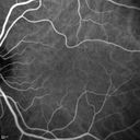
Acute Macular Neuroretinopathy - (AMN) - Spot in Vision OS - 40 Year old Female699 x angesehenICG angiogram is normal
|
|

Birdshot Chorioretinitis - Retinal Vasculitis - HLA A29 Positive 698 x angesehen64-year-old woman three or four year’s fluctuations in her vision. She mostly notices them in bright light her vision glares out and she has difficulty seeing. PMHx: malaria in 1948 that was treated. She was told at one point she could possibly have had Familial Mediterranean fever.
She does intermittently get rashes around her arms. She has arthritis in her knees and ankles.
VISUAL ACUITY: OD 20/40, OS 20/50 - HLA - A29 Positive
|
|

Punctate Inner Choroidopathy - Recurrent CNVM 4 years after initial treatment Left Eye 698 x angesehenJune 7, 2012. This pleasant 35-year-old woman noticed decreasing vision in the left eye recently. She returns now noticing the vision in the left eye is declining. OD 20/50, OS 20/80. OS: Vertical C/D ratio is 0.3. There is a scar just inferonasal to the fovea, which has grown some towards the fovea with a little blood just under the fovea.
|
|
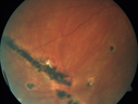
Multifocal Choroiditis - Linear Scars - Schlaegel lines698 x angesehen66 year old woman with macular pucker right eye. Images show typical linear peripheral scars for MFC or POHS - this is unilateral.
|
|
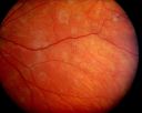
Acute Annular Outer Retinopathy - VA loss 2 days Right Eye Only - 25 Year Old Woman- AZOOR - White Line Around Fresh Lesions697 x angesehenInitial visit (June 11, 2011): 2 days of vision loss right eye. Wedge suddenly of VA loss OD, too dardkto see through. No flashes or floaters. VA 20/20 OU.
|
|

Punctate Inner Choroidopathy - PIC and Choroidal Neovascular Membrane - Right Eye - Fluorescein Angiogram697 x angesehen34-year-old woman with a change in vision in the right eye about eight days ago. Her vision was hazy and she saw waviness in the vision. She does have a history of aches and pains and she was diagnosed with Sjogren’s and 2 months ago she had some fevers and some sinus drainage and that went on for about two months. She is a moderate myope. OD 20/25, OS 20/20.
|
|

Multifocal Choroiditis and Subretinal Fibrosis - 32 yo Female New Lesion OS695 x angesehen32-year-old woman vision loss in the right eye associated with macular scarring and multifocal choroiditis in 1999 with new vision loss in left eye: OD 20/400, OS 20/50.
|
|
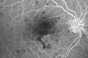
Punctate Inner Choroidopathy - PIC and Choroidal Neovascular Membrane - Right Eye - Fluorescein Angiogram695 x angesehen34-year-old woman with a change in vision in the right eye about eight days ago. Her vision was hazy and she saw waviness in the vision. She does have a history of aches and pains and she was diagnosed with Sjogren’s and 2 months ago she had some fevers and some sinus drainage and that went on for about two months. She is a moderate myope. OD 20/25, OS 20/20.
|
|

Acute Zonal Occult Outer Retinopathy with Severe Peripheral Vision Loss Left Eye 694 x angesehen 58-year-old man developed a peripheral vision problem about 25 years ago. OD 20/30, OS 20/40.
|
|

Punctate Inner Choroidopathy - Recurrent CNVM 4 years after initial treatment Left Eye 694 x angesehenJune 7, 2012. This pleasant 35-year-old woman noticed decreasing vision in the left eye recently. She returns now noticing the vision in the left eye is declining. OD 20/50, OS 20/80. OS: Vertical C/D ratio is 0.3. There is a scar just inferonasal to the fovea, which has grown some towards the fovea with a little blood just under the fovea.
|
|
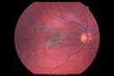
Acute Posterior Multifocal Placoid Pigment Epitheliopathy - 4 years after onset693 x angesehen4 years after presentation - vision 20/80 OD 20/16 OS and there is subretinal fibrosis
|
|

Serpiginous choroiditis692 x angesehenInactive scars in both eyes
|
|

Psuedo-retinitis Pigmentosa - Bone Spicules One Eye - Probably Acute Zonal Occult Outer Retinopathy (AZOOR)692 x angesehen65-year-old woman has pseudoretinitis pigmentosa in the right eye only, most likely from acute zonal occult outer retinopathy. OD 20/25, OS 20/25
|
|

Punctate Inner Choroidopathy - Recurrent CNVM 4 years after initial treatment Left Eye - FAF691 x angesehenJune 7, 2012. This pleasant 35-year-old woman noticed decreasing vision in the left eye recently. She returns now noticing the vision in the left eye is declining. OD 20/50, OS 20/80. OS: Vertical C/D ratio is 0.3. There is a scar just inferonasal to the fovea, which has grown some towards the fovea with a little blood just under the fovea.
|
|

Multifocal Choroiditis and Subretinal Fibrosis - 32 yo Female New Lesion OS689 x angesehen32-year-old woman vision loss in the right eye associated with macular scarring and multifocal choroiditis in 1999 with new vision loss in left eye: OD 20/400, OS 20/50.
|
|

Normal Fellow Eye689 x angesehen65-year-old woman has pseudoretinitis pigmentosa in the right eye only, most likely from acute zonal occult outer retinopathy. OD 20/25, OS 20/25
|
|

Punctate Inner Choroidopathy - Recurrent CNVM 4 years after initial treatment Left Eye - Fluorescein Angiogram688 x angesehenJune 7, 2012. This pleasant 35-year-old woman noticed decreasing vision in the left eye recently. She returns now noticing the vision in the left eye is declining. OD 20/50, OS 20/80. OS: Vertical C/D ratio is 0.3. There is a scar just inferonasal to the fovea, which has grown some towards the fovea with a little blood just under the fovea.
|
|

Uveitis and Cystoid Macular Edema - Probably Sarcoidosis688 x angesehenVA 20/63 OD 20/50 OS - 3 months vision loss - second episode by history of uveitis
|
|

Multiple Evanescent White Dot Syndrome - Acute Idiopathic Blind Spot Enlargement687 x angesehen33-year-old woman one week ago she awoke and saw a red spots flashing out of her right eye. She notices them to the right side, which is the temporal visual field out of the right eye, then they changed to bursts of white lights without colors and then she notices straight ahead a couple spots. She has headaches in the back of her head. OD 20/20, OS 20/20
|
|

Acute Posterior Multifocal Placoid Pigment Epitheliopathy - APMPPE - Indocyanine Green Angiogram687 x angesehen 25-year-old man with one and half weeks ago he woke up with night chills. Also around that time his joints started hurting him. He also has headaches, although the headache at this point is not severe. His vision has been changing for about the last week and a half. He sees some spots in his vision and they are blurred spots. They are not in the central vision.
VISUAL ACUITY: OD 20/32, OS 20/32
|
|

Uveitis and Cystoid Macular Edema - Probably Sarcoidosis684 x angesehenVA 20/63 OD 20/50 OS - 3 months vision loss - second episode by history of uveitis
|
|

Birdshot Chorioretinitis - Retinal Vasculitis - HLA A29 Positive 683 x angesehen64-year-old woman three or four year’s fluctuations in her vision. She mostly notices them in bright light her vision glares out and she has difficulty seeing. PMHx: malaria in 1948 that was treated. She was told at one point she could possibly have had Familial Mediterranean fever.
She does intermittently get rashes around her arms. She has arthritis in her knees and ankles.
VISUAL ACUITY: OD 20/40, OS 20/50 - HLA - A29 Positive
|
|
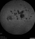
Punctate Inner Choroidopathy - Recurrent CNVM 4 years after initial treatment Left Eye - Indocyanine Green Angiogram683 x angesehenJune 7, 2012. This pleasant 35-year-old woman noticed decreasing vision in the left eye recently. She returns now noticing the vision in the left eye is declining. OD 20/50, OS 20/80. OS: Vertical C/D ratio is 0.3. There is a scar just inferonasal to the fovea, which has grown some towards the fovea with a little blood just under the fovea.
|
|
| 1298 Dateien auf 13 Seite(n) |
 |
3 | |
|
|
|
|
|
|
|
|


