Most viewed - Non-infectious Posterior Uveitis
|

Multifocal Choroiditis - Linear Scars - Schlaegel lines618 views66 year old woman with macular pucker right eye. Images show typical linear peripheral scars for MFC or POHS - this is unilateral.
|
|

Serpiginous choroiditis617 viewsInactive scars in both eyes
|
|
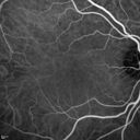
Acute Macular Neuroretinopathy - (AMN) - Spot in Vision OS - 40 Year old Female615 views
|
|

Uveitis and Cystoid Macular Edema - Probably Sarcoidosis614 viewsVA 20/63 OD 20/50 OS - 3 months vision loss - second episode by history of uveitis
|
|

Multifocal Choroidits Vitiligo ( Voght- Kayanagi-Harada Syndrome )612 views36-year old patient with decreased vision in both eyes with fatigue for over a year. VA 20/25-OD, 20/60 -OS. Patient was treated with Avastin and oral prednisone.
|
|
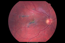
Acute Posterior Multifocal Placoid Pigment Epitheliopathy - 4 years after onset612 views4 years after presentation - vision 20/80 OD 20/16 OS and there is subretinal fibrosis
|
|
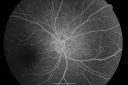
Multiple Evanescent White Dot Syndrome - Acute Idiopathic Blind Spot Enlargement611 views33-year-old woman one week ago she awoke and saw a red spots flashing out of her right eye. She notices them to the right side, which is the temporal visual field out of the right eye, then they changed to bursts of white lights without colors and then she notices straight ahead a couple spots. She has headaches in the back of her head. OD 20/20, OS 20/20
|
|

Sarcoidosis Multifocal Choroiditis611 views66 year old man with dense cataracts and recurrent uveitis. Images show multifocal choroidal granulomas from sarcoidosis more in the right eye than the left eye.
|
|

Acute Zonal Occult Outer Retinopathy with Severe Peripheral Vision Loss Left Eye 610 views 58-year-old man developed a peripheral vision problem about 25 years ago. OD 20/30, OS 20/40.
|
|

Birdshot Chorioretinitis - Retinal Vasculitis - HLA A29 Positive 610 views64-year-old woman three or four year’s fluctuations in her vision. She mostly notices them in bright light her vision glares out and she has difficulty seeing. PMHx: malaria in 1948 that was treated. She was told at one point she could possibly have had Familial Mediterranean fever.
She does intermittently get rashes around her arms. She has arthritis in her knees and ankles.
VISUAL ACUITY: OD 20/40, OS 20/50 - HLA - A29 Positive
|
|
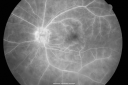
Birdshot Chorioretinitis - Retinal Vasculitis - HLA A29 Positive 609 views64-year-old woman three or four year’s fluctuations in her vision. She mostly notices them in bright light her vision glares out and she has difficulty seeing. PMHx: malaria in 1948 that was treated. She was told at one point she could possibly have had Familial Mediterranean fever.
She does intermittently get rashes around her arms. She has arthritis in her knees and ankles.
VISUAL ACUITY: OD 20/40, OS 20/50 - HLA - A29 Positive
|
|

Multiple Evanescent White Dot Syndrome - Acute Idiopathic Blind Spot Enlargement608 views33-year-old woman one week ago she awoke and saw a red spots flashing out of her right eye. She notices them to the right side, which is the temporal visual field out of the right eye, then they changed to bursts of white lights without colors and then she notices straight ahead a couple spots. She has headaches in the back of her head. OD 20/20, OS 20/20
|
|

Acute Annular Outer Retinopathy for 3 Months - 25 YO Female Late ICG607 views25-year-old female who, about three months ago, put on her sunglasses and noticed a smudge in the vision and at that point she noticed the right eye was hazy. She had a cold a week or two before the vision loss, but she didn’t have any fever. She has no focal neurological problems in her past history. VA 20/20 OU
|
|

Recurrent MEWDS Left Eye - Teenage Girl607 viewsPatient presented in 2014 (first images) 2 months following vision loss left eye. Vision went from 20/60 - 20/20. OCT showed outer retinal irregularities which normalized. Then 3 years later in 2017 she had full blown MEWDS. She presented acutely and all the images are typical. Both episodes where in the same time of year, about 3 months after her flu shot for sports.
|
|
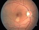
Choroidal Neovascular Membrane - Multifocal Scars - PIC vs MFC vs OHS 606 views
|
|

Acute Posterior Multifocal Placoid Pigment Epitheliopathy - 4 years after onset606 views4 years after presentation - vision 20/80 OD 20/16 OS and there is subretinal fibrosis
|
|

Multiple Evanescent White Dot Syndrome - Atypical Yellow Foveal Spot Right Eye605 views12-year-old had decreased vision in the left eye from reported amblyopia since around the year 2000. His right eye was doing fine until two days ago, when he had sudden severe vision loss. He sees a blurred spot in the middle of the vision in the right eye wherever he looks.
OD 20/60 with eccentric movement, OS 20/200. Pinhole 20/70. IOP: OD 14, OS 10.
|
|

Punctate Inner Choroidopathy - Recurrent CNVM 4 years after initial treatment Left Eye - Indocyanine Green Angiogram605 viewsJune 7, 2012. This pleasant 35-year-old woman noticed decreasing vision in the left eye recently. She returns now noticing the vision in the left eye is declining. OD 20/50, OS 20/80. OS: Vertical C/D ratio is 0.3. There is a scar just inferonasal to the fovea, which has grown some towards the fovea with a little blood just under the fovea.
|
|

Multifocal Choroiditis - Linear Scars - Schlaegel lines605 views66 year old woman with macular pucker right eye. Images show typical linear peripheral scars for MFC or POHS - this is unilateral.
|
|
Uveitis and Cystoid Macular Edema - Probably Sarcoidosis600 viewsVA 20/63 OD 20/50 OS - 3 months vision loss - second episode by history of uveitis
|
|
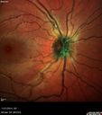
Recurrent MEWDS Left Eye - Teenage Girl600 viewsPatient presented in 2014 (first images) 2 months following vision loss left eye. Vision went from 20/60 - 20/20. OCT showed outer retinal irregularities which normalized. Then 3 years later in 2017 she had full blown MEWDS. She presented acutely and all the images are typical. Both episodes where in the same time of year, about 3 months after her flu shot for sports.
|
|

AMN 3 years later600 viewsShe still sees the blind spot one she looks for it.
|
|

Serpiginous choroiditis598 viewsInactive scars in both eyes
|
|
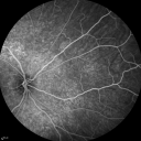
Acute Annular Outer Retinopathy for 3 Months - 25 YO Female FA598 views25-year-old female who, about three months ago, put on her sunglasses and noticed a smudge in the vision and at that point she noticed the right eye was hazy. She had a cold a week or two before the vision loss, but she didn’t have any fever. She has no focal neurological problems in her past history. VA 20/20 OU
|
|
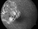
Serpiginous choroiditis596 viewsInactive scars in both eyes
|
|

Punctate Inner Choroidopathy - Recurrent CNVM 4 years after initial treatment Left Eye - Fluorescein Angiogram596 viewsJune 7, 2012. This pleasant 35-year-old woman noticed decreasing vision in the left eye recently. She returns now noticing the vision in the left eye is declining. OD 20/50, OS 20/80. OS: Vertical C/D ratio is 0.3. There is a scar just inferonasal to the fovea, which has grown some towards the fovea with a little blood just under the fovea.
|
|

Birdshot Chorioretinitis - Retinal Vasculitis - HLA A29 Positive 595 views64-year-old woman three or four year’s fluctuations in her vision. She mostly notices them in bright light her vision glares out and she has difficulty seeing. PMHx: malaria in 1948 that was treated. She was told at one point she could possibly have had Familial Mediterranean fever.
She does intermittently get rashes around her arms. She has arthritis in her knees and ankles.
VISUAL ACUITY: OD 20/40, OS 20/50 - HLA - A29 Positive
|
|

Punctate Inner Choroidopathy - Recurrent CNVM 4 years after initial treatment Left Eye - Indocyanine Green Angiogram595 viewsJune 7, 2012. This pleasant 35-year-old woman noticed decreasing vision in the left eye recently. She returns now noticing the vision in the left eye is declining. OD 20/50, OS 20/80. OS: Vertical C/D ratio is 0.3. There is a scar just inferonasal to the fovea, which has grown some towards the fovea with a little blood just under the fovea.
|
|

Recurrent MEWDS Left Eye - Teenage Girl595 viewsPatient presented in 2014 (first images) 2 months following vision loss left eye. Vision went from 20/60 - 20/20. OCT showed outer retinal irregularities which normalized. Then 3 years later in 2017 she had full blown MEWDS. She presented acutely and all the images are typical. Both episodes where in the same time of year, about 3 months after her flu shot for sports.
|
|

Normal Fellow Eye594 views65-year-old woman has pseudoretinitis pigmentosa in the right eye only, most likely from acute zonal occult outer retinopathy. OD 20/25, OS 20/25
|
|

Acute Macular Neuroretinopathy - (AMN) - Spot in Vision OS - 40 Year old Female594 viewsFluorescein Angiogram is normal
|
|

Acute Posterior Multifocal Placoid Pigment Epitheliopathy - 4 years after onset594 views4 years after presentation - vision 20/80 OD 20/16 OS and there is subretinal fibrosis
|
|

Serpiginous choroiditis593 viewsInactive scars in both eyes
|
|
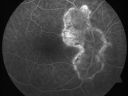
Serpiginous choroiditis593 viewsInactive scars in both eyes
|
|

Acute Posterior Multifocal Placoid Pigment Epitheliopathy - 4 years after onset593 views4 years after presentation - vision 20/80 OD 20/16 OS and there is subretinal fibrosis
|
|
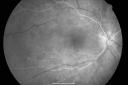
Birdshot Chorioretinitis - Retinal Vasculitis - HLA A29 Positive 592 views64-year-old woman three or four year’s fluctuations in her vision. She mostly notices them in bright light her vision glares out and she has difficulty seeing. PMHx: malaria in 1948 that was treated. She was told at one point she could possibly have had Familial Mediterranean fever.
She does intermittently get rashes around her arms. She has arthritis in her knees and ankles.
VISUAL ACUITY: OD 20/40, OS 20/50 - HLA - A29 Positive
|
|

Acute Macular Neuroretinopathy - (AMN) - Spot in Vision OS - 40 Year old Female591 viewsFluorescein Angiogram is normal
|
|

Punctate Inner Choroidopathy - Recurrent CNVM 4 years after initial treatment Left Eye 590 viewsJune 7, 2012. This pleasant 35-year-old woman noticed decreasing vision in the left eye recently. She returns now noticing the vision in the left eye is declining. OD 20/50, OS 20/80. OS: Vertical C/D ratio is 0.3. There is a scar just inferonasal to the fovea, which has grown some towards the fovea with a little blood just under the fovea.
|
|

Punctate Inner Choroidopathy - Recurrent CNVM 4 years after initial treatment Left Eye - Fluorescein Angiogram590 viewsJune 7, 2012. This pleasant 35-year-old woman noticed decreasing vision in the left eye recently. She returns now noticing the vision in the left eye is declining. OD 20/50, OS 20/80. OS: Vertical C/D ratio is 0.3. There is a scar just inferonasal to the fovea, which has grown some towards the fovea with a little blood just under the fovea.
|
|

Punctate Inner Choroidopathy - Recurrent CNVM 4 years after initial treatment Left Eye - Indocyanine Green Angiogram590 viewsJune 7, 2012. This pleasant 35-year-old woman noticed decreasing vision in the left eye recently. She returns now noticing the vision in the left eye is declining. OD 20/50, OS 20/80. OS: Vertical C/D ratio is 0.3. There is a scar just inferonasal to the fovea, which has grown some towards the fovea with a little blood just under the fovea.
|
|

Acute Posterior Multifocal Placoid Pigment Epitheliopathy - 4 years after onset589 views4 years after presentation - vision 20/80 OD 20/16 OS and there is subretinal fibrosis
|
|

Serpiginous choroiditis588 viewsInactive scars in both eyes
|
|

Punctate Inner Choroidopathy - Recurrent CNVM 4 years after initial treatment Left Eye - Fluorescein Angiogram587 viewsJune 7, 2012. This pleasant 35-year-old woman noticed decreasing vision in the left eye recently. She returns now noticing the vision in the left eye is declining. OD 20/50, OS 20/80. OS: Vertical C/D ratio is 0.3. There is a scar just inferonasal to the fovea, which has grown some towards the fovea with a little blood just under the fovea.
|
|

Multiple Evanescent White Dot Syndrome - Acute Idiopathic Blind Spot Enlargement585 views33-year-old woman one week ago she awoke and saw a red spots flashing out of her right eye. She notices them to the right side, which is the temporal visual field out of the right eye, then they changed to bursts of white lights without colors and then she notices straight ahead a couple spots. She has headaches in the back of her head. OD 20/20, OS 20/20
|
|

Acute Annular Outer Retinopathy for 3 Months - 25 YO Female Fundus Photograph585 views25-year-old female who, about three months ago, put on her sunglasses and noticed a smudge in the vision and at that point she noticed the right eye was hazy. She had a cold a week or two before the vision loss, but she didn’t have any fever. She has no focal neurological problems in her past history. VA 20/20 OU
|
|
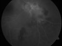
Serpiginouse Choroiditis (Chorioretinitis) - Acute Right Eye - Old Left Eye VA 20/25 OD , 20/50 OS584 views63-year-old woman has serpiginous choroiditis (date - March 2011). The right eye has not been previously involved, and then she noticed new onset floaters in the right eye for the last two weeks. Her vision in the right eye is hazy because of that.
VISUAL ACUITY: Vision OD is 20/25, OS is 20/50
|
|

Birdshot Chorioretinitis - Retinal Vasculitis - HLA A29 Positive 583 views64-year-old woman three or four year’s fluctuations in her vision. She mostly notices them in bright light her vision glares out and she has difficulty seeing. PMHx: malaria in 1948 that was treated. She was told at one point she could possibly have had Familial Mediterranean fever.
She does intermittently get rashes around her arms. She has arthritis in her knees and ankles.
VISUAL ACUITY: OD 20/40, OS 20/50 - HLA - A29 Positive
|
|

Acute Annular Outer Retinopathy for 3 Months - 25 YO Female Fundus Photograph583 views25-year-old female who, about three months ago, put on her sunglasses and noticed a smudge in the vision and at that point she noticed the right eye was hazy. She had a cold a week or two before the vision loss, but she didn’t have any fever. She has no focal neurological problems in her past history. VA 20/20 OU
|
|
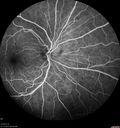
Birdshot Chorioretinitis - Fresh Presentation582 viewsUltawidefield ICG shows best the multifocal lesions. 61 year old woman with floaters for less than 6 months. HLA-A29 Positive
|
|

Sarcoidosis Multifocal Choroiditis582 views66 year old man with dense cataracts and recurrent uveitis. Images show multifocal choroidal granulomas from sarcoidosis more in the right eye than the left eye.
|
|

Multiple Evanescent White Dot Syndrome - Acute Idiopathic Blind Spot Enlargement581 views33-year-old woman one week ago she awoke and saw a red spots flashing out of her right eye. She notices them to the right side, which is the temporal visual field out of the right eye, then they changed to bursts of white lights without colors and then she notices straight ahead a couple spots. She has headaches in the back of her head. OD 20/20, OS 20/20
|
|

Normal Fellow Eye581 views65-year-old woman has pseudoretinitis pigmentosa in the right eye only, most likely from acute zonal occult outer retinopathy. OD 20/25, OS 20/25
|
|

Acute Annular Outer Retinopathy for 3 Months - 25 YO Female Fellow Normal Eye581 views25-year-old female who, about three months ago, put on her sunglasses and noticed a smudge in the vision and at that point she noticed the right eye was hazy. She had a cold a week or two before the vision loss, but she didn’t have any fever. She has no focal neurological problems in her past history. VA 20/20 OU
|
|

Acute Zonal Occult Outer Retinopathy with Severe Peripheral Vision Loss Left Eye 580 views 58-year-old man developed a peripheral vision problem about 25 years ago. OD 20/30, OS 20/40.
|
|

Acute Annular Outer Retinopathy for 3 Months - 25 YO Female Fluorescein Angiogram580 views25-year-old female who, about three months ago, put on her sunglasses and noticed a smudge in the vision and at that point she noticed the right eye was hazy. She had a cold a week or two before the vision loss, but she didn’t have any fever. She has no focal neurological problems in her past history. VA 20/20 OU
|
|

Multiple Evanescent White Dot Syndrome - Atypical Yellow Foveal Spot Right Eye579 views12-year-old had decreased vision in the left eye from reported amblyopia since around the year 2000. His right eye was doing fine until two days ago, when he had sudden severe vision loss. He sees a blurred spot in the middle of the vision in the right eye wherever he looks.
OD 20/60 with eccentric movement, OS 20/200. Pinhole 20/70. IOP: OD 14, OS 10.
|
|

Birdshot Chorioretinitis - Retinal Vasculitis - HLA A29 Positive 578 views64-year-old woman three or four year’s fluctuations in her vision. She mostly notices them in bright light her vision glares out and she has difficulty seeing. PMHx: malaria in 1948 that was treated. She was told at one point she could possibly have had Familial Mediterranean fever.
She does intermittently get rashes around her arms. She has arthritis in her knees and ankles.
VISUAL ACUITY: OD 20/40, OS 20/50 - HLA - A29 Positive
|
|

Uveitis and Cystoid Macular Edema - Probably Sarcoidosis578 viewsVA 20/63 OD 20/50 OS - 3 months vision loss - second episode by history of uveitis
|
|

Birdshot Chorioretinitis - Retinal Vasculitis - HLA A29 Positive 577 views64-year-old woman three or four year’s fluctuations in her vision. She mostly notices them in bright light her vision glares out and she has difficulty seeing. PMHx: malaria in 1948 that was treated. She was told at one point she could possibly have had Familial Mediterranean fever.
She does intermittently get rashes around her arms. She has arthritis in her knees and ankles.
VISUAL ACUITY: OD 20/40, OS 20/50 - HLA - A29 Positive
|
|
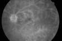
Birdshot Chorioretinitis - Retinal Vasculitis - HLA A29 Positive 577 views64-year-old woman three or four year’s fluctuations in her vision. She mostly notices them in bright light her vision glares out and she has difficulty seeing. PMHx: malaria in 1948 that was treated. She was told at one point she could possibly have had Familial Mediterranean fever.
She does intermittently get rashes around her arms. She has arthritis in her knees and ankles.
VISUAL ACUITY: OD 20/40, OS 20/50 - HLA - A29 Positive
|
|
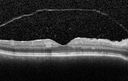
Birdshot Chorioretintis and PAMM577 viewsPatient presented with paracentral acute middle maculopathy as the presenting symptom of birdshot. This case was published. RETINAL CASES & BRIEF REPORTS 10:151–153, 2016.
|
|
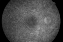
Multiple Evanescent White Dot Syndrome - Atypical Yellow Foveal Spot Right Eye576 views12-year-old had decreased vision in the left eye from reported amblyopia since around the year 2000. His right eye was doing fine until two days ago, when he had sudden severe vision loss. He sees a blurred spot in the middle of the vision in the right eye wherever he looks.
OD 20/60 with eccentric movement, OS 20/200. Pinhole 20/70. IOP: OD 14, OS 10.
|
|

Recurrent MEWDS Left Eye - Teenage Girl576 viewsPatient presented in 2014 (first images) 2 months following vision loss left eye. Vision went from 20/60 - 20/20. OCT showed outer retinal irregularities which normalized. Then 3 years later in 2017 she had full blown MEWDS. She presented acutely and all the images are typical. Both episodes where in the same time of year, about 3 months after her flu shot for sports.
|
|

Sarcoid Uveitis574 viewsPatient comes in with 6-month follow up for uveitis. VA is 20/20, both eyes. RPE peripheral scarring and punched out lesions visible. Stable. Pred Forte as needed for flare ups.
|
|

Sarcoidosis Multifocal Choroiditis574 views66 year old man with dense cataracts and recurrent uveitis. Images show multifocal choroidal granulomas from sarcoidosis more in the right eye than the left eye.
|
|

Multiple Evanescent White Dot Syndrome - Atypical Yellow Foveal Spot Right Eye573 views12-year-old had decreased vision in the left eye from reported amblyopia since around the year 2000. His right eye was doing fine until two days ago, when he had sudden severe vision loss. He sees a blurred spot in the middle of the vision in the right eye wherever he looks.
OD 20/60 with eccentric movement, OS 20/200. Pinhole 20/70. IOP: OD 14, OS 10.
|
|

Acute Zonal Occult Outer Retinopathy with Severe Peripheral Vision Loss Left Eye 573 views 58-year-old man developed a peripheral vision problem about 25 years ago. OD 20/30, OS 20/40.
|
|

Acute Annular Outer Retinopathy for 3 Months - 25 YO Female Fellow Normal Eye572 views25-year-old female who, about three months ago, put on her sunglasses and noticed a smudge in the vision and at that point she noticed the right eye was hazy. She had a cold a week or two before the vision loss, but she didn’t have any fever. She has no focal neurological problems in her past history. VA 20/20 OU
|
|

Acute Annular Outer Retinopathy for 3 Months - 25 YO Female FA571 views25-year-old female who, about three months ago, put on her sunglasses and noticed a smudge in the vision and at that point she noticed the right eye was hazy. She had a cold a week or two before the vision loss, but she didn’t have any fever. She has no focal neurological problems in her past history. VA 20/20 OU
|
|

Acute Annular Outer Retinopathy for 3 Months - 25 YO Female FA571 views25-year-old female who, about three months ago, put on her sunglasses and noticed a smudge in the vision and at that point she noticed the right eye was hazy. She had a cold a week or two before the vision loss, but she didn’t have any fever. She has no focal neurological problems in her past history. VA 20/20 OU
|
|

Recurrent MEWDS Left Eye - Teenage Girl571 viewsPatient presented in 2014 (first images) 2 months following vision loss left eye. Vision went from 20/60 - 20/20. OCT showed outer retinal irregularities which normalized. Then 3 years later in 2017 she had full blown MEWDS. She presented acutely and all the images are typical. Both episodes where in the same time of year, about 3 months after her flu shot for sports.
|
|

Acute Annular Outer Retinopathy for 3 Months - 25 YO Female Fellow Normal Eye570 views25-year-old female who, about three months ago, put on her sunglasses and noticed a smudge in the vision and at that point she noticed the right eye was hazy. She had a cold a week or two before the vision loss, but she didn’t have any fever. She has no focal neurological problems in her past history. VA 20/20 OU
|
|

Sarcoidosis Multifocal Choroiditis570 views66 year old man with dense cataracts and recurrent uveitis. Images show multifocal choroidal granulomas from sarcoidosis more in the right eye than the left eye.
|
|

Punctate Inner Choroidopathy - New CNVM right eye570 views42 year old female with recent vision loss in the right eye - Right eye is 20/80 - Vision improved to 20/16 with Ranabizumab injections over 3 months
|
|
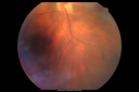
Birdshot Chorioretinitis - Retinal Vasculitis - HLA A29 Positive 569 views64-year-old woman three or four year’s fluctuations in her vision. She mostly notices them in bright light her vision glares out and she has difficulty seeing. PMHx: malaria in 1948 that was treated. She was told at one point she could possibly have had Familial Mediterranean fever.
She does intermittently get rashes around her arms. She has arthritis in her knees and ankles.
VISUAL ACUITY: OD 20/40, OS 20/50 - HLA - A29 Positive
|
|

Acute Annular Outer Retinopathy for 3 Months - 25 YO Female InfraRed Image569 views25-year-old female who, about three months ago, put on her sunglasses and noticed a smudge in the vision and at that point she noticed the right eye was hazy. She had a cold a week or two before the vision loss, but she didn’t have any fever. She has no focal neurological problems in her past history. VA 20/20 OU
|
|

Acute Posterior Multifocal Placoid Pigment Epitheliopathy - 4 years after onset568 views4 years after presentation - vision 20/80 OD 20/16 OS and there is subretinal fibrosis
|
|

Acute Posterior Multifocal Placoid Pigment Epitheliopathy - 4 years after onset567 views4 years after presentation - vision 20/80 OD 20/16 OS and there is subretinal fibrosis
|
|

Birdshot Chorioretinitis - Retinal Vasculitis - HLA A29 Positive 566 views64-year-old woman three or four year’s fluctuations in her vision. She mostly notices them in bright light her vision glares out and she has difficulty seeing. PMHx: malaria in 1948 that was treated. She was told at one point she could possibly have had Familial Mediterranean fever.
She does intermittently get rashes around her arms. She has arthritis in her knees and ankles.
VISUAL ACUITY: OD 20/40, OS 20/50 - HLA - A29 Positive
|
|
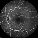
Acute Macular Neuroretinopathy - (AMN) - Spot in Vision OS - 40 Year old Female565 viewsFluorescein Angiogram is normal
|
|

Multifocal Choroiditis Panuveitis - New Choroidal Neovascular Membrane Right Eye565 views74 year old female had vitrectomy elsewhere for possible ocular lymphoma - 5 years of shimmery vision changes then 3 weeks ago noticed vision loss right eye.
|
|

Multiple Evanescent White Dot Syndrome - Acute Idiopathic Blind Spot Enlargement564 views33-year-old woman one week ago she awoke and saw a red spots flashing out of her right eye. She notices them to the right side, which is the temporal visual field out of the right eye, then they changed to bursts of white lights without colors and then she notices straight ahead a couple spots. She has headaches in the back of her head. OD 20/20, OS 20/20
|
|

Punctate Inner Choroidopathy - Recurrent CNVM 4 years after initial treatment Left Eye - Indocyanine Green Angiogram564 viewsJune 7, 2012. This pleasant 35-year-old woman noticed decreasing vision in the left eye recently. She returns now noticing the vision in the left eye is declining. OD 20/50, OS 20/80. OS: Vertical C/D ratio is 0.3. There is a scar just inferonasal to the fovea, which has grown some towards the fovea with a little blood just under the fovea.
|
|

Birdshot Chorioretinitis - Fresh Presentation564 viewsUltawidefield ICG shows best the multifocal lesions. 61 year old woman with floaters for less than 6 months. HLA-A29 Positive
|
|

Acute Zonal Occult Outer Retinopathy with Severe Peripheral Vision Loss Left Eye 562 views 58-year-old man developed a peripheral vision problem about 25 years ago. OD 20/30, OS 20/40.
|
|

Acute Annular Outer Retinopathy for 3 Months - 25 YO Female Spectral Domain Optical Coherence Tomgraphy562 views25-year-old female who, about three months ago, put on her sunglasses and noticed a smudge in the vision and at that point she noticed the right eye was hazy. She had a cold a week or two before the vision loss, but she didn’t have any fever. She has no focal neurological problems in her past history. VA 20/20 OU
|
|

Birdshot Chorioretinitis - Retinal Vasculitis - HLA A29 Positive 561 views64-year-old woman three or four year’s fluctuations in her vision. She mostly notices them in bright light her vision glares out and she has difficulty seeing. PMHx: malaria in 1948 that was treated. She was told at one point she could possibly have had Familial Mediterranean fever.
She does intermittently get rashes around her arms. She has arthritis in her knees and ankles.
VISUAL ACUITY: OD 20/40, OS 20/50 - HLA - A29 Positive
|
|
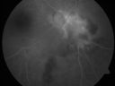
Serpiginouse Choroiditis (Chorioretinitis) - Acute Right Eye - Old Left Eye VA 20/25 OD , 20/50 OS560 views63-year-old woman has serpiginous choroiditis (date - March 2011). The right eye has not been previously involved, and then she noticed new onset floaters in the right eye for the last two weeks. Her vision in the right eye is hazy because of that.
VISUAL ACUITY: Vision OD is 20/25, OS is 20/50
|
|

Choroidal Neovascular Membrane - Multifocal Scars - PIC vs MFC vs OHS 559 views
|
|

Acute Annular Outer Retinopathy for 3 Months - 25 YO Female FA557 views25-year-old female who, about three months ago, put on her sunglasses and noticed a smudge in the vision and at that point she noticed the right eye was hazy. She had a cold a week or two before the vision loss, but she didn’t have any fever. She has no focal neurological problems in her past history. VA 20/20 OU
|
|

Choroidal Neovascular Membrane - Multifocal Scars - PIC vs MFC vs OHS 557 views
|
|

Recurrent MEWDS Left Eye - Teenage Girl557 viewsPatient presented in 2014 (first images) 2 months following vision loss left eye. Vision went from 20/60 - 20/20. OCT showed outer retinal irregularities which normalized. Then 3 years later in 2017 she had full blown MEWDS. She presented acutely and all the images are typical. Both episodes where in the same time of year, about 3 months after her flu shot for sports.
|
|

Acute Annular Outer Retinopathy for 3 Months - 25 YO Female Fundus Photograph556 views25-year-old female who, about three months ago, put on her sunglasses and noticed a smudge in the vision and at that point she noticed the right eye was hazy. She had a cold a week or two before the vision loss, but she didn’t have any fever. She has no focal neurological problems in her past history. VA 20/20 OU
|
|
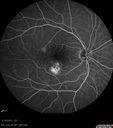
Choroidal Neovascular Membrane - Multifocal Scars - PIC vs MFC vs OHS 555 views
|
|

Recurrent MEWDS Left Eye - Teenage Girl555 viewsPatient presented in 2014 (first images) 2 months following vision loss left eye. Vision went from 20/60 - 20/20. OCT showed outer retinal irregularities which normalized. Then 3 years later in 2017 she had full blown MEWDS. She presented acutely and all the images are typical. Both episodes where in the same time of year, about 3 months after her flu shot for sports.
|
|

Choroidal Neovascular Membrane - Multifocal Scars - PIC vs MFC vs OHS 553 views
|
|

Choroidal Neovascular Membrane - Multifocal Scars - PIC vs MFC vs OHS 552 views
|
|

Acute Posterior Multifocal Placoid Pigment Epitheliopathy - 4 years after onset552 views4 years after presentation - vision 20/80 OD 20/16 OS and there is subretinal fibrosis
|
|

Multifocal Choroiditis Panuveitis with Subretinal Fibrosis Acute Phase551 views36 year old latin american female 3 weeks of vision loss and new headaches. MRI was normal. VA initially 20/32 OD; 20/20 OS Final vision was 20/63 OD; 20/20 OS
|
|

AZOOR - unilateral pseudo-retinitis pigmentosa551 views74 year old woman with unilateral vision loss 40 years ago.
|
|
| 1298 files on 13 page(s) |
 |
|
|
5 | |
|
|
|
|
|
|


