am meisten angesehen - Non-infectious Posterior Uveitis
|

Multiple Evanescent White Dot Syndrome - Atypical Yellow Foveal Spot Right Eye548 x angesehen12-year-old had decreased vision in the left eye from reported amblyopia since around the year 2000. His right eye was doing fine until two days ago, when he had sudden severe vision loss. He sees a blurred spot in the middle of the vision in the right eye wherever he looks.
OD 20/60 with eccentric movement, OS 20/200. Pinhole 20/70. IOP: OD 14, OS 10.
|
|

Acute Posterior Multifocal Placoid Pigment Epitheliopathy - 4 years after onset548 x angesehen4 years after presentation - vision 20/80 OD 20/16 OS and there is subretinal fibrosis
|
|
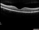
Recurrent MEWDS Left Eye - Teenage Girl548 x angesehenPatient presented in 2014 (first images) 2 months following vision loss left eye. Vision went from 20/60 - 20/20. OCT showed outer retinal irregularities which normalized. Then 3 years later in 2017 she had full blown MEWDS. She presented acutely and all the images are typical. Both episodes where in the same time of year, about 3 months after her flu shot for sports.
|
|
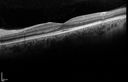
Acute Posterior Multifocal Placoid Pigment Epitheliopathy - 4 years after onset547 x angesehen4 years after presentation - vision 20/80 OD 20/16 OS and there is subretinal fibrosis
|
|
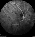
Sarcoidosis Multifocal Choroiditis547 x angesehen66 year old man with dense cataracts and recurrent uveitis. Images show multifocal choroidal granulomas from sarcoidosis more in the right eye than the left eye.
|
|

Acute Posterior Multifocal Placoid Pigment Epitheliopathy - 4 years after onset546 x angesehen4 years after presentation - vision 20/80 OD 20/16 OS and there is subretinal fibrosis
|
|

Multifocal Choroiditis Panuveitis with Subretinal Fibrosis Acute Phase546 x angesehen36 year old latin american female 3 weeks of vision loss and new headaches. MRI was normal. VA initially 20/32 OD; 20/20 OS Final vision was 20/63 OD; 20/20 OS
|
|

Multifocal Choroiditis Panuveitis with Subretinal Fibrosis Acute Phase546 x angesehen36 year old latin american female 3 weeks of vision loss and new headaches. MRI was normal. VA initially 20/32 OD; 20/20 OS Final vision was 20/63 OD; 20/20 OS
|
|

Acute Annular Outer Retinopathy for 3 Months - 25 YO Female Spectral Domain Optical Coherence Tomgraphy545 x angesehen25-year-old female who, about three months ago, put on her sunglasses and noticed a smudge in the vision and at that point she noticed the right eye was hazy. She had a cold a week or two before the vision loss, but she didn’t have any fever. She has no focal neurological problems in her past history. VA 20/20 OU
|
|

Multifocal Choroidits Vitiligo ( Voght- Kayanagi-Harada Syndrome )545 x angesehen36-year old patient with decreased vision in both eyes with fatigue for over a year. VA 20/25-OD, 20/60 -OS. Patient was treated with Avastin and oral prednisone.
|
|

Recurrent MEWDS Left Eye - Teenage Girl545 x angesehenPatient presented in 2014 (first images) 2 months following vision loss left eye. Vision went from 20/60 - 20/20. OCT showed outer retinal irregularities which normalized. Then 3 years later in 2017 she had full blown MEWDS. She presented acutely and all the images are typical. Both episodes where in the same time of year, about 3 months after her flu shot for sports.
|
|

Multiple Evanescent White Dot Syndrome - Atypical Yellow Foveal Spot Right Eye544 x angesehen12-year-old had decreased vision in the left eye from reported amblyopia since around the year 2000. His right eye was doing fine until two days ago, when he had sudden severe vision loss. He sees a blurred spot in the middle of the vision in the right eye wherever he looks.
OD 20/60 with eccentric movement, OS 20/200. Pinhole 20/70. IOP: OD 14, OS 10.
|
|

Sarcoidosis Multifocal Choroiditis544 x angesehen66 year old man with dense cataracts and recurrent uveitis. Images show multifocal choroidal granulomas from sarcoidosis more in the right eye than the left eye.
|
|

Acute Annular Outer Retinopathy for 3 Months - 25 YO Female Fundus Photograph543 x angesehen25-year-old female who, about three months ago, put on her sunglasses and noticed a smudge in the vision and at that point she noticed the right eye was hazy. She had a cold a week or two before the vision loss, but she didn’t have any fever. She has no focal neurological problems in her past history. VA 20/20 OU
|
|
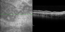
Acute Annular Outer Retinopathy for 3 Months - 25 YO Female Spectral Domain Optical Coherence Tomgraphy543 x angesehen25-year-old female who, about three months ago, put on her sunglasses and noticed a smudge in the vision and at that point she noticed the right eye was hazy. She had a cold a week or two before the vision loss, but she didn’t have any fever. She has no focal neurological problems in her past history. VA 20/20 OU
|
|

Multifocal Choroiditis and Subretinal Fibrosis with new Subretinal Neovascular Membrane543 x angesehen26 year old African American Girl Saw me at age 10 (16 years ago) with acute multifocal choroiditis and panuveitis which evolved into subretinal fibrosis. She was initially treated with 3 years of methotrexate. Vision ended up 20/20 in both eyes. Her eyes have been inactive since and she was lost to follow-up.
A week ago she noticed sudden decreased vision in the right eye. Within the past few weeks there are more floaters in the right eye.
VA OD: sc6’/200 PHNI
VA OS: sc20/16
|
|

Acute Annular Outer Retinopathy for 3 Months - 25 YO Female Spectral Domain Optical Coherence Tomgraphy542 x angesehen25-year-old female who, about three months ago, put on her sunglasses and noticed a smudge in the vision and at that point she noticed the right eye was hazy. She had a cold a week or two before the vision loss, but she didn’t have any fever. She has no focal neurological problems in her past history. VA 20/20 OU
|
|

Multifocal Choroidits Vitiligo ( Voght- Kayanagi-Harada Syndrome )542 x angesehen36-year old patient with decreased vision in both eyes with fatigue for over a year. VA 20/25-OD, 20/60 -OS. Patient was treated with Avastin and oral prednisone.
|
|

Acute Posterior Multifocal Placoid Pigment Epitheliopathy - 4 years after onset541 x angesehen4 years after presentation - vision 20/80 OD 20/16 OS and there is subretinal fibrosis
|
|

Birdshot Chorioretinitis - Fresh Presentation541 x angesehenUltawidefield ICG shows best the multifocal lesions. 61 year old woman with floaters for less than 6 months. HLA-A29 Positive
|
|

Multifocal Choroiditis Panuveitis with Subretinal Fibrosis Acute Phase541 x angesehen36 year old latin american female 3 weeks of vision loss and new headaches. MRI was normal. VA initially 20/32 OD; 20/20 OS Final vision was 20/63 OD; 20/20 OS
|
|

Acute Annular Outer Retinopathy for 3 Months - 25 YO Female Fellow Normal Eye540 x angesehen25-year-old female who, about three months ago, put on her sunglasses and noticed a smudge in the vision and at that point she noticed the right eye was hazy. She had a cold a week or two before the vision loss, but she didn’t have any fever. She has no focal neurological problems in her past history. VA 20/20 OU
|
|

Choroidal Neovascular Membrane - Multifocal Scars - PIC vs MFC vs OHS 540 x angesehen
|
|

Choroidal Neovascular Membrane - Multifocal Scars - PIC vs MFC vs OHS 540 x angesehen
|
|

Acute Posterior Multifocal Placoid Pigment Epitheliopathy - 4 years after onset540 x angesehen4 years after presentation - vision 20/80 OD 20/16 OS and there is subretinal fibrosis
|
|

Recurrent MEWDS Left Eye - Teenage Girl540 x angesehenPatient presented in 2014 (first images) 2 months following vision loss left eye. Vision went from 20/60 - 20/20. OCT showed outer retinal irregularities which normalized. Then 3 years later in 2017 she had full blown MEWDS. She presented acutely and all the images are typical. Both episodes where in the same time of year, about 3 months after her flu shot for sports.
|
|

Acute Zonal Occult Outer Retinopathy with Severe Peripheral Vision Loss Left Eye 539 x angesehen 58-year-old man developed a peripheral vision problem about 25 years ago. OD 20/30, OS 20/40.
|
|

Acute Annular Outer Retinopathy for 3 Months - 25 YO Female InfraRed Image539 x angesehen25-year-old female who, about three months ago, put on her sunglasses and noticed a smudge in the vision and at that point she noticed the right eye was hazy. She had a cold a week or two before the vision loss, but she didn’t have any fever. She has no focal neurological problems in her past history. VA 20/20 OU
|
|

Acute Annular Outer Retinopathy for 3 Months - 25 YO Female Fellow Normal Eye539 x angesehen25-year-old female who, about three months ago, put on her sunglasses and noticed a smudge in the vision and at that point she noticed the right eye was hazy. She had a cold a week or two before the vision loss, but she didn’t have any fever. She has no focal neurological problems in her past history. VA 20/20 OU
|
|
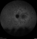
Recurrent MEWDS Left Eye - Teenage Girl539 x angesehenPatient presented in 2014 (first images) 2 months following vision loss left eye. Vision went from 20/60 - 20/20. OCT showed outer retinal irregularities which normalized. Then 3 years later in 2017 she had full blown MEWDS. She presented acutely and all the images are typical. Both episodes where in the same time of year, about 3 months after her flu shot for sports.
|
|

Recurrent MEWDS Left Eye - Teenage Girl539 x angesehenPatient presented in 2014 (first images) 2 months following vision loss left eye. Vision went from 20/60 - 20/20. OCT showed outer retinal irregularities which normalized. Then 3 years later in 2017 she had full blown MEWDS. She presented acutely and all the images are typical. Both episodes where in the same time of year, about 3 months after her flu shot for sports.
|
|

Acute Annular Outer Retinopathy for 3 Months - 25 YO Female Fluorescein Angiogram538 x angesehen25-year-old female who, about three months ago, put on her sunglasses and noticed a smudge in the vision and at that point she noticed the right eye was hazy. She had a cold a week or two before the vision loss, but she didn’t have any fever. She has no focal neurological problems in her past history. VA 20/20 OU
|
|

Choroidal Neovascular Membrane - Multifocal Scars - PIC vs MFC vs OHS 538 x angesehen
|
|

Recurrent MEWDS Left Eye - Teenage Girl538 x angesehenPatient presented in 2014 (first images) 2 months following vision loss left eye. Vision went from 20/60 - 20/20. OCT showed outer retinal irregularities which normalized. Then 3 years later in 2017 she had full blown MEWDS. She presented acutely and all the images are typical. Both episodes where in the same time of year, about 3 months after her flu shot for sports.
|
|

537 x angesehenPatient comes in with 6-month follow up for uveitis. VA is 20/20, both eyes. RPE peripheral scarring and punched out lesions visible. Stable. Pred Forte as needed for flare ups.
|
|

Recurrent MEWDS Left Eye - Teenage Girl537 x angesehenPatient presented in 2014 (first images) 2 months following vision loss left eye. Vision went from 20/60 - 20/20. OCT showed outer retinal irregularities which normalized. Then 3 years later in 2017 she had full blown MEWDS. She presented acutely and all the images are typical. Both episodes where in the same time of year, about 3 months after her flu shot for sports.
|
|
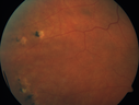
Multifocal Choroiditis - Linear Scars - Schlaegel lines537 x angesehen66 year old woman with macular pucker right eye. Images show typical linear peripheral scars for MFC or POHS - this is unilateral.
|
|

Birdshot Chorioretintis and PAMM537 x angesehenPatient presented with paracentral acute middle maculopathy as the presenting symptom of birdshot. This case was published. RETINAL CASES & BRIEF REPORTS 10:151–153, 2016.
|
|

Multifocal Choroidits Vitiligo ( Voght- Kayanagi-Harada Syndrome )535 x angesehen36-year old patient with decreased vision in both eyes with fatigue for over a year. VA 20/25-OD, 20/60 -OS. Patient was treated with Avastin and oral prednisone.
|
|

Birdshot Chorioretinitis - Retinal Vasculitis - HLA A29 Positive 534 x angesehen64-year-old woman three or four year’s fluctuations in her vision. She mostly notices them in bright light her vision glares out and she has difficulty seeing. PMHx: malaria in 1948 that was treated. She was told at one point she could possibly have had Familial Mediterranean fever.
She does intermittently get rashes around her arms. She has arthritis in her knees and ankles.
VISUAL ACUITY: OD 20/40, OS 20/50 - HLA - A29 Positive
|
|

Birdshot Chorioretinitis - Retinal Vasculitis - HLA A29 Positive 533 x angesehen64-year-old woman three or four year’s fluctuations in her vision. She mostly notices them in bright light her vision glares out and she has difficulty seeing. PMHx: malaria in 1948 that was treated. She was told at one point she could possibly have had Familial Mediterranean fever.
She does intermittently get rashes around her arms. She has arthritis in her knees and ankles.
VISUAL ACUITY: OD 20/40, OS 20/50 - HLA - A29 Positive
|
|

Multifocal Choroiditis - Linear Scars - Schlaegel lines533 x angesehen66 year old woman with macular pucker right eye. Images show typical linear peripheral scars for MFC or POHS - this is unilateral.
|
|

Sarcoidosis Multifocal Choroiditis533 x angesehen66 year old man with dense cataracts and recurrent uveitis. Images show multifocal choroidal granulomas from sarcoidosis more in the right eye than the left eye.
|
|

Recurrent MEWDS Left Eye - Teenage Girl532 x angesehenPatient presented in 2014 (first images) 2 months following vision loss left eye. Vision went from 20/60 - 20/20. OCT showed outer retinal irregularities which normalized. Then 3 years later in 2017 she had full blown MEWDS. She presented acutely and all the images are typical. Both episodes where in the same time of year, about 3 months after her flu shot for sports.
|
|

Multifocal Choroiditis - Linear Scars - Schlaegel lines532 x angesehen66 year old woman with macular pucker right eye. Images show typical linear peripheral scars for MFC or POHS - this is unilateral.
|
|

Multifocal Choroiditis Panuveitis - New Choroidal Neovascular Membrane Right Eye532 x angesehen74 year old female had vitrectomy elsewhere for possible ocular lymphoma - 5 years of shimmery vision changes then 3 weeks ago noticed vision loss right eye.
|
|

AZOOR - unilateral pseudo-retinitis pigmentosa532 x angesehen74 year old woman with unilateral vision loss 40 years ago.
|
|

Sarcoidosis Multifocal Choroiditis531 x angesehen66 year old man with dense cataracts and recurrent uveitis. Images show multifocal choroidal granulomas from sarcoidosis more in the right eye than the left eye.
|
|

Multifocal Choroiditis Panuveitis with Subretinal Fibrosis Acute Phase531 x angesehen36 year old latin american female 3 weeks of vision loss and new headaches. MRI was normal. VA initially 20/32 OD; 20/20 OS Final vision was 20/63 OD; 20/20 OS
|
|

Birdshot Chorioretinitis - Fresh Presentation530 x angesehenUltawidefield ICG shows best the multifocal lesions. 61 year old woman with floaters for less than 6 months. HLA-A29 Positive
|
|

Recurrent MEWDS Left Eye - Teenage Girl530 x angesehenPatient presented in 2014 (first images) 2 months following vision loss left eye. Vision went from 20/60 - 20/20. OCT showed outer retinal irregularities which normalized. Then 3 years later in 2017 she had full blown MEWDS. She presented acutely and all the images are typical. Both episodes where in the same time of year, about 3 months after her flu shot for sports.
|
|
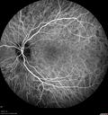
Recurrent MEWDS Left Eye - Teenage Girl530 x angesehenPatient presented in 2014 (first images) 2 months following vision loss left eye. Vision went from 20/60 - 20/20. OCT showed outer retinal irregularities which normalized. Then 3 years later in 2017 she had full blown MEWDS. She presented acutely and all the images are typical. Both episodes where in the same time of year, about 3 months after her flu shot for sports.
|
|

Multifocal Choroiditis Panuveitis with Subretinal Fibrosis Acute Phase530 x angesehen36 year old latin american female 3 weeks of vision loss and new headaches. MRI was normal. VA initially 20/32 OD; 20/20 OS Final vision was 20/63 OD; 20/20 OS
|
|

AZOOR - unilateral pseudo-retinitis pigmentosa530 x angesehen74 year old woman with unilateral vision loss 40 years ago.
|
|

Birdshot Chorioretintis and PAMM530 x angesehenPatient presented with paracentral acute middle maculopathy as the presenting symptom of birdshot. This case was published. RETINAL CASES & BRIEF REPORTS 10:151–153, 2016.
|
|

Acute Annular Outer Retinopathy for 3 Months - 25 YO Female Fundus Photograph529 x angesehen25-year-old female who, about three months ago, put on her sunglasses and noticed a smudge in the vision and at that point she noticed the right eye was hazy. She had a cold a week or two before the vision loss, but she didn’t have any fever. She has no focal neurological problems in her past history. VA 20/20 OU
|
|

Acute Annular Outer Retinopathy for 3 Months - 25 YO Female Early ICT529 x angesehen25-year-old female who, about three months ago, put on her sunglasses and noticed a smudge in the vision and at that point she noticed the right eye was hazy. She had a cold a week or two before the vision loss, but she didn’t have any fever. She has no focal neurological problems in her past history. VA 20/20 OU
|
|

Birdshot Chorioretinitis - Fresh Presentation529 x angesehenUltawidefield ICG shows best the multifocal lesions. 61 year old woman with floaters for less than 6 months. HLA-A29 Positive
|
|
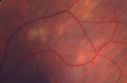
Sarcoidosis Multifocal Choroiditis529 x angesehen66 year old man with dense cataracts and recurrent uveitis. Images show multifocal choroidal granulomas from sarcoidosis more in the right eye than the left eye.
|
|

Recurrent MEWDS Left Eye - Teenage Girl528 x angesehenPatient presented in 2014 (first images) 2 months following vision loss left eye. Vision went from 20/60 - 20/20. OCT showed outer retinal irregularities which normalized. Then 3 years later in 2017 she had full blown MEWDS. She presented acutely and all the images are typical. Both episodes where in the same time of year, about 3 months after her flu shot for sports.
|
|

Multifocal Choroiditis Panuveitis with Subretinal Fibrosis Acute Phase528 x angesehen36 year old latin american female 3 weeks of vision loss and new headaches. MRI was normal. VA initially 20/32 OD; 20/20 OS Final vision was 20/63 OD; 20/20 OS
|
|

Acute Annular Outer Retinopathy for 3 Months - 25 YO Female Spectral Domain Optical Coherence Tomgraphy527 x angesehen25-year-old female who, about three months ago, put on her sunglasses and noticed a smudge in the vision and at that point she noticed the right eye was hazy. She had a cold a week or two before the vision loss, but she didn’t have any fever. She has no focal neurological problems in her past history. VA 20/20 OU
|
|

Acute Annular Outer Retinopathy for 3 Months - 25 YO Female Late FA527 x angesehen25-year-old female who, about three months ago, put on her sunglasses and noticed a smudge in the vision and at that point she noticed the right eye was hazy. She had a cold a week or two before the vision loss, but she didn’t have any fever. She has no focal neurological problems in her past history. VA 20/20 OU
|
|

Multiple Evanescent White Dot Syndrome - Atypical Yellow Foveal Spot Right Eye526 x angesehen12-year-old had decreased vision in the left eye from reported amblyopia since around the year 2000. His right eye was doing fine until two days ago, when he had sudden severe vision loss. He sees a blurred spot in the middle of the vision in the right eye wherever he looks.
OD 20/60 with eccentric movement, OS 20/200. Pinhole 20/70. IOP: OD 14, OS 10.
|
|

Acute Annular Outer Retinopathy for 3 Months - 25 YO Female Fundus Photograph526 x angesehen25-year-old female who, about three months ago, put on her sunglasses and noticed a smudge in the vision and at that point she noticed the right eye was hazy. She had a cold a week or two before the vision loss, but she didn’t have any fever. She has no focal neurological problems in her past history. VA 20/20 OU
|
|

Recurrent MEWDS Left Eye - Teenage Girl526 x angesehenPatient presented in 2014 (first images) 2 months following vision loss left eye. Vision went from 20/60 - 20/20. OCT showed outer retinal irregularities which normalized. Then 3 years later in 2017 she had full blown MEWDS. She presented acutely and all the images are typical. Both episodes where in the same time of year, about 3 months after her flu shot for sports.
|
|
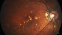
Punctate Inner Choroidopathy - New CNVM right eye526 x angesehen42 year old female with recent vision loss in the right eye - Right eye is 20/80 - Vision improved to 20/16 with Ranabizumab injections over 3 months
|
|
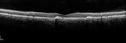
Acute Posterior Multifocal Placoid Pigment Epitheliopathy - 4 years after onset525 x angesehen4 years after presentation - vision 20/80 OD 20/16 OS and there is subretinal fibrosis
|
|
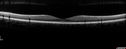
Recurrent MEWDS Left Eye - Teenage Girl525 x angesehenPatient presented in 2014 (first images) 2 months following vision loss left eye. Vision went from 20/60 - 20/20. OCT showed outer retinal irregularities which normalized. Then 3 years later in 2017 she had full blown MEWDS. She presented acutely and all the images are typical. Both episodes where in the same time of year, about 3 months after her flu shot for sports.
|
|

Multiple Evanescent White Dot Syndrome - Acute Idiopathic Blind Spot Enlargement524 x angesehen33-year-old woman one week ago she awoke and saw a red spots flashing out of her right eye. She notices them to the right side, which is the temporal visual field out of the right eye, then they changed to bursts of white lights without colors and then she notices straight ahead a couple spots. She has headaches in the back of her head. OD 20/20, OS 20/20
|
|

Acute Annular Outer Retinopathy for 3 Months - 25 YO Female InfraRed Image524 x angesehen25-year-old female who, about three months ago, put on her sunglasses and noticed a smudge in the vision and at that point she noticed the right eye was hazy. She had a cold a week or two before the vision loss, but she didn’t have any fever. She has no focal neurological problems in her past history. VA 20/20 OU
|
|
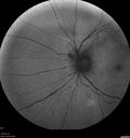
Birdshot Chorioretinitis - Fresh Presentation523 x angesehenUltawidefield ICG shows best the multifocal lesions. 61 year old woman with floaters for less than 6 months. HLA-A29 Positive
|
|

Multifocal Choroiditis Panuveitis with Subretinal Fibrosis Acute Phase523 x angesehen36 year old latin american female 3 weeks of vision loss and new headaches. MRI was normal. VA initially 20/32 OD; 20/20 OS Final vision was 20/63 OD; 20/20 OS
|
|
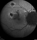
Multifocal Choroiditis Panuveitis - New Choroidal Neovascular Membrane Right Eye522 x angesehen74 year old female had vitrectomy elsewhere for possible ocular lymphoma - 5 years of shimmery vision changes then 3 weeks ago noticed vision loss right eye.
|
|

Multifocal Choroiditis Panuveitis with Subretinal Fibrosis Acute Phase522 x angesehen36 year old latin american female 3 weeks of vision loss and new headaches. MRI was normal. VA initially 20/32 OD; 20/20 OS Final vision was 20/63 OD; 20/20 OS
|
|

Acute Annular Outer Retinopathy for 3 Months - 25 YO Female InfraRed Image521 x angesehen25-year-old female who, about three months ago, put on her sunglasses and noticed a smudge in the vision and at that point she noticed the right eye was hazy. She had a cold a week or two before the vision loss, but she didn’t have any fever. She has no focal neurological problems in her past history. VA 20/20 OU
|
|

Multifocal Choroiditis - Linear Scars - Schlaegel lines521 x angesehen66 year old woman with macular pucker right eye. Images show typical linear peripheral scars for MFC or POHS - this is unilateral.
|
|

Sarcoidosis Multifocal Choroiditis521 x angesehen66 year old man with dense cataracts and recurrent uveitis. Images show multifocal choroidal granulomas from sarcoidosis more in the right eye than the left eye.
|
|
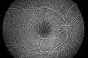
Multiple Evanescent White Dot Syndrome - Atypical Yellow Foveal Spot Right Eye520 x angesehen12-year-old had decreased vision in the left eye from reported amblyopia since around the year 2000. His right eye was doing fine until two days ago, when he had sudden severe vision loss. He sees a blurred spot in the middle of the vision in the right eye wherever he looks.
OD 20/60 with eccentric movement, OS 20/200. Pinhole 20/70. IOP: OD 14, OS 10.
|
|

Choroidal Neovascular Membrane - Multifocal Scars - PIC vs MFC vs OHS 520 x angesehen
|
|

Recurrent MEWDS Left Eye - Teenage Girl520 x angesehenPatient presented in 2014 (first images) 2 months following vision loss left eye. Vision went from 20/60 - 20/20. OCT showed outer retinal irregularities which normalized. Then 3 years later in 2017 she had full blown MEWDS. She presented acutely and all the images are typical. Both episodes where in the same time of year, about 3 months after her flu shot for sports.
|
|

Acute Annular Outer Retinopathy for 3 Months - 25 YO Female Spectral Domain Optical Coherence Tomgraphy519 x angesehen25-year-old female who, about three months ago, put on her sunglasses and noticed a smudge in the vision and at that point she noticed the right eye was hazy. She had a cold a week or two before the vision loss, but she didn’t have any fever. She has no focal neurological problems in her past history. VA 20/20 OU
|
|

Choroidal Neovascular Membrane - Multifocal Scars - PIC vs MFC vs OHS 519 x angesehen
|
|

Multifocal Choroiditis Panuveitis with Subretinal Fibrosis Acute Phase519 x angesehen36 year old latin american female 3 weeks of vision loss and new headaches. MRI was normal. VA initially 20/32 OD; 20/20 OS Final vision was 20/63 OD; 20/20 OS
|
|

Multiple Evanescent White Dot Syndrome - Atypical Yellow Foveal Spot Right Eye518 x angesehen12-year-old had decreased vision in the left eye from reported amblyopia since around the year 2000. His right eye was doing fine until two days ago, when he had sudden severe vision loss. He sees a blurred spot in the middle of the vision in the right eye wherever he looks.
OD 20/60 with eccentric movement, OS 20/200. Pinhole 20/70. IOP: OD 14, OS 10.
|
|

Choroidal Neovascular Membrane - Multifocal Scars - PIC vs MFC vs OHS 518 x angesehen
|
|

Sarcoidosis Multifocal Choroiditis518 x angesehen66 year old man with dense cataracts and recurrent uveitis. Images show multifocal choroidal granulomas from sarcoidosis more in the right eye than the left eye.
|
|
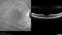
Recurrent MEWDS Left Eye - Teenage Girl517 x angesehenPatient presented in 2014 (first images) 2 months following vision loss left eye. Vision went from 20/60 - 20/20. OCT showed outer retinal irregularities which normalized. Then 3 years later in 2017 she had full blown MEWDS. She presented acutely and all the images are typical. Both episodes where in the same time of year, about 3 months after her flu shot for sports.
|
|

Acute Annular Outer Retinopathy for 3 Months - 25 YO Female Fundus Photograph516 x angesehen25-year-old female who, about three months ago, put on her sunglasses and noticed a smudge in the vision and at that point she noticed the right eye was hazy. She had a cold a week or two before the vision loss, but she didn’t have any fever. She has no focal neurological problems in her past history. VA 20/20 OU
|
|

Recurrent MEWDS Left Eye - Teenage Girl516 x angesehenPatient presented in 2014 (first images) 2 months following vision loss left eye. Vision went from 20/60 - 20/20. OCT showed outer retinal irregularities which normalized. Then 3 years later in 2017 she had full blown MEWDS. She presented acutely and all the images are typical. Both episodes where in the same time of year, about 3 months after her flu shot for sports.
|
|

Multifocal Choroiditis - Linear Scars - Schlaegel lines516 x angesehen66 year old woman with macular pucker right eye. Images show typical linear peripheral scars for MFC or POHS - this is unilateral.
|
|
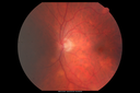
Sarcoidosis Multifocal Choroiditis516 x angesehen66 year old man with dense cataracts and recurrent uveitis. Images show multifocal choroidal granulomas from sarcoidosis more in the right eye than the left eye.
|
|

Birdshot Chorioretintis and PAMM516 x angesehenPatient presented with paracentral acute middle maculopathy as the presenting symptom of birdshot. This case was published. RETINAL CASES & BRIEF REPORTS 10:151–153, 2016.
|
|

Choroidal Neovascular Membrane - Multifocal Scars - PIC vs MFC vs OHS 514 x angesehen
|
|
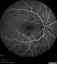
Choroidal Neovascular Membrane - Multifocal Scars - PIC vs MFC vs OHS 514 x angesehen
|
|

Choroidal Neovascular Membrane - Multifocal Scars - PIC vs MFC vs OHS 514 x angesehen
|
|

AZOOR - unilateral pseudo-retinitis pigmentosa514 x angesehen74 year old woman with unilateral vision loss 40 years ago.
|
|

Multifocal Choroiditis and Subretinal Fibrosis with new Subretinal Neovascular Membrane514 x angesehen26 year old African American Girl Saw me at age 10 (16 years ago) with acute multifocal choroiditis and panuveitis which evolved into subretinal fibrosis. She was initially treated with 3 years of methotrexate. Vision ended up 20/20 in both eyes. Her eyes have been inactive since and she was lost to follow-up.
A week ago she noticed sudden decreased vision in the right eye. Within the past few weeks there are more floaters in the right eye.
VA OD: sc6’/200 PHNI
VA OS: sc20/16
|
|

Choroidal Neovascular Membrane - Multifocal Scars - PIC vs MFC vs OHS 513 x angesehen
|
|

Choroidal Neovascular Membrane - Multifocal Scars - PIC vs MFC vs OHS 513 x angesehen
|
|
| 1298 Dateien auf 13 Seite(n) |
 |
|
|
|
6 | |
|
|
|
|
|


