Most viewed - Tumors, Masses, Pigmented Lesions
|

Primary Ocular Lymphoma - 051812 597 views
|
|

Choroidal Melanoma Medium595 views33-year-old man with a history of decreased in the right eye. He had blurring in the eye about a year ago. He reports that it improved spontaneously until it was “nearly resolvedâ€. He said he had some persistent distortion, but that over the past couple of weeks he has noticed this dark spot recurring in the right eye, the left eye is unaffected. He does not have any flashes, floaters or change in his peripheral visual acuity. 20/80 OD, 20/16 OS. No improvement with pinhole. IOP: 12 OD, 11 OS.
POSTERIOR SEGMENT EXAMINATION: Shows a clear view with no PVD in either eye. C/D ratio is 0.2 OU. Large retinal vessels are unremarkable. The macula of the right eye has a brown choroidal lesion with pigment overlying the surface and orange spots along its lateral surface. The left macula is normal.
B-SCAN: B-scan ecography of the ultrasound of the right eye shows a mass about 5 mm high with a moderate to low internal reflectivity on A-scan echogram. There is serous fluid inferiorly on the echogram
|
|

Medium Choroidal Melanoma Left Eye 595 views
|
|
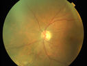
Primary Ocular Lymphoma - 051812 594 views
|
|

Ocular Lymphoma - non-Hodgkins - B-Cell594 viewsMultifocal sub-RPE lesions in the left eye of a patient with history of Non-hodgkins B-cell Lymphoma last active 2 years ago
|
|

Melanosis Oculi and Choroidal Melanoma594 views88 year old female with 2.5 mm elevated choroidal melanoma and melanosis oculi. She also has geographic atrophy from AMD. The right eye is the better eye with 20/40 vision. After observation with growth, brachytherapy was done.
|
|

Retinal Capillary Hemangioma - Endophytic and Exophytic593 views46 year old female with normal vision - her father died of pancreatic cancer
|
|

Choroidal Metastases - Both Eyes - Exudative Retinal Detachment592 views58-year-old man smoke a pack of cigarettes a day. He has had some ribcage pain for about the last two weeks. He also had a sinus infection for about two weeks, and he has also had hazy vision on the superior visual field in the left eye for about the last two weeks. vision is 20/25 in both eyes
|
|

Ocular Lymphoma - non-Hodgkins - B-Cell592 viewsMultifocal sub-RPE lesions in the left eye of a patient with history of Non-hodgkins B-cell Lymphoma last active 2 years ago
|
|
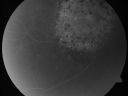
Pigmented Choroidal Mass Right Eye - Melanoma vs. Nevus vs. Unilateral Melanocytic Proliferation - Initial Visit591 views71-year-old man is vision has been bothering him for about a month. He does not notice one eye particularly worse than the other. OD is 20/40, OS is 20/25 - Initial Visit
|
|

Choroidal Melanoma - Exudative Retinal Detachment 82 Year Old Man 590 views82-year-old man who had 3 falls 2 weeks ago. After the falls he started checking his vision and noticed there was a veil over the left eye, which he had seen for about two weeks in the superior visual field. OD 20/25, OS 20/40
(patient was medically never well enough for brachytherapy and died 6 months later from heart disease)
|
|

Choroidal Metastases - Both Eyes - Exudative Retinal Detachment590 views58-year-old man smoke a pack of cigarettes a day. He has had some ribcage pain for about the last two weeks. He also had a sinus infection for about two weeks, and he has also had hazy vision on the superior visual field in the left eye for about the last two weeks. vision is 20/25 in both eyes
|
|

Retinal Capillary Hemangioma - Endophytic and Exophytic590 views46 year old female with normal vision - her father died of pancreatic cancer
|
|
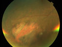
Primary Ocular Lymphoma - 051812 589 views
|
|

Choroidal Melanoma (Treated)589 viewsFundus photography shows choroidal melanoma at 2-o'clock superiorily. Patient had a radioactive implant back in August 2011.
|
|

Radiation Retinopathy and Macular Pucker 4 years following Brachytherapy (Plaque) for Nasal Melanoma588 views67-year-old woman has a macular pucker in the left eye. She had radiation for a melanoma in that eye 4 years aog. She had cataract surgery 2 years ago and a posterior sub-Tenon Kenalog injection in 7 months ago. Her vision in the left eye is hazy and she has trouble seeing out of it. VA OS 20/70.
|
|

Pigmented Choroidal Mass Right Eye - Melanoma vs. Nevus vs. Unilateral Melanocytic Proliferation - Initial Visit588 views71-year-old man is vision has been bothering him for about a month. He does not notice one eye particularly worse than the other. OD is 20/40, OS is 20/25 - Initial Visit
|
|

Ocular Lymphoma - non-Hodgkins - B-Cell - Right eye has equatorial drusen588 viewsMultifocal sub-RPE lesions in the left eye of a patient with history of Non-hodgkins B-cell Lymphoma last active 2 years ago
|
|

Choroidal Metastases - Both Eyes - Exudative Retinal Detachment587 views58-year-old man smoke a pack of cigarettes a day. He has had some ribcage pain for about the last two weeks. He also had a sinus infection for about two weeks, and he has also had hazy vision on the superior visual field in the left eye for about the last two weeks. vision is 20/25 in both eyes
|
|

Pigmented Choroidal Mass Right Eye - Melanoma vs. Nevus vs. Unilateral Melanocytic Proliferation - 4 Month Follow-Up Images586 views71-year-old man his vision 4 month follow-up. Vision is 20/60 and patient sees flashing lights. Lesion has grown slightly in the superior half of the macula.
|
|
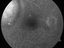
Choroidal Metastases - Both Eyes - Exudative Retinal Detachment586 views58-year-old man smoke a pack of cigarettes a day. He has had some ribcage pain for about the last two weeks. He also had a sinus infection for about two weeks, and he has also had hazy vision on the superior visual field in the left eye for about the last two weeks. vision is 20/25 in both eyes
|
|
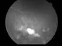
Uveitis and Retinal Whitening - 04-27-12 - Prior to PPV OD - Possible Primary Ocular Lymphoma 586 views
|
|

Choroidal Nevus - Macula - Flat586 views37 year old female with 5 year stable nevus Left eye - OCT shows flat nevus (EDI scan)
|
|

Choroidal Melanoma (Medium) Exudative Retinal Detachment 585 views87-year-old man noticed a new onset floater a few months ago. OD is 20/40, OS is 20/30
|
|

Choroidal Metastases - Both Eyes - Exudative Retinal Detachment585 views58-year-old man smoke a pack of cigarettes a day. He has had some ribcage pain for about the last two weeks. He also had a sinus infection for about two weeks, and he has also had hazy vision on the superior visual field in the left eye for about the last two weeks. vision is 20/25 in both eyes
|
|
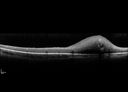
Retinal Capillary Hemangioma - Endophytic and Exophytic585 views46 year old female with normal vision - her father died of pancreatic cancer
|
|

Primary Ocular Lymphoma - White Mass Left Eye - 112712 584 views
|
|

Medium Choroidal Melanoma Left Eye 584 views
|
|

583 views33-year-old man with a history of decreased in the right eye. He had blurring in the eye about a year ago. He reports that it improved spontaneously until it was “nearly resolvedâ€. He said he had some persistent distortion, but that over the past couple of weeks he has noticed this dark spot recurring in the right eye, the left eye is unaffected. He does not have any flashes, floaters or change in his peripheral visual acuity. 20/80 OD, 20/16 OS. No improvement with pinhole. IOP: 12 OD, 11 OS.
POSTERIOR SEGMENT EXAMINATION: Shows a clear view with no PVD in either eye. C/D ratio is 0.2 OU. Large retinal vessels are unremarkable. The macula of the right eye has a brown choroidal lesion with pigment overlying the surface and orange spots along its lateral surface. The left macula is normal.
B-SCAN: B-scan ecography of the ultrasound of the right eye shows a mass about 5 mm high with a moderate to low internal reflectivity on A-scan echogram. There is serous fluid inferiorly on the echogram
|
|

Peripapillary Lesion Hemangioma or CNVM MID FA583 views67-year-old man with a history of a peripapillary lesion in the right eye.
|
|

Pigmented Choroidal Mass Right Eye - Melanoma vs. Nevus vs. Unilateral Melanocytic Proliferation - Initial Visit583 views71-year-old man is vision has been bothering him for about a month. He does not notice one eye particularly worse than the other. OD is 20/40, OS is 20/25 - Initial Visit
|
|
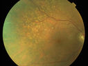
Primary Ocular Lymphoma - 051812 583 views
|
|

Choroidal Osteoma 13 Year Old583 views13 year old with 6 months of vision loss right eye and vision of 20/40
|
|

582 views
|
|

Primary Ocular Lymphoma - 051812 582 views
|
|

Choroidal Osteoma 13 Year Old582 views13 year old with 6 months of vision loss right eye and vision of 20/40
|
|

Metastatic Non Small Cell Lung Cancer to Choroid - Before External radiation582 views
|
|

Choroidal-Melanoma with Retinal Hemorrhages581 viewsPatient comes in with blurry vision in his inferior visual field in his right eye. Fundus photography shows choroidal melanoma superiorly at 12-o'clock. Retinal hemorrhages scattered around the periphery.
|
|

Primary Ocular Lymphoma - 020513 - FUndus Autofluorescence580 views
|
|
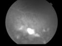
Uveitis and Retinal Whitening - 04-27-12 - Prior to PPV OD - Possible Primary Ocular Lymphoma 579 views
|
|

Choroidal Melanoma - Exudative Retinal Detachment 82 Year Old Man 578 views82-year-old man who had 3 falls 2 weeks ago. After the falls he started checking his vision and noticed there was a veil over the left eye, which he had seen for about two weeks in the superior visual field. OD 20/25, OS 20/40
(patient was medically never well enough for brachytherapy and died 6 months later from heart disease)
|
|

Choroidal Nevus - Macula - Flat578 views37 year old female with 5 year stable nevus Left eye - OCT shows flat nevus (EDI scan)
|
|

577 views
|
|

Pigmented Choroidal Mass Right Eye - Melanoma vs. Nevus vs. Unilateral Melanocytic Proliferation - 4 Month Follow-Up Images577 views71-year-old man his vision 4 month follow-up. Vision is 20/60 and patient sees flashing lights. Lesion has grown slightly in the superior half of the macula.
|
|

Pigmented Choroidal Mass Right Eye - Melanoma vs. Nevus vs. Unilateral Melanocytic Proliferation - Initial Visit577 views71-year-old man is vision has been bothering him for about a month. He does not notice one eye particularly worse than the other. OD is 20/40, OS is 20/25 - Initial Visit
|
|

Pigmented Choroidal Mass Right Eye - Melanoma vs. Nevus vs. Unilateral Melanocytic Proliferation - Initial Visit577 views71-year-old man is vision has been bothering him for about a month. He does not notice one eye particularly worse than the other. OD is 20/40, OS is 20/25 - Initial Visit71-year-old man is vision has been bothering him for about a month. He does not notice one eye particularly worse than the other. OD is 20/40, OS is 20/25 - Initial Visit
|
|

Ocular Lymphoma - non-Hodgkins - B-Cell577 viewsMultifocal sub-RPE lesions in the left eye of a patient with history of Non-hodgkins B-cell Lymphoma last active 2 years ago
|
|

576 views
|
|

Choroidal Melanoma (Medium) Exudative Retinal Detachment 576 views87-year-old man noticed a new onset floater a few months ago. OD is 20/40, OS is 20/30
|
|

Primary Ocular Lymphoma - 051812 576 views
|
|

Primary Ocular Lymphoma - White Mass Left Eye - 112712 576 views
|
|
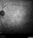
Primary Ocular Lymphoma - 020513 575 views
|
|

574 views
|
|

Retinal Capillary Hemangioma - Endophytic and Exophytic574 views46 year old female with normal vision - her father died of pancreatic cancer
|
|

Choroidal Melanoma Medium572 views33-year-old man with a history of decreased in the right eye. He had blurring in the eye about a year ago. He reports that it improved spontaneously until it was “nearly resolvedâ€. He said he had some persistent distortion, but that over the past couple of weeks he has noticed this dark spot recurring in the right eye, the left eye is unaffected. He does not have any flashes, floaters or change in his peripheral visual acuity. 20/80 OD, 20/16 OS. No improvement with pinhole. IOP: 12 OD, 11 OS.
POSTERIOR SEGMENT EXAMINATION: Shows a clear view with no PVD in either eye. C/D ratio is 0.2 OU. Large retinal vessels are unremarkable. The macula of the right eye has a brown choroidal lesion with pigment overlying the surface and orange spots along its lateral surface. The left macula is normal.
B-SCAN: B-scan ecography of the ultrasound of the right eye shows a mass about 5 mm high with a moderate to low internal reflectivity on A-scan echogram. There is serous fluid inferiorly on the echogram
|
|

Peripapillary Lesion Hemangioma or CNVM LATE FA571 views67-year-old man with a history of a peripapillary lesion in the right eye. VA 20/400. Worsening vision.
|
|

Choroidal Melanoma - Exudative Retinal Detachment 82 Year Old Man 571 views82-year-old man who had 3 falls 2 weeks ago. After the falls he started checking his vision and noticed there was a veil over the left eye, which he had seen for about two weeks in the superior visual field. OD 20/25, OS 20/40
(patient was medically never well enough for brachytherapy and died 6 months later from heart disease)
|
|

Pigmented Choroidal Mass Right Eye - Melanoma vs. Nevus vs. Unilateral Melanocytic Proliferation - 4 Month Follow-Up Images571 views71-year-old man his vision 4 month follow-up. Vision is 20/60 and patient sees flashing lights. Lesion has grown slightly in the superior half of the macula.
|
|

Pigmented Choroidal Mass Right Eye - Melanoma vs. Nevus vs. Unilateral Melanocytic Proliferation - 4 Month Follow-Up Images571 views71-year-old man his vision 4 month follow-up. Vision is 20/60 and patient sees flashing lights. Lesion has grown slightly in the superior half of the macula.
|
|

Pigmented Choroidal Mass Right Eye - Melanoma vs. Nevus vs. Unilateral Melanocytic Proliferation -4 Month Follow-Up Images570 views71-year-old man his vision 4 month follow-up. Vision is 20/60 and patient sees flashing lights. Lesion has grown slightly in the superior half of the macula.
|
|

Primary Ocular Lymphoma - 051812 569 views
|
|
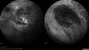
Medium Choroidal Melanoma Left Eye 569 views
|
|

Pigmented Choroidal Mass Right Eye - Melanoma vs. Nevus vs. Unilateral Melanocytic Proliferation - 4 Month Follow-Up Images 567 views71-year-old man his vision 4 month follow-up. Vision is 20/60 and patient sees flashing lights. Lesion has grown slightly in the superior half of the macula.
|
|

Choroidal Melanoma Medium566 views33-year-old man with a history of decreased in the right eye. He had blurring in the eye about a year ago. He reports that it improved spontaneously until it was “nearly resolvedâ€. He said he had some persistent distortion, but that over the past couple of weeks he has noticed this dark spot recurring in the right eye, the left eye is unaffected. He does not have any flashes, floaters or change in his peripheral visual acuity. 20/80 OD, 20/16 OS. No improvement with pinhole. IOP: 12 OD, 11 OS.
POSTERIOR SEGMENT EXAMINATION: Shows a clear view with no PVD in either eye. C/D ratio is 0.2 OU. Large retinal vessels are unremarkable. The macula of the right eye has a brown choroidal lesion with pigment overlying the surface and orange spots along its lateral surface. The left macula is normal.
B-SCAN: B-scan ecography of the ultrasound of the right eye shows a mass about 5 mm high with a moderate to low internal reflectivity on A-scan echogram. There is serous fluid inferiorly on the echogram
|
|

Ocular Lymphoma - non-Hodgkins - B-Cell - OCT shows multifocal sub-RPE masses 566 viewsMultifocal sub-RPE lesions in the left eye of a patient with history of Non-hodgkins B-cell Lymphoma last active 2 years ago
|
|

Choroidal Melanoma (Medium) Exudative Retinal Detachment 565 views87-year-old man noticed a new onset floater a few months ago. OD is 20/40, OS is 20/30
|
|

Uveal Choroidal Melanoma565 viewsPatient comes in for evaluation on a Choroidal Melanoma in the right eye. VA was 20/25 in both eyes. The melanoma is in the temporal aspect of the right eye. It measured at 0.7mm elevated after doing a BSCAN Ultrasound.
|
|
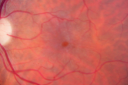
Radiation Retinopathy and Macular Pucker 4 years following Brachytherapy (Plaque) for Nasal Melanoma564 views67-year-old woman has a macular pucker in the left eye. She had radiation for a melanoma in that eye 4 years aog. She had cataract surgery 2 years ago and a posterior sub-Tenon Kenalog injection in 7 months ago. Her vision in the left eye is hazy and she has trouble seeing out of it. VA OS 20/70.
|
|
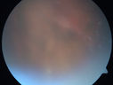
Primary Ocular Lymphoma - White Mass Left Eye - 112712 564 views
|
|

Choroidal Metastases - Both Eyes - Exudative Retinal Detachment563 views58-year-old man smoke a pack of cigarettes a day. He has had some ribcage pain for about the last two weeks. He also had a sinus infection for about two weeks, and he has also had hazy vision on the superior visual field in the left eye for about the last two weeks. vision is 20/25 in both eyes
|
|

Retinal Capillary Hemangioma - Endophytic and Exophytic563 views46 year old female with normal vision - her father died of pancreatic cancer
|
|

Pigmented Choroidal Mass Right Eye - Melanoma vs. Nevus vs. Unilateral Melanocytic Proliferation - Initial Visit562 views71-year-old man is vision has been bothering him for about a month. He does not notice one eye particularly worse than the other. OD is 20/40, OS is 20/25 - Initial Visit
|
|

Retinal Capillary Hemangioma - Endophytic and Exophytic562 views46 year old female with normal vision - her father died of pancreatic cancer
|
|

Retinal Capillary Hemangioma - Endophytic and Exophytic561 views46 year old female with normal vision - her father died of pancreatic cancer
|
|

560 views
|
|

Uveal Choroidal Melanoma560 viewsPatient comes in for evaluation on a Choroidal Melanoma in the right eye. VA was 20/25 in both eyes. The melanoma is in the temporal aspect of the right eye. It measured at 0.7mm elevated after doing a BSCAN Ultrasound.
|
|

Pigmented Choroidal Mass Right Eye - Melanoma vs. Nevus vs. Unilateral Melanocytic Proliferation - 4 Month Follow-Up Images559 views71-year-old man his vision 4 month follow-up. Vision is 20/60 and patient sees flashing lights. Lesion has grown slightly in the superior half of the macula.
|
|

Medium Choroidal Melanoma Left Eye 558 views
|
|

558 views
|
|

Choroidal Metastases - Both Eyes - Exudative Retinal Detachment556 views58-year-old man smoke a pack of cigarettes a day. He has had some ribcage pain for about the last two weeks. He also had a sinus infection for about two weeks, and he has also had hazy vision on the superior visual field in the left eye for about the last two weeks. vision is 20/25 in both eyes
|
|
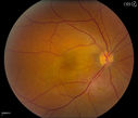
Cavernous Choroidal Hemangioma556 viewsPatient presents with Cavernous Choroidal Hemangioma in the right eye. VA is 20/50 after manifest refraction. Patient also notices decreased vision with shadow close to her central vision in the right eye. Patient will undergo photodynamic therapy in the right eye.
|
|

Choroidal Melanoma - Exudative Retinal Detachment 82 Year Old Man 554 views82-year-old man who had 3 falls 2 weeks ago. After the falls he started checking his vision and noticed there was a veil over the left eye, which he had seen for about two weeks in the superior visual field. OD 20/25, OS 20/40
(patient was medically never well enough for brachytherapy and died 6 months later from heart disease)
|
|

Radiation Retinopathy and Macular Pucker 4 years following Brachytherapy (Plaque) for Nasal Melanoma554 views67-year-old woman has a macular pucker in the left eye. She had radiation for a melanoma in that eye 4 years aog. She had cataract surgery 2 years ago and a posterior sub-Tenon Kenalog injection in 7 months ago. Her vision in the left eye is hazy and she has trouble seeing out of it. VA OS 20/70.
|
|

Pigmented Choroidal Mass Right Eye - Melanoma vs. Nevus vs. Unilateral Melanocytic Proliferation - Initial Visit554 views71-year-old man is vision has been bothering him for about a month. He does not notice one eye particularly worse than the other. OD is 20/40, OS is 20/25 - Initial Visit
|
|

Choroidal Melanoma - Exudative Retinal Detachment 82 Year Old Man 552 views82-year-old man who had 3 falls 2 weeks ago. After the falls he started checking his vision and noticed there was a veil over the left eye, which he had seen for about two weeks in the superior visual field. OD 20/25, OS 20/40
(patient was medically never well enough for brachytherapy and died 6 months later from heart disease)
|
|

Pigmented Choroidal Mass Right Eye - Melanoma vs. Nevus vs. Unilateral Melanocytic Proliferation - Initial Visit552 views71-year-old man is vision has been bothering him for about a month. He does not notice one eye particularly worse than the other. OD is 20/40, OS is 20/25 - Initial Visit
|
|

Medium Choroidal Melanoma Left Eye 552 views
|
|

Ciliary Body Melanoma552 views71-year old male who was recently diagnosed with a large ciliary body melanoma that is pushing into the anterior chamber of the left eye. Patient is going to proceed with proton therapy.
|
|

Choroidal Melanoma - Exudative Retinal Detachment 82 Year Old Man 551 views82-year-old man who had 3 falls 2 weeks ago. After the falls he started checking his vision and noticed there was a veil over the left eye, which he had seen for about two weeks in the superior visual field. OD 20/25, OS 20/40
(patient was medically never well enough for brachytherapy and died 6 months later from heart disease)
|
|

Choroidal Metastases - Both Eyes - Exudative Retinal Detachment551 views58-year-old man smoke a pack of cigarettes a day. He has had some ribcage pain for about the last two weeks. He also had a sinus infection for about two weeks, and he has also had hazy vision on the superior visual field in the left eye for about the last two weeks. vision is 20/25 in both eyes
|
|

Retinal Capillary Hemangioma due to Von Hipple-Lindau Syndrome551 viewsA 47-year old female comes in for a second opinion for retinal capillary Hemangioma in the left eye. VA was HM (hand motion) in the left eye. She has had multiple procedures done for the left eye including, Retinal Detachment surgery, YAG Laser PI, YAG Laser Capsulotomy, and Cataract surgery.
|
|
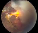
Retinal Capillary Hemangioma due to Von Hipple-Lindau Syndrome551 viewsA 47-year old female comes in for a second opinion for retinal capillary Hemangioma in the left eye. VA was HM (hand motion) in the left eye. She has had multiple procedures done for the left eye including, Retinal Detachment surgery, YAG Laser PI, YAG Laser Capsulotomy, and Cataract surgery.
|
|

Medium Choroidal Melanoma Left Eye 551 views
|
|

Primary Ocular Lymphoma - 051812 550 views
|
|

Ocular Lymphoma - non-Hodgkins - B-Cell - Right eye has equatorial drusen549 viewsMultifocal sub-RPE lesions in the left eye of a patient with history of Non-hodgkins B-cell Lymphoma last active 2 years ago
|
|

Radiation Retinopathy and Macular Pucker 4 years following Brachytherapy (Plaque) for Nasal Melanoma548 views67-year-old woman has a macular pucker in the left eye. She had radiation for a melanoma in that eye 4 years aog. She had cataract surgery 2 years ago and a posterior sub-Tenon Kenalog injection in 7 months ago. Her vision in the left eye is hazy and she has trouble seeing out of it. VA OS 20/70.
|
|

Pigmented Choroidal Mass Right Eye - Melanoma vs. Nevus vs. Unilateral Melanocytic Proliferation - Initial Visit548 views71-year-old man is vision has been bothering him for about a month. He does not notice one eye particularly worse than the other. OD is 20/40, OS is 20/25 - Initial Visit
|
|

Medium Choroidal Melanoma Left Eye 548 views
|
|
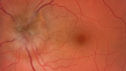
Melanocytoma - Lightly Pigmented548 views42 year old female with very mild visual field and vision loss in the left eye: 20/20 OU
|
|

Radiation Retinopathy and Macular Pucker 4 years following Brachytherapy (Plaque) for Nasal Melanoma546 views67-year-old woman has a macular pucker in the left eye. She had radiation for a melanoma in that eye 4 years aog. She had cataract surgery 2 years ago and a posterior sub-Tenon Kenalog injection in 7 months ago. Her vision in the left eye is hazy and she has trouble seeing out of it. VA OS 20/70.
|
|
| 1098 files on 11 page(s) |
 |
|
4 | |
|
|
|
|
|
|


