Top rated - Retinal Vascular Disease
|

Lupus Retinopathy835 viewsFemale patient comes in for eval on Lupus Retinopathy. Has poor vision in the right eye. VA is hand motion in the right eye. Fundus photos show fibrosis along the temporal arcades and narrowing of the arteries. No macular edema found.    
(2 votes)
|
|

Proliferative Diabetic Retinopathy1004 viewsFundus photography shows severe fibrosis and arterial narrowing. Peripheral laser scars in both eyes. VA is 20/40, right eye and 20/50, left eye.     
(2 votes)
|
|

Diabetic Retinopathy with CME894 viewsSecond opinion for diabetic retinopathy. VA is 20/30, right eye, 20/50, left eye. Patient has mild swelling in the macula of the right eye. No evidence of CME but does have a moderate cataract in the left eye.     
(2 votes)
|
|

Fibrosis Traction797 viewsYoung female patient with severe diabetes. Patients VA is 20/400 in the right eye. Fundus photos show a lot of fibrosis traction throughout the macula area. Fovea seems flat. Patient will undergo surgery for peeling of traction.     
(2 votes)
|
|
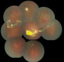
coats disease; exudative retinitis; retinal telangiectasis; leber multiple miliary aneurysm disease1484 viewsclassic coats    
(2 votes)
|
|

HTN Retinopathy w/ cotton wool spots and hemorrhages937 views23 year old african american male w/ visual acuity of 20/20    
(2 votes)
|
|

BRVO548 views42 year old female VA 20/25
Branch Retinal Vein Occlusion - Hot Spot on Vein at Occlusion Site    
(2 votes)
|
|

Central Retinal Artery Occlusion less than 24 hours - 69 year old man VA light perception508 views    
(2 votes)
|
|
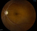
Retinal Artery Occlusion1140 viewsPatient comes in for eval on artery occlusions in both eyes. VA is 20/400, right eye and NLP, left eye. Fundus photos show paniretinal scars in the right eye with arterial narrowing and the left eye has arterial narrowing as well.    
(1 votes)
|
|

Vitreo-Traction due to Diabetic Retinopathy929 viewsPatient comes in for follow up on her diabetes. VA is 20/30 in the right eye. Fundus photos shows thick traction nasally to the optic nerve forming a ring around the macula. Will follow up in 6-months.    
(1 votes)
|
|
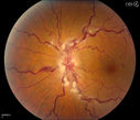
Stasis Retinopathy, Central Retinal Vein Occlusion799 viewsYoung male who complains of decreased vision in the left eye. VA is 20/20 in the left eye. Fundus photo shows mild central retinal vein occlusion in the left eye.     
(1 votes)
|
|

Diabetic Retinopathy1054 viewsAnnual diabetic exam. VA 20/30, right eye and 20/50, left eye. Fundus photos shows hemorrhages with cotton wool spots. Exudates are visible around the macula in the right eye. Scattered hemorrhages in the left eye.    
(1 votes)
|
|

Central Retinal Vein Occlusion948 viewsPatient comes in with mild blurred vision in the right eye. Fundus exam shows CRVO with scattered retinal hemorrhages in the right eye.    
(1 votes)
|
|

Optic Nerve Edema with Retinal Hemorrhages.709 viewsFemale patient diagnosed with lupus a year ago, comes in with spot and decreased vision in the right eye. VA is 20/400 in the right eye and 20/40, in the left eye. Fundus photography shows multiple retinal hemorrhages and possible bilateral papilledema.     
(1 votes)
|
|
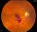
Branch Retinal Vein Occlusion775 viewsPatient comes in for eval on retinal hemorrhage in the right eye. VA is 20/30 in the right eye. Fundus photography shows branch retinal vein occlusion inferior of the macula with retinal hemorrhage present. Will reevaluate in 2-months.    
(1 votes)
|
|

Macular Hemorrhage762 viewsPatient comes in with central vision loss in the right eye. Fundus photography shows a macular hemorrhage in the central portion of the fovea. Cotton wool spots are present around the optic nerve and a little hemorrhage present inferiorily at 6-o'clock.    
(1 votes)
|
|

Background Diabetic Retinopathy1441 viewsPatient with diabetes for over 11-years comes in with blurred vision. Blood sugar control is very poor. VA 20/50- right eye, 20/25-left eye. Fundus exam shows hard exudates with Circinate Rings and edema. Micro-aneurysms and retinal hemorrhage are present in both eyes. Patient will come back for laser treatment.     
(1 votes)
|
|

Central Arterial Occlusion with Embolism Present704 viewsPatient comes in with CRAO in the right eye. VA was hand motion. Fundus photo shows white veil over the retina with 2-emboli in a branch artery temporal in the right eye. She will be evaluated for emboli and followed up in a month    
(1 votes)
|
|

Macular Pre-Retinal Hemorrhage 872 viewsPatient complains of loss of vision in his left eye. Patient is diabetic. VA was 20/20, right eye and 20/150, left eye with no improvement pinhole. Fundus exam reveals very large pre retinal hemorrhage in the left eye. Embolism located inferior, nasally just off the optic nerve in the left eye. Patient underwent PRP for treatment of the hemorrhage.    
(1 votes)
|
|

Prominent Posterior Hyaloid with Background Diabetic Retinopathy897 viewsPatient comes in for follow up on her Diabetic Retinopathy and glaucoma. Patient's VA was 20/30 in the left eye. Fundus exam presents a Posterior Hyaloid with hemorrhage inferiorily. Patient will be seen again in 6-months for follow up.     
(1 votes)
|
|

Post-Op Vitrectomy with Membrane Stripping and Laser693 viewsPatient had surgery to help clear up some vision in the left eye. Pre-op VA was count fingers at 1-ft. Post op VA was 20/200 in the left eye. Patient will return in 3-months for follow-up.    
(1 votes)
|
|

Hypertensive Retinopathy729 viewsPatient comes in complaining of spots in vision in both eyes. VA was 20/25 - right eye and 20/20- left eye. Fundus exam reveals little hemorrhages with cotton wool spots due to hypertension and anemia.     
(1 votes)
|
|

Nonproliferative Diabetic Retinopathy7358 views65-year old female with diabetes. Has had cataract surgery in the left eye with VA 20/25. She has had laser in the past. Fundus examination shows microaneurysms with retinal hemorrahages and exudates in the left eye.     
(1 votes)
|
|

Macroaneurysm FP945 viewsMacroaneurysm with Subretinal Hemorrhage and ME in OS of an Elderly Female    
(1 votes)
|
|

48.2 seconds317 viewsCorresponding FA for Cilioretinal artery occlusion with early venous stasis retinopathy     
(1 votes)
|
|
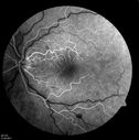
14.5 seconds720 viewsCorresponding FA for Cilioretinal artery occlusion with early venous stasis retinopathy     
(1 votes)
|
|

Central Retinal Vein Occlusion667 views    
(1 votes)
|
|

Angiogram of PDR969 views    
(1 votes)
|
|

Extensive PRP scarring767 views64 year old male with extensive PRP scarring due to diabetic retinopathy.    
(1 votes)
|
|

Purtscher's Retinopathy714 views19 year old female after pregnancy    
(1 votes)
|
|

Purtscher's Retinopathy498 views19 year old female after pregnancy    
(1 votes)
|
|

Purtscher's Retinopathy682 views19 year old female after pregnancy    
(1 votes)
|
|

hypertension retinopathy w/ disc edema and CRVO897 views64 year old white male w/ a vision of 20/200 which eventually improved to 20/60 without treatment    
(1 votes)
|
|

TRD w/ fibrovascular proliferation and sub retinal PVR OD1604 views41 year old african american female with tractional retinal detachment, fibrovascular proliferation, old vitreous hemorrhage, disc neovascularization, and proliferative vitreoretinopathy. Pt has a vision of 20/200    
(1 votes)
|
|

Sickle Cell1087 views    
(1 votes)
|
|
|
|
|
|

Diabetic Macular Edema369 views63 yr old Male
VA 20/20 OD 20/25 OS
No complaints
    
(1 votes)
|
|

Diabetic Macular Edema362 views63 yr old Male
VA 20/20 OD 20/25 OS
No complaints
    
(1 votes)
|
|

CRVO (retinal thrombophlebitis)401 views42 yr old Female c/o floaters
20/25 VA
Labs normal    
(1 votes)
|
|

CRVO (retinal thrombophlebitis)506 views42 yr old female
20/25 VA
Labs normal    
(1 votes)
|
|

Proliferative Sickle Retinopathy - Hemoglobin SC725 views53-year-old woman with a new spider-like floater in the right eye for about six days. She sees one big floater and another little one. She does have hemoglobin SC hemoglobinopathy. VISUAL ACUITY: Vision OD is 20/20; OS is 20/50, PH is 20/20.     
(1 votes)
|
|

Chronic - Old Diabetic Tractional Retinal Detachment Left Eye694 views77-year-old woman Vision OD is 20/40, OS is 7/200OD: Vertical C/D ratio is 0.3. There is panretinal laser. The macula is flat and attached.
OS: Vertical C/D ratio is 0.3. There is panretinal laser. There is a tractional retinal detachment with preretinal fibrosis nasal to the optic nerves.
    
(1 votes)
|
|

Ruptured Retinal Arterial Macroaneurysm - Submacular Hemorrhage - Vision 20/400 never recovered - Hypertension 205/94772 views    
(1 votes)
|
|

Central Retinal Artery Occlusion less than 24 hours - 69 year old man VA light perception458 views    
(1 votes)
|
|

Central Retinal Artery Occlusion less than 24 hours - 69 year old man VA light perception648 views    
(1 votes)
|
|
|

Proliferative Diabetic Retinopathy - Vitreous Hemorrhage and Tractional Retinal Detachment Left Eye586 views52-year-old man has been diabetic for nineteen years developed substantial vision loss over the last month or two. OD 20/70, OS 20/200.    
(1 votes)
|
|

Diabetic Vitreous Hemorrhage Left Eye538 views35-year-old man diabetic for the last 25 years. His hemoglobin A1C has been running between 10 and 11. He was doing fine and then when he woke up from sleeping last night he noticed sudden vision loss in his left eye OD 20/30, OS 20/200.    
(1 votes)
|
|

Proliferative Diabetic Retinopathy both eyes Type I Diabetic for 25 years437 views47-year-old woman with diabetes for 25 years and decreased vision for 3 months. OD 20/40, OS 20/40    
(1 votes)
|
|

366 views49-year-old woman has central retinal vein occlusion right eye with fluctuating vision for a few month now with OD 20/100. IOP: OD 20    
(1 votes)
|
|

Diabetic Retinopathy - Non-Perfusion - Possible Central Retinal Vein Occlusion508 views68-year-old woman OD 20/70, OS 20/200 CYSTOID MACULAR EDEMA, POSSIBLE EITHER OCULAR ISCHEMIC SYNDROME IN THE RIGHT EYE OR OCCULT CENTRAL RETINAL VEIN OCCLUSION IN THE RIGHT EYE,S EVERE NON-PerfUSION WITH DIABETIC RETINOPATHY IN THE LEFT EYE.    
(1 votes)
|
|
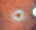
Central Retinal Artery Occlusion - 2 months old by history1289 views77-year-old man was doing fine. His left eye was a little worse than the right eye, and then in the 2 months ago he had sudden severe vision loss in the right eye. Vision OD is 3/200; OS is 20/60. IOP: OD 30, OS 15.
OD: Vertical C/D ratio is 0.4. The macula is white from ischemia. The arteries are narrow.
Photos confirm clinical findings.
OCT SCAN: OCT scan of the right eye shows retinal thickening, consistent with macular edema associated with a central retinal artery occlusion. The left eye has severe inferior retinal thinning, consistent with an old hemicentral retinal artery occlusion in the left eye. The fovea in the left eye is a little bit thin but not too bad.
IMPRESSION: 1. CENTRAL RETINAL ARTERY OCCLUSION RIGHT EYE
2 OLD HEMICENTRAL RETINAL ARTERY OCCLUSION LEFT EYE
3 DIABETES WITHOUT RETINOPATHY
4 ELEVATED INTRAOCULAR PRESSURE RIGHT EYE STATUS POST INJECTION RIGHT EYE A WEEK AGO
DISCUSSION: I explained to the patient that the right eye unfortunately has had a central retinal artery and unfortunately we do not have anything to treat that. His vision will probably improve some over time. He needs to be watched though. It is possible he is developing rubeotic glaucoma, although it is probable the intraocular pressure is high from the injection last week. I asked him to restart the Alphagan, to stop the other two drops, and to return here in three weeks to make sure his pressure is more acceptable.
At this point, having lost almost all the retinal circulation in the right eye and half of the retinal circulation in the left eye, he only has half a retina in the left eye he is working off of. I told him it is very important he keep seeing you and anything that can be done to keep his blood pressure low and his cholesterol reasonable would be helpful to what remains of his retinal circulation.
    
(1 votes)
|
|

branch retinal vein occlusion - fundus photo778 views70 year old woman with 20/80 vision from a branch retinal vein occlusion and macular edema. She has had laser treatment and intravitreal steroids. Her vision with further laser improved to 20/50.    
(1 votes)
|
|

branch retinal vein occlusion - oct line scan609 views70 year old woman with 20/80 vision from a branch retinal vein occlusion and macular edema. She has had laser treatment and intravitreal steroids. Her vision with further laser improved to 20/50.    
(1 votes)
|
|

Subhyloid Hem with PDR1151 views    
(3 votes)
|
|

Central Retinal Vein Occlusion with Macular Edema669 viewsPatient notices decreased vision in the left eye. VA is 20/60, left eye. Fundus and Fluorescence Angiogram shows CRVO in the left eye with scattered hemorrhages throughout the retina.    
(2 votes)
|
|

PDR with subhyloid hem941 viewsPDR with subhyloid hem    
(2 votes)
|
|

Proliferative Diabetic Retinopathy with Macular Hole2536 viewsPatitent with diabetes diagnosed 12 years ago and low visual acuity.     
(5 votes)
|
|

Central Retinal Artery Occlusion857 viewsCentral Retinal Artery Occlusion with cilio artery perfusion.     
(1 votes)
|
|

Juvenile Onset Diabetic - Proliferative Diabetic retinopathy with NVE and NVD3652 viewsProliferative Diabetic Retinopathy with Neovascularization of the Disc and Neovascularization Elsewhere.
James L. Perron, C.R.A.    
(1 votes)
|
|

Retinal Arterial Macroaneurysm - Recurrent Hemorrhage 6 months post laser1368 views6 months post laser: Her vision had improved, but then three weeks ago it worsened again. She is not on any blood thinners. She wasn’t doing any heavy lifting or straining.
VISUAL ACUITY: OD 3/200    
(1 votes)
|
|

Hemicentral Retinal Vein Occlusion Vision 20/60 897 views84-year-old man has a hemicentral retinal vein occlusion in the right eye. His vision has declined since then.
VISUAL ACUITY: Vision OD is 20/60.    
(1 votes)
|
|

Retinal Arterial Macroaneurysm - Increased Swelling after Laser 492 views65-year-old woman was seen in the office on January 12, 2011. She has noticed decreased vision in the right eye for the last two or three weeks. She has had some discomfort in both eyes as well. She has a history of low blood pressure, but she does have high cholesterol.
VISUAL ACUITY: OD 20/60    
(1 votes)
|
|

Retinal Arterial Macroaneurysm - Increased Swelling after Laser 732 views65-year-old woman was seen in the office on January 12, 2011. She has noticed decreased vision in the right eye for the last two or three weeks. She has had some discomfort in both eyes as well. She has a history of low blood pressure, but she does have high cholesterol.
VISUAL ACUITY: OD 20/60    
(1 votes)
|
|

Proliferative Diabetic Retinopathy both Eyes Neovascularization of the Disc1172 views63-year-old woman has been diabetic for twenty one years who sees new floaters in the right eye. OD 20/40, OS 20/40.    
(1 votes)
|
|

Diabetic Patient with Macular Edema and Blood Pressure 200/95749 views49-year-old decreasing vision over the last year. OD is 20/80, OS 20/80. blood pressure which was 200/95.
    
(1 votes)
|
|

Focal Laser for diabetic macular edema553 views66 year old female with diabetes for 45 years. VA 20/20 OU. Extrafoveal edema in the left eye treated with light focal laser.    
(1 votes)
|
|

Central Retinal Artery Occlusion1332 views60 year old male with LP vision due to extensive blood flow loss.    
(4 votes)
|
|

Retinal Vasculitis due to Lupus (color)2049 viewsSystemic Lupus Erythematosis - Vasculitis    
(3 votes)
|
|

Hemi-retinal Vein Occlusion1212 views81 year old female seen in 2009 for a Hemi-retinal vein Occlusion OS with vision of 20/25.     
(3 votes)
|
|

Purtscher's Retinopathy - Motor Vehicle Accident3404 viewsNerve Fiber Layer Inflammation and retinal hemorrhages due to severe trauma. Patient was a teenage boy that got into a car accident.     
(1 votes)
|
|

Retinal Arterial Macroaneurysm - Increased Swelling after Laser 672 viewsVisit 3 - 4 months post laser:
on May 23, 2011. This pleasant 66-year-old woman had a leaky arterial macroaneurysm I lasered in January. She developed more swelling subsequently, but an angiogram showed the lesion was closed. Since then she has noticed her vision poor in the eye.
VISUAL ACUITY: OD 20/200
    
(1 votes)
|
|

80 Year old man with 3 day history of vision loss right eye. Vision 4/200.365 views    
(1 votes)
|
|

Proliferative Diabetic Retinopathy in patient with Moyamoya Disease394 views28-year-old womandiabetic since age nine, and she also has had multiple strokes. OD is 20/30, OS is 20/30.    
(1 votes)
|
|

Inferotemporal and Inferonasal BRAO1260 viewsBranch Retinal Artery Occlusions - Multiple    
(1 votes)
|
|

Central Retinal Vein Occlusion Recurrent Edema 6 weeks after Lucentis - Nonperfusion Temporally566 views66-year-old woman has a central retinal vein occlusion in the right eye with macular edema. She had intravitreal Lucentis treatment six weeks ago and she has noticed over the last week or two her vision is declining.
VISUAL ACUITY: OD 20/70, OS 20/30.     
(1 votes)
|
|

Chronic - Old Diabetic Tractional Retinal Detachment Left Eye573 views77-year-old woman Vision OD is 20/40, OS is 7/200OD: Vertical C/D ratio is 0.3. There is panretinal laser. The macula is flat and attached.
OS: Vertical C/D ratio is 0.3. There is panretinal laser. There is a tractional retinal detachment with preretinal fibrosis nasal to the optic nerves.
    
(1 votes)
|
|

Old Branch Retinal Vein Occlusion- Severe CME - Pucker - Diabetic Map OCT579 views61-year old man has diabetic retinopathy in the right eye. He also had a macular pucker and macular edema. I did a vitrectomy on September 5th. His vision was initially improving. His vision in the right eye seems to be a little more foggy.
VISUAL ACUITY: OD: 20/200    
(1 votes)
|
|

Mild Clinically Significant Diabetic Macular Edema846 views58-year-old woman diabetic for 20 years who has diabetic macular edema in both eyes. She has had waxing and waning edema in the past and I thought it might improve on its own but it did not. She notices her vision is still a little hazy.
VISUAL ACUITY: Vision OD is 20/30, OS is 20/25.     
(1 votes)
|
|

Proliferative Diabetic Retinopathy - Vitreous Hemorrhage and Tractional Retinal Detachment Left Eye840 views52-year-old man has been diabetic for nineteen years developed substantial vision loss over the last month or two. OD 20/70, OS 20/200.    
(1 votes)
|
|
|
|
|
|