Les mieux notées - Retina Vascular Occlusions
|

BRVOvu 818 fois42 year old female VA 20/25
Branch Retinal Vein Occlusion - Hot Spot on Vein at Occlusion Site
(2 votes)
|
|

Central Retinal Artery Occlusion less than 24 hours - 69 year old man VA light perceptionvu 826 fois
(2 votes)
|
|
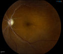
Retinal Artery Occlusionvu 1670 foisPatient comes in for eval on artery occlusions in both eyes. VA is 20/400, right eye and NLP, left eye. Fundus photos show paniretinal scars in the right eye with arterial narrowing and the left eye has arterial narrowing as well.
(1 votes)
|
|
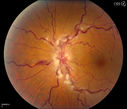
Stasis Retinopathy, Central Retinal Vein Occlusionvu 1244 foisYoung male who complains of decreased vision in the left eye. VA is 20/20 in the left eye. Fundus photo shows mild central retinal vein occlusion in the left eye.
(1 votes)
|
|

Central Retinal Vein Occlusionvu 1322 foisPatient comes in with mild blurred vision in the right eye. Fundus exam shows CRVO with scattered retinal hemorrhages in the right eye.
(1 votes)
|
|
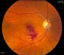
Branch Retinal Vein Occlusionvu 1109 foisPatient comes in for eval on retinal hemorrhage in the right eye. VA is 20/30 in the right eye. Fundus photography shows branch retinal vein occlusion inferior of the macula with retinal hemorrhage present. Will reevaluate in 2-months.
(1 votes)
|
|

Central Arterial Occlusion with Embolism Presentvu 1026 foisPatient comes in with CRAO in the right eye. VA was hand motion. Fundus photo shows white veil over the retina with 2-emboli in a branch artery temporal in the right eye. She will be evaluated for emboli and followed up in a month
(1 votes)
|
|

48.2 secondsvu 554 foisCorresponding FA for Cilioretinal artery occlusion with early venous stasis retinopathy
(1 votes)
|
|
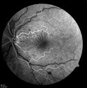
14.5 secondsvu 1048 foisCorresponding FA for Cilioretinal artery occlusion with early venous stasis retinopathy
(1 votes)
|
|

Central Retinal Vein Occlusionvu 964 fois
(1 votes)
|
|
|
|
|
|

CRVO (retinal thrombophlebitis)vu 570 fois42 yr old Female c/o floaters
20/25 VA
Labs normal
(1 votes)
|
|

CRVO (retinal thrombophlebitis)vu 718 fois42 yr old female
20/25 VA
Labs normal
(1 votes)
|
|

Central Retinal Artery Occlusion less than 24 hours - 69 year old man VA light perceptionvu 714 fois
(1 votes)
|
|

Central Retinal Artery Occlusion less than 24 hours - 69 year old man VA light perceptionvu 918 fois
(1 votes)
|
|

vu 618 fois49-year-old woman has central retinal vein occlusion right eye with fluctuating vision for a few month now with OD 20/100. IOP: OD 20
(1 votes)
|
|

Central Retinal Artery Occlusion - 2 months old by historyvu 1802 fois77-year-old man was doing fine. His left eye was a little worse than the right eye, and then in the 2 months ago he had sudden severe vision loss in the right eye. Vision OD is 3/200; OS is 20/60. IOP: OD 30, OS 15.
OD: Vertical C/D ratio is 0.4. The macula is white from ischemia. The arteries are narrow.
Photos confirm clinical findings.
OCT SCAN: OCT scan of the right eye shows retinal thickening, consistent with macular edema associated with a central retinal artery occlusion. The left eye has severe inferior retinal thinning, consistent with an old hemicentral retinal artery occlusion in the left eye. The fovea in the left eye is a little bit thin but not too bad.
IMPRESSION: 1. CENTRAL RETINAL ARTERY OCCLUSION RIGHT EYE
2 OLD HEMICENTRAL RETINAL ARTERY OCCLUSION LEFT EYE
3 DIABETES WITHOUT RETINOPATHY
4 ELEVATED INTRAOCULAR PRESSURE RIGHT EYE STATUS POST INJECTION RIGHT EYE A WEEK AGO
DISCUSSION: I explained to the patient that the right eye unfortunately has had a central retinal artery and unfortunately we do not have anything to treat that. His vision will probably improve some over time. He needs to be watched though. It is possible he is developing rubeotic glaucoma, although it is probable the intraocular pressure is high from the injection last week. I asked him to restart the Alphagan, to stop the other two drops, and to return here in three weeks to make sure his pressure is more acceptable.
At this point, having lost almost all the retinal circulation in the right eye and half of the retinal circulation in the left eye, he only has half a retina in the left eye he is working off of. I told him it is very important he keep seeing you and anything that can be done to keep his blood pressure low and his cholesterol reasonable would be helpful to what remains of his retinal circulation.
(1 votes)
|
|

branch retinal vein occlusion - fundus photovu 1075 fois70 year old woman with 20/80 vision from a branch retinal vein occlusion and macular edema. She has had laser treatment and intravitreal steroids. Her vision with further laser improved to 20/50.
(1 votes)
|
|
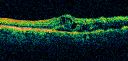
branch retinal vein occlusion - oct line scanvu 843 fois70 year old woman with 20/80 vision from a branch retinal vein occlusion and macular edema. She has had laser treatment and intravitreal steroids. Her vision with further laser improved to 20/50.
(1 votes)
|
|

Central Retinal Vein Occlusion with Macular Edemavu 981 foisPatient notices decreased vision in the left eye. VA is 20/60, left eye. Fundus and Fluorescence Angiogram shows CRVO in the left eye with scattered hemorrhages throughout the retina.
(2 votes)
|
|

Central Retinal Artery Occlusionvu 1082 foisCentral Retinal Artery Occlusion with cilio artery perfusion. 
(1 votes)
|
|
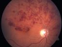
Hemicentral Retinal Vein Occlusion Vision 20/60 vu 1246 fois84-year-old man has a hemicentral retinal vein occlusion in the right eye. His vision has declined since then.
VISUAL ACUITY: Vision OD is 20/60.
(1 votes)
|
|

Central Retinal Artery Occlusionvu 1750 fois60 year old male with LP vision due to extensive blood flow loss.
(4 votes)
|
|

Hemi-retinal Vein Occlusionvu 1587 fois81 year old female seen in 2009 for a Hemi-retinal vein Occlusion OS with vision of 20/25.
(3 votes)
|
|

80 Year old man with 3 day history of vision loss right eye. Vision 4/200.vu 574 fois
(1 votes)
|
|

Inferotemporal and Inferonasal BRAOvu 1625 foisBranch Retinal Artery Occlusions - Multiple
(1 votes)
|
|

Central Retinal Vein Occlusion Recurrent Edema 6 weeks after Lucentis - Nonperfusion Temporallyvu 771 fois66-year-old woman has a central retinal vein occlusion in the right eye with macular edema. She had intravitreal Lucentis treatment six weeks ago and she has noticed over the last week or two her vision is declining.
VISUAL ACUITY: OD 20/70, OS 20/30.
(1 votes)
|
|

Old Branch Retinal Vein Occlusion- Severe CME - Pucker - Diabetic Map OCTvu 812 fois61-year old man has diabetic retinopathy in the right eye. He also had a macular pucker and macular edema. I did a vitrectomy on September 5th. His vision was initially improving. His vision in the right eye seems to be a little more foggy.
VISUAL ACUITY: OD: 20/200
(1 votes)
|
|
|
|
|
|


