am besten bewertet - Tumors, Masses, Pigmented Lesions
|

Retinoblastoma2049 x angesehenYoung male who has a retinal tumor inferior with a calcium core in the left eye. This is a regressed retinoblastoma. 
(3 Bewertungen)
|
|

Red Free Hemangioma left eye2068 x angesehen
(3 Bewertungen)
|
|

Metastatic Choroidal Lesion1135 x angesehenYoung female with choroidal lesion with subretinal fluid in the left eye. VA is finger count in the left eye.
(2 Bewertungen)
|
|

Choroidal Melanoma1162 x angesehenPatient comes in with spot in vision in the right eye. Fundus photography shows medium size melanoma adjacent to the optic nerve and measures 13mm across horizontally and 6mm diameter in thickness. Patient will undergo proton beam therapy.
(2 Bewertungen)
|
|

Choroidal Melanoma with Radiation Retinopathy1289 x angesehenPatient comes with follow up on Choroidal Melanoma, Right Eye that was treated back in June of 2009 with Radioactive Implant. Vein Occlusion is also present with VA - Hand Motion. Hemorrhages visible with hard exudates from the Radiation Retinopathy.
(2 Bewertungen)
|
|

Choroidal Melanoma1600 x angesehen
(2 Bewertungen)
|
|

Choroidal Melanoma751 x angesehenChoroidal Melanoma with Retinal Detachment present inferior.
(2 Bewertungen)
|
|

Retinocytoma - Retinoma - Regressed Retinoblastoma 43 year old male incidental finding 1431 x angesehen
(2 Bewertungen)
|
|

Treated Choroidal Melanoma1076 x angesehenFollow up patient for melanoma in the right eye that was treated with proton beam therapy. VA is 20/50 in the right eye. Fundus photo shows scattered hemorrhages inferiorly with tumor temporal inferior at 8-o'clock. Patient will be followed up in 6-months
(1 Bewertungen)
|
|

Melanoma of Optic Nerve1140 x angesehenPatient comes in for evaluation on Optic Nerve Melanoma in the right eye. Patient's VA is 20/40 in the right eye. Fundus photograph reveals melanoma invading optic nerve and ultrasound reveals behind the optic nerve as well.
(1 Bewertungen)
|
|

Treated Melanoma931 x angesehenTreated melanoma in the right eye. VA is light perception in the right eye.
(1 Bewertungen)
|
|

Choroidal Osteoma1460 x angesehenPatient with decreased vision within the past 2-years. VA 20/100 in the right eye. Fundus photo shows atrophic scar temporal to the macula in the right eye.
(1 Bewertungen)
|
|

Choroidal Nevus1084 x angesehenPatient comes in with pigmented spot in the right eye. VA is 20/25. Fundus photography shows elevated choroidal nevus inferiorily in the right eye. Will be reevaluated in 3-months...
(1 Bewertungen)
|
|

Choroidal Melanoma1109 x angesehenFemale patient comes in with a visual defect in the right eye. VA is 20/40 in the right eye. Fundus photography shows choroidal melanoma with retinal detachment temporally at 3-o'clock in the right eye.
(1 Bewertungen)
|
|

Choroidal Melanoma with Sub Retinal Fluid1189 x angesehenFundus photography shows choroidal melanoma superiorily between 11-12-o'clock in the left eye. VA is 20/150 in the left eye.
(1 Bewertungen)
|
|

Optic Nerve Tumor1039 x angesehenYoung male patient with a tumor present on the optic nerve in the right eye. VA is hand motion in the right eye
(1 Bewertungen)
|
|

Choroidal Metastasis Tumor 1089 x angesehenPatient that a history of breast cancer. She has a metastasis tumor superiority in the right eye. VA was 20/20, right eye. Patient underwent radiation and chemotherapy and mass has not changed in size for the past year.
(1 Bewertungen)
|
|

Ciliary Body Melanoma925 x angesehen71-year old male who was recently diagnosed with a large ciliary body melanoma that is pushing into the anterior chamber of the left eye. Patient is going to proceed with proton therapy.
(1 Bewertungen)
|
|

Choroidal Tumor consistent with Uveal Melanoma1178 x angesehenPatient comes in for second opinion for 2-melanomas in the right eye. Patient's VA is 20/20, both eyes. Fundus exam reveals melanoma superior to the macula which measured at 0.7-mm thick. Second melanoma is nasally which measured at 4.1-mm thick and has low internal reflectivity. Patient will be followed up with MRI and ocular protocol.
(1 Bewertungen)
|
|

Uveal Choroidal Melanoma1028 x angesehenPatient comes in for evaluation on a Choroidal Melanoma in the right eye. VA was 20/25 in both eyes. The melanoma is in the temporal aspect of the right eye. It measured at 0.7mm elevated after doing a BSCAN Ultrasound.
(1 Bewertungen)
|
|

CHOROIDAL Melanoma1206 x angesehen
(1 Bewertungen)
|
|

Choroidal Malignant Melanoma - mid phase FA626 x angesehenmid-phase of FA at 5 minutes 10 seconds (Choroidal malignant melanoma status post plaque radiotherapy OD in 1986, patient also has a submacular hemorrhage OD, and radiation retinopathy OD.)
(1 Bewertungen)
|
|

Choroidal Malignant Melanoma - early phase FA806 x angesehenearly phase of FA at 23 seconds (Choroidal malignant melanoma status post plaque radiotherapy OD in 1986, patient also has a submacular hemorrhage OD, and radiation retinopathy OD.)
(1 Bewertungen)
|
|

Choroidal Malignant Melanoma - Montage677 x angesehenChoroidal malignant melanoma status post plaque radiotherapy OD in 1986, patient also has a submacular hemorrhage OD, and radiation retinopathy OD.
(1 Bewertungen)
|
|

Choroidal Melanoma with Retinal Detachment1098 x angesehen57 year old female who underwent proton beam therapy for Choroidal Melanoma. VA was 20/400 at post op visit.
(1 Bewertungen)
|
|

Choroidal Melanoma with Retinal Detachment1401 x angesehen57 year old female who underwent proton beam therapy for Choroidal Melanoma. VA was 20/400 at post op visit.
(1 Bewertungen)
|
|
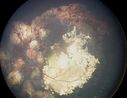
Retinoblastoma1845 x angesehen
(1 Bewertungen)
|
|

Metastasis- Tumor with Sub-retinal Fluid Present 713 x angesehen
(1 Bewertungen)
|
|
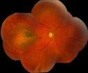
Choroidal Melanoma695 x angesehen
(1 Bewertungen)
|
|
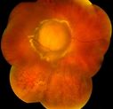
Choroidal Melanoma734 x angesehen
(1 Bewertungen)
|
|

Choroidal Melanoma796 x angesehenNotice the orange strands presently on the tumor.
(1 Bewertungen)
|
|
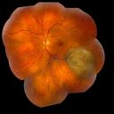
Choroidal Melanoma755 x angesehen
(1 Bewertungen)
|
|

Choroidal Melanoma647 x angesehen
(1 Bewertungen)
|
|
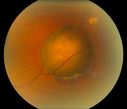
Choroidal Melanoma750 x angesehen
(1 Bewertungen)
|
|

Choroidal Melanoma753 x angesehen
(1 Bewertungen)
|
|

Choroidal Melanoma921 x angesehenPost op Radioactive Implant with hard exudates surrounding the Melanoma with hemorrhage present.
(1 Bewertungen)
|
|

Choroidal Nevus with Overlying Drusen1715 x angesehen48-year-old woman Vision OD is 20/16, OS is 20/20.
(1 Bewertungen)
|
|

Retinocytoma - Retinoma - Regressed Retinoblastoma 43 year old male incidental finding 749 x angesehen
(1 Bewertungen)
|
|
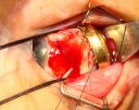
Radioactive Plaque Placement for Choroidal Melanoma Plaque Placed but not Sutured to Eye Yet1303 x angesehen
(1 Bewertungen)
|
|
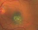
Macular Choroidal Nevus1740 x angesehen78-year-old woman has choroidal nevus in the macula of the left eye. You asked her to come in here for an evaluation and nevus was noted in the past about 7 years ago. She has not noticed any change in vision in that eye.
VISUAL ACUITY: OU 20/30.
(1 Bewertungen)
|
|

Choroidal Melanoma Medium758 x angesehen33-year-old man with a history of decreased in the right eye. He had blurring in the eye about a year ago. He reports that it improved spontaneously until it was “nearly resolvedâ€. He said he had some persistent distortion, but that over the past couple of weeks he has noticed this dark spot recurring in the right eye, the left eye is unaffected. He does not have any flashes, floaters or change in his peripheral visual acuity. 20/80 OD, 20/16 OS. No improvement with pinhole. IOP: 12 OD, 11 OS.
POSTERIOR SEGMENT EXAMINATION: Shows a clear view with no PVD in either eye. C/D ratio is 0.2 OU. Large retinal vessels are unremarkable. The macula of the right eye has a brown choroidal lesion with pigment overlying the surface and orange spots along its lateral surface. The left macula is normal.
B-SCAN: B-scan ecography of the ultrasound of the right eye shows a mass about 5 mm high with a moderate to low internal reflectivity on A-scan echogram. There is serous fluid inferiorly on the echogram
(1 Bewertungen)
|
|

Chorodial Melanoma1388 x angesehen80 year old male came in for retinal evaluation and presented with a melanoma.
(4 Bewertungen)
|
|

Melanoma1685 x angesehenYoung male with large Melanoma
(6 Bewertungen)
|
|
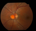
Optic Nerve Tumor3902 x angesehen21 year old male: Presents with a history of Von Hippel-Lindau Syndrome, diagnosed in 2005 affecting his left Optic Nerve, Brain, Spine, Kidney and Pancreas. He has undergone Laser for Retinal Hemangioma measuring 2/3-4/5DD -vs_ 1/3DD when originally diagnosed. However his visual acuity remains good 20/25. Patient has also undergone Neurosurgery in 2005 and Spinal cord in 2006. Recent MRI of spinal cord hemangioma showed stable tumors.
(6 Bewertungen)
|
|
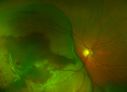
Large Choroidal Melanoma with Retinal Detachment1016 x angesehenPatient notices a decline in her vision for the past 3-weeks in her right eye. VA was light perception in the right eye. Fundus photo shows a very large melanoma temporal to the optic nerve and retinal detachment inferiorly. Patient will under go proton beam therapy.
(2 Bewertungen)
|
|

Melanoma - Large Ciliary Body Photo shows tumor visible throught the pupil969 x angesehen
(2 Bewertungen)
|
|

Melanoma - Large Ciliary Body A scan shows medium to low internal reflectivity844 x angesehen
(1 Bewertungen)
|
|

Melanoma1647 x angesehen40 Year old male
(5 Bewertungen)
|
|
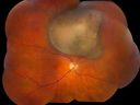
Choroidal Melanoma1470 x angesehen
(2 Bewertungen)
|
|
|
|
|
| 49 Dateien auf 1 Seite(n) |
|


