Top rated - Macular Diseases - Acquired
|
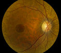
Full Thickness Macular Hole1178 viewsFull Thickness Macular Hole
(2 votes)
|
|

Central Serous Retinopathy1095 viewsYoung male who comes in for follow up on CSR in the left eye. Patient had focal laser treatment 1-month ago. There are still spots of leaking and proceeded with another focal laser treatment. Notice the top of the hyperfluorescent area.
(2 votes)
|
|
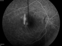
arteriovenous phase, acute recurrent CSR 796 viewsAngiogram focused on disc illustrates anterior displacement of the macula. Smokestack sign doesn't get any better than this.
(2 votes)
|
|

fellow eye with subclinical areas of leakage781 views
(2 votes)
|
|

Solar retinopathy829 viewsA 27 year old African American male presented with a complaint of poor vision in both eyes for several months.
PMH: none
Medications: none
BCVA 20/40 OU
Slit-lamp exam: normal OU
On further questioning, he enjoys staring at the sun for 3-4 hours each day.
(2 votes)
|
|

Macular Pucker1307 viewsPatient comes in for followup on macular pucker, right eye. VA is 20/50 in the right eye...Fundus photo shows prominent pucker centrally pulling temporally in the right eye.
(1 votes)
|
|

Epiretinal Membrane1075 viewsPatient with a history of pars planitis comes in with decreased vision in the left eye. VA is 20/30 in the left eye. Fundus photos show a rather large epiretinal membrane covering the optic nerve in the left eye.
(1 votes)
|
|

Macular Hole with Peripheral Laser Scars1195 viewsDiabetic patient comes in with flashes and floaters in both eyes. Patient's VA is 20/400, right eye and 20/25, left eye. Fundus exam presents with large macular hole with diabetic changes in the retina. Laser scars in the peripheral.
(1 votes)
|
|

Retinal Pigment Epithelial Detachment with no Sub-Retinal Fluid...1451 viewsA 38-year old male who comes in with blurred vision in the left eye. VA is 20/30. Notices a defect inferior of his central vision. Did an Fluorescence Angiogram to determine an RPE with no sub retinal fluid. Also OCT confirms. Patient was injected with Avastin.
(1 votes)
|
|

Epiretinal Membrane1354 viewsEpiretinal Membrane
(1 votes)
|
|
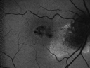
Juxtafoveal Telangiectasis (MacTel) and Asymptomatic Extrafoveal Juxtapapillary Choroidal Neovascular Membrane Fundus Autofluorescence814 views84-year-old woman has juxtafoveal retinal telangiectasis in both eyes. The right eye has an asymptomatic extra foveal juxtapapillary choroidal neovascular membrane. OD 20/400, OS 20/63.
(1 votes)
|
|

Juxtafoveal Telangiectasis - Stage 5 Left Eye - New Blood and Neovascularization MacTel 658 views60-year-old man has juxtafoveal retinal telangiectasis recent vision loss OD 20/50, OS 20/70 : Hemorrhage OS and new SRNVM
(1 votes)
|
|

Prefoveal Opercula after Aborted Stage 2 Macular Hole - Present for 1 Year - VA 20/20 - OCT920 viewsThis pleasant 61-year-old man had a small Stage 2 macular hole, which spontaneously close 12 months ago. He is still bothered by a pre-foveal opercula. OD 20/20.
(1 votes)
|
|

Plaquenil Toxicity - Bull's Eye Maculopathy - 25 Years Therapy - Fundus Autofluorescence1268 views73-year-old woman has been on Plaquenil for twenty five years. Plaquenil has now been stopped for 6 months. VA continues to decline as does visual function - 20/80 OD , 20/40 OS
(1 votes)
|
|

Plaquenil Toxicity - Bull's Eye Maculopathy - 25 Years Therapy - OCT Line Scan868 views73-year-old woman has been on Plaquenil for twenty five years. Plaquenil has now been stopped for 6 months. VA continues to decline as does visual function - 20/80 OD , 20/40 OS
(1 votes)
|
|

Plaquenil Toxicity - Bull's Eye Maculopathy - 25 Years Therapy - Fundus Autofluorescence829 views73-year-old woman has been on Plaquenil for twenty five years. Plaquenil has now been stopped for 6 months. VA continues to decline as does visual function - 20/80 OD , 20/40 OS
(1 votes)
|
|

Central Serous Retinopathy Acute - Indocyanine Green Angiogram - Leakage Choriocapillaris 1625 views42-year-old man was seen in the office on October 5, 2011. He had noticed starting in August after a course of antibiotic and steroids, that he developed new spots in his vision in the right eye. He may have had an episode like this sometime in the past. He did take steroids a few years ago and his vision did change at that time, but then returned.
VISUAL ACUITY: OD 20/32, OS 20/32
(1 votes)
|
|

Central Serous Retinopathy Acute - Fundus Autofluorescence1322 views42-year-old man was seen in the office on October 5, 2011. He had noticed starting in August after a course of antibiotic and steroids, that he developed new spots in his vision in the right eye. He may have had an episode like this sometime in the past. He did take steroids a few years ago and his vision did change at that time, but then returned.
VISUAL ACUITY: OD 20/32, OS 20/32
(1 votes)
|
|

Central Serous Retinopathy Acute - Fundus Autofluorescence1490 views42-year-old man was seen in the office on October 5, 2011. He had noticed starting in August after a course of antibiotic and steroids, that he developed new spots in his vision in the right eye. He may have had an episode like this sometime in the past. He did take steroids a few years ago and his vision did change at that time, but then returned.
VISUAL ACUITY: OD 20/32, OS 20/32
(1 votes)
|
|

Central Serous Retinopathy Acute - Fundus Autofluorescence1222 views42-year-old man was seen in the office on October 5, 2011. He had noticed starting in August after a course of antibiotic and steroids, that he developed new spots in his vision in the right eye. He may have had an episode like this sometime in the past. He did take steroids a few years ago and his vision did change at that time, but then returned.
VISUAL ACUITY: OD 20/32, OS 20/32
(1 votes)
|
|

Juxtafoveal Telangiectasis - MacTel - Group 2a - Stage IV - Pigment Plaque 89 Year Old Man - Nevus921 views89-year-old man has stage IV juxtafoveal retinal telangiectasis in both eyes with background diabetic retinopathy. His vision is stable since he was here last which is about 6 months ago. OD 20/50, OS 20/70.
(1 votes)
|
|

Juxtafoveal Telangiectasis - MacTel - Retinal Telangiectasis - Both Eyes - Crystals903 views61-year-old man has idiopathic juxtafoveal retinal telangiectasis type II in both eyes OD is 20/40, OS is 20/25
(1 votes)
|
|

Juxtafoveal Telangiectasis - MacTel - Retinal Telangiectasis - Both Eyes - Crystals1487 views61-year-old man has idiopathic juxtafoveal retinal telangiectasis type II in both eyes OD is 20/40, OS is 20/25
(1 votes)
|
|

Juxtfoveal Telangiectasis Group 2a - Type II - MacTel - also Juxtapapillary Asymptomatic CNVM Right Eye743 views82-year-old woman who has juxtafoveal retinal telangiectasis in both eyes. She recently had cataract surgery in the right eye and the vision in the right eye is still limited. OD 20/100, OS 20/40.
(1 votes)
|
|

Juxtafoveal Telangiectasis and Newly Decreasing Vision678 views70-year-old woman followed for 10 years with IJFT with newly decreasing vision OU OD: 20/160; OD: 20/80
(1 votes)
|
|

Macular Pucker in eye following giant retinal tear RD repair pre-op Macular Photo Pucker741 viewsnow that the bubble is gone, but he is starting to notice distortion of central vision. He had a giant retinal tear retinal detachment repair emergently done on November 8, 2007, and subsequent laser on November 15th.
VISUAL ACUITY: Vision OD 20/400.
6 weeks post-op VA 20/40
(1 votes)
|
|

late phase mushroom cloud, acute CSR1625 viewsFocus is on the peri-foveal vessels illustrates the extremely bullous RD from this mushroom cloud of leakage. The RD was substantially improved after 2 weeks of oral rifampin. Visual acuity improved from 20/200 to 20/70 OD.
(5 votes)
|
|

Vitreomacular Traction - Closing Macular Hole - 20/40 Vision954 views75-year-old woman has a small macular hole in the right eye. It looked to be spontaneously closing, so I cancelled her surgery and I have been watching it. Her last visit was three months ago. She feels her vision is a little better and she is seeing some floaters.
VISUAL ACUITY: Vision OD is 20/40
(1 votes)
|
|

Chronic Central Serous Retinopathy treated with Photodynamic Laser and Visudyne820 views48-year-old man has had waxing and waning vision in the left eye for the last year from central serous retinopathy. OD is 20/16, OS is 20/16 - 2 months post PDT therapy with Visudyne.
(1 votes)
|
|
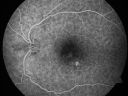
Chronic Central Serous Retinopathy treated with Photodynamic Laser and Visudyne653 views48-year-old man has had waxing and waning vision in the left eye for the last year from central serous retinopathy. OD is 20/16, OS is 20/30.
(1 votes)
|
|

Chronic Central Serous Retinopathy treated with Photodynamic Laser and Visudyne723 views48-year-old man has had waxing and waning vision in the left eye for the last year from central serous retinopathy. OD is 20/16, OS is 20/30.
(1 votes)
|
|

Acute Central Serous Chorioretinopathy with Symptoms in Hazy Vision Both Eyes722 views48-year-old man with hazy vision in the right eye for about three days. He has a little more of a headache than usual. He is under a fair amount of stress. He does not take any steroids that he knows of. He does use a nasal spray, which he is not sure if that is a steroid spray or not. He is going to call us with that medicine. I checked his blood pressure today is was 115/95.
VISUAL ACUITY: OD 20/15, OS 20/25
(1 votes)
|
|

Chronic Pseudophakic Cystoid Macular Edema in a Patient with Diabetes757 views74-year-old woman has chronic cystoid macular edema in the left eye. She has had vision loss intermittently in that eye for 2 years, the right eye, unfortunately, lost vision from a central retinal vein occlusion in 6 years ago.
(1 votes)
|
|

Group 2a Juxtafoveal Retinal Telangiectasis (type 2)683 views58-year-old man decreased vision right eye and not better since cataract surgery one year ago. OD 20/40, OS 20/40.
(1 votes)
|
|

Macular Pucker in eye following giant retinal tear RD repair - photo of tear765 views 42-year old man has noticed his vision is changing now that the bubble is gone, but he is starting to notice distortion of central vision. He had a giant retinal tear retinal detachment repair emergently done on November 8, 2007, and subsequent laser on November 15th.
VISUAL ACUITY: Vision OD 20/400.
6 weeks post-op VA 20/40
(1 votes)
|
|

Central Serous Retinopathy835 viewsRed Free photo
(1 votes)
|
|
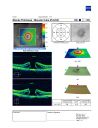
Large mac hole OCT2257 views76 yof with large mac hole
VA: CF at 3ft
chance of spontaneous closure low; surgery option available
(3 votes)
|
|

Macular Pucker - Diabetic - Severe non-foveal Edema OS848 views
(1 votes)
|
|

Idiopathic Central Serous Retinopathy - Bilateral - Acute 683 views49-year-old man decreasing vision in the left eye for one week. He is not taking any steroid or steroid-related products. His blood pressure as far as he knows is under control.
VISUAL ACUITY: OD 20/30, OS 20/400
(1 votes)
|
|

Group 2a Juxtafoveal Retinal Telangiectasis (type 2)634 views58-year-old man decreased vision right eye and not better since cataract surgery one year ago. OD 20/40, OS 20/40.
(1 votes)
|
|

Idiopathic Cystoid Macular Edema - CME - Responded to Subtenons Kenalog - Color Photo Showing Foveal Cysts1038 views49-year-old man had a flu two weeks ago the right eye has been throbbing and his vision in the right eye has been foggy. OD 20/80, OS 20/40. IOP: 14 OU.
(1 votes)
|
|

Juxtafoveal Telangiectasis - MacTel - Retinal Telangiectasis - Both Eyes - Crystals1272 views61-year-old man has idiopathic juxtafoveal retinal telangiectasis type II in both eyes OD is 20/40, OS is 20/25
(1 votes)
|
|
|
|
|
|


