|
|
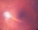
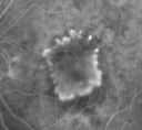
Retina Gallery is a resource for teaching and learning through exchange of interesting retina cases and the sharing of high quality images of retinal diseases. You can download images from the site without registering.
|
| Retina Gallery has been live since October 20, 2010. As an independent site, RetinaGallery.com is and will always be free, and this site is and will always be open to ALL retina physicians and allied personnel. MORE |
| Category |
|
|
 | ChoroidChoroidal Folds, Sclerochoroidal Calcification, Choroidal Atrophy |
|
|
 | Congenital AnomaliesPhakomatoses, Hamartoma, Coloboma, Myelinated Nerve Fiber Layer, Persistent Fetal Vasculature (PHPV) |
|
|
 | DystrophiesRetinal - Macular - RPE - Choroidal: Best's Disease, Retinitis Pigmentosa, Stargart's Macular Dystrophy, X-linked retinoschisis |
|
|
 | Iatrogenic DiseasesComplications of surgery - anterior segment (lens fragments, dislocated IOL, choroidal hemorrhage) or posterior segment (retinal injury, vitrectomy or scleral buckle complications). Also complications of laser surgery. Side effects of ocular or systemic medications. |
|
|
|
|  |  | Macular Degeneration - Age Related, Myopic, Choroidal Neovascular MembranesAge-Related Macular Degeneration (AMD) - Dry, Wet, Atrophic, Drusen ..., Angioid Myopic Macular Degeneration, Pattern Dystrophy, Other CNVM (Angioid Streaks, Idiopathic, Optic Nerve Drusen) |
|
|
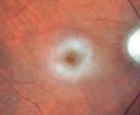
| |  | Macular Diseases - Acquiredcentral serous retinopathy, cystoid macular edema, macular hole, macular pucker, juxtafoveal telangiectasis (MacTel), toxic retinopathy |
|
|
|
|
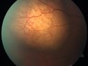
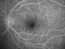 | Normal ExamplesPhotos, Fluorescein Angiograms, OCT's, Ultrasounds - Normal Examples |
|
|
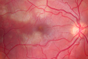
 | Optic Nerve Diseaseanterior ischemic optic neuropathy, drusen, myopic nerves, glaucoma, optic atrophy, papilledema, trauma, etc |
|
|
|
|
|
| |  | Infectious UveitisToxoplasmosis, Toxocariasis, Cytomegalovirus Retinitis, Acute Retinal Necrosis, Progressive Outer Retinal Necrosis, Diffuse Unilateral Subacute Neuroretinitis (nematode), Fundgal Endophthalmitis, Ocular Histoplasmosis Syndrome |
|
|
|
|
| |  | Paraneoplastic / Auto-immune ChorioretinopathyAuto-immune retinopathy, Bilateral diffuse melanocytic proliferation, Cancer associated retinopathy, Melanoma associated retinopathy, acute exudative polymorphous paraneoplastic retinopathy |
|
|
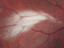
 | Retinal DetachmentRhegmatogenous Retinal Detachment, Retinopathy of Prematurity, Complex Retinal Detachment with Proliferative Vitreoretinopathy, Retinal Tear, Lattice Degeneration, and Retinoschisis |
|
|
|
| |  | Diabetic RetinopathyAll manifestations of diabetic retinopathy, proliferative, background, macular edema, non-perfusion, ischemia, hemorrhage, retinal detachment, surgical pre and post-op images. |
|
|
| |  | MacroaneurysmArterial Macroaneurysm, Venous Macroaneurysm, Coats Disease |
|
|
|
|
|
|
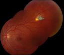
| Tumors, Masses, Pigmented LesionsAll eye tumors, choroidal, retinal, iris ... Melanoma, Retinoblastoma, Hemangioma, Osteoma, Nevus, Sclerochoroidal calcification
|
|
|
|
|
|
|
|
 | TraumaChoroidal Rupture, Sclopteria Chorioretinitis, Etc. |
|
|
 | VitreousVitreous hemorrhage, asteroid hyalosis, other vitreous disease |
|
|
| Retina Gallery User GalleryThis category contains albums that belong to Retina Gallery users who do not include their albums in the other specified categories on the website. |
|
|
 | Best Images - 2012 - 2014These are excellent images from 2012 - 2014 during which there was a monthly best image award contest on this website. |
|
|
| 17,980 files in 1,421 albums and 50 categories with 72 comments viewed 9,308,815 times |