Bilateral Diffuse Uveal Melanocytic Proliferation
|
|
80-year-old man was seen in the office on May 11, 2009. He has had cataract surgery done in December of 2008 at an eye doctors office and then he had several intravitreal injections by another retina specialist. He feels his vision is still hazy in both eyes and comes in because of that.
Although he did not initially volunteer history of cancer, he does have a history of recently being evaluated for possible carcinoma. He has been and still is a smoker.
His weight has decreased over the last year from about 170 pounds to about 160 pounds.
He was evaluated for pelvic lymphadenopathy and a biopsy was inconclusive and then the pelvic lymphadenopathy spontaneously cleared. He has had a PET scan in the past and there have been some possible lesions in his lungs, but a referral to a lung specialist was unrewarding and that the doctor felt there was nothing there that could definitely be biopsied. He feels well, but is concerned about his poor vision.
VISUAL ACUITY: OD 20/80, OS 20/80. IOP: OD 9, OS 6.
SLIT EXAMINATION: There is a posterior chamber intraocular lens is in good position in both eyes. There is a growth on the superior margin on the right upper lid.
EXTENDED OPHTHALMOSCOPY:
OD: Vertical C/D ratio is 0.3. There is diffuse inner regular brown hyperpigmented spots around the macula.
OS: Vertical C/D ratio is 0.3. There is posterior vitreous separation. There is diffuse brown hyperpigmented spots around the macula and there was one well-formed lesion just inferior to the fovea a disc and a half diameter across, which is elevated.
OCT SCAN: The OCT scan shows irregular elevation of the pigment epithelium in both eyes with no intraretinal or subretinal fluid. Photos confirm clinical findings.
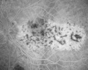
FLUORESCEIN ANGIOGRAPHY: Fluorescein angiography shows stippled areas of hyper and hypofluorescence in the macula of each eye.
ULTRASOUND: Ultrasound shows diffuse mild choroidal thickening in both eyes. The left eye does show elevation to about a half a millimeter in the area where there is a brown choroidal lesion.
IMPRESSION:
1. BILATERAL DIFFUSE UVEAL MELANOCYTIC PROLIFERATION
DISCUSSION: I explained to the patient and I spoke with his oncologist as well,
that he does have what looks to be paraneoplastic syndrome. This sometimes can be accompanied by cataracts and his rapidly advancing cataracts combined with the pigmentation in both eyes are consistent with the diagnosis. I asked him to return for a check in six weeks, unfortunately there is nothing at this point that I know of that can be done to help improve his vision. I asked him to see you back regularly and hopefully if cancer is ultimately diagnosed in him, the appropriate treatment can be pursued, which may help his symptoms systemically and may also alleviate some of his eye problems.
|

Bilateral Diffuse Uveal Melanocytic Proliferation - BDUMP - Paraneoplastic Syndrome1184 views80-year-old man vision loss for one year. He died about one year after these photos from Metastatic Poorly Differentiated Large Cell Carcinoma of unknown primary. He was a smoker.     
(0 votes)
|
|

Bilateral Diffuse Uveal Melanocytic Proliferation - BDUMP - Paraneoplastic Syndrome1097 views80-year-old man vision loss for one year. He died about one year after these photos from Metastatic Poorly Differentiated Large Cell Carcinoma of unknown primary. He was a smoker.     
(0 votes)
|
|

Bilateral Diffuse Uveal Melanocytic Proliferation - BDUMP - Paraneoplastic Syndrome1075 views80-year-old man vision loss for one year. He died about one year after these photos from Metastatic Poorly Differentiated Large Cell Carcinoma of unknown primary. He was a smoker.     
(1 votes)
|
|

Bilateral Diffuse Uveal Melanocytic Proliferation - BDUMP - Paraneoplastic Syndrome890 views80-year-old man vision loss for one year. He died about one year after these photos from Metastatic Poorly Differentiated Large Cell Carcinoma of unknown primary. He was a smoker.     
(0 votes)
|
|

Bilateral Diffuse Uveal Melanocytic Proliferation - BDUMP - Paraneoplastic Syndrome1778 views80-year-old man vision loss for one year. He died about one year after these photos from Metastatic Poorly Differentiated Large Cell Carcinoma of unknown primary. He was a smoker.     
(0 votes)
|
|

Bilateral Diffuse Uveal Melanocytic Proliferation - BDUMP - Paraneoplastic Syndrome713 views80-year-old man vision loss for one year. He died about one year after these photos from Metastatic Poorly Differentiated Large Cell Carcinoma of unknown primary. He was a smoker.     
(0 votes)
|
|

Bilateral Diffuse Uveal Melanocytic Proliferation - BDUMP - Paraneoplastic Syndrome799 views80-year-old man vision loss for one year. He died about one year after these photos from Metastatic Poorly Differentiated Large Cell Carcinoma of unknown primary. He was a smoker.     
(0 votes)
|
|

Bilateral Diffuse Uveal Melanocytic Proliferation - BDUMP - Paraneoplastic Syndrome857 views80-year-old man vision loss for one year. He died about one year after these photos from Metastatic Poorly Differentiated Large Cell Carcinoma of unknown primary. He was a smoker.     
(0 votes)
|
|

Bilateral Diffuse Uveal Melanocytic Proliferation - BDUMP - Paraneoplastic Syndrome731 views80-year-old man vision loss for one year. He died about one year after these photos from Metastatic Poorly Differentiated Large Cell Carcinoma of unknown primary. He was a smoker.     
(0 votes)
|
|

Bilateral Diffuse Uveal Melanocytic Proliferation - BDUMP - Paraneoplastic Syndrome915 views80-year-old man vision loss for one year. He died about one year after these photos from Metastatic Poorly Differentiated Large Cell Carcinoma of unknown primary. He was a smoker.     
(2 votes)
|
|

Bilateral Diffuse Uveal Melanocytic Proliferation - BDUMP - Paraneoplastic Syndrome765 views80-year-old man vision loss for one year. He died about one year after these photos from Metastatic Poorly Differentiated Large Cell Carcinoma of unknown primary. He was a smoker.     
(0 votes)
|
|

Bilateral Diffuse Uveal Melanocytic Proliferation - BDUMP - Paraneoplastic Syndrome1034 views80-year-old man vision loss for one year. He died about one year after these photos from Metastatic Poorly Differentiated Large Cell Carcinoma of unknown primary. He was a smoker.     
(0 votes)
|
|

Bilateral Diffuse Uveal Melanocytic Proliferation - BDUMP - Paraneoplastic Syndrome882 views80-year-old man vision loss for one year. He died about one year after these photos from Metastatic Poorly Differentiated Large Cell Carcinoma of unknown primary. He was a smoker.     
(0 votes)
|
|

Bilateral Diffuse Uveal Melanocytic Proliferation - BDUMP - Paraneoplastic Syndrome639 views80-year-old man vision loss for one year. He died about one year after these photos from Metastatic Poorly Differentiated Large Cell Carcinoma of unknown primary. He was a smoker.     
(0 votes)
|
|

Bilateral Diffuse Uveal Melanocytic Proliferation - BDUMP - Paraneoplastic Syndrome711 views80-year-old man vision loss for one year. He died about one year after these photos from Metastatic Poorly Differentiated Large Cell Carcinoma of unknown primary. He was a smoker.     
(0 votes)
|
|

Bilateral Diffuse Uveal Melanocytic Proliferation - BDUMP - Paraneoplastic Syndrome649 views80-year-old man vision loss for one year. He died about one year after these photos from Metastatic Poorly Differentiated Large Cell Carcinoma of unknown primary. He was a smoker.     
(0 votes)
|
|

Bilateral Diffuse Uveal Melanocytic Proliferation - BDUMP - Paraneoplastic Syndrome600 views80-year-old man vision loss for one year. He died about one year after these photos from Metastatic Poorly Differentiated Large Cell Carcinoma of unknown primary. He was a smoker.     
(0 votes)
|
|

Bilateral Diffuse Uveal Melanocytic Proliferation - BDUMP - Paraneoplastic Syndrome688 views80-year-old man vision loss for one year. He died about one year after these photos from Metastatic Poorly Differentiated Large Cell Carcinoma of unknown primary. He was a smoker.     
(0 votes)
|
|

Bilateral Diffuse Uveal Melanocytic Proliferation - BDUMP - Paraneoplastic Syndrome743 views80-year-old man vision loss for one year. He died about one year after these photos from Metastatic Poorly Differentiated Large Cell Carcinoma of unknown primary. He was a smoker.     
(0 votes)
|
|

Bilateral Diffuse Uveal Melanocytic Proliferation - BDUMP - Paraneoplastic Syndrome730 views80-year-old man vision loss for one year. He died about one year after these photos from Metastatic Poorly Differentiated Large Cell Carcinoma of unknown primary. He was a smoker.     
(0 votes)
|
|

Bilateral Diffuse Uveal Melanocytic Proliferation - BDUMP - Paraneoplastic Syndrome873 views80-year-old man vision loss for one year. He died about one year after these photos from Metastatic Poorly Differentiated Large Cell Carcinoma of unknown primary. He was a smoker.     
(0 votes)
|
|
|
|
|
|
|
80-year-old man was seen in the office on May 11, 2009. He has had cataract surgery done in December of 2008 at an eye doctors office and then he had several intravitreal injections by another retina specialist. He feels his vision is still hazy in both eyes and comes in because of that.
Although he did not initially volunteer history of cancer, he does have a history of recently being evaluated for possible carcinoma. He has been and still is a smoker.
His weight has decreased over the last year from about 170 pounds to about 160 pounds.
He was evaluated for pelvic lymphadenopathy and a biopsy was inconclusive and then the pelvic lymphadenopathy spontaneously cleared. He has had a PET scan in the past and there have been some possible lesions in his lungs, but a referral to a lung specialist was unrewarding and that the doctor felt there was nothing there that could definitely be biopsied. He feels well, but is concerned about his poor vision.
VISUAL ACUITY: OD 20/80, OS 20/80. IOP: OD 9, OS 6.
SLIT EXAMINATION: There is a posterior chamber intraocular lens is in good position in both eyes. There is a growth on the superior margin on the right upper lid.
EXTENDED OPHTHALMOSCOPY:
OD: Vertical C/D ratio is 0.3. There is diffuse inner regular brown hyperpigmented spots around the macula.
OS: Vertical C/D ratio is 0.3. There is posterior vitreous separation. There is diffuse brown hyperpigmented spots around the macula and there was one well-formed lesion just inferior to the fovea a disc and a half diameter across, which is elevated.
OCT SCAN: The OCT scan shows irregular elevation of the pigment epithelium in both eyes with no intraretinal or subretinal fluid. Photos confirm clinical findings.
FLUORESCEIN ANGIOGRAPHY: Fluorescein angiography shows stippled areas of hyper and hypofluorescence in the macula of each eye.
ULTRASOUND: Ultrasound shows diffuse mild choroidal thickening in both eyes. The left eye does show elevation to about a half a millimeter in the area where there is a brown choroidal lesion.
IMPRESSION:
1. BILATERAL DIFFUSE UVEAL MELANOCYTIC PROLIFERATION
DISCUSSION: I explained to the patient and I spoke with his oncologist as well,
that he does have what looks to be paraneoplastic syndrome. This sometimes can be accompanied by cataracts and his rapidly advancing cataracts combined with the pigmentation in both eyes are consistent with the diagnosis. I asked him to return for a check in six weeks, unfortunately there is nothing at this point that I know of that can be done to help improve his vision. I asked him to see you back regularly and hopefully if cancer is ultimately diagnosed in him, the appropriate treatment can be pursued, which may help his symptoms systemically and may also alleviate some of his eye problems.