|
|

Leucoma vascolarizzato577 views67 female failed PKP outcome11/14/14 at 08:18jessiecooper: nice photo!
|
|

393 views10/22/14 at 10:35jessiecooper: Looks like Telangiectasia,
|
|

Chorodial Melanoma1002 views80 year old male came in for retinal evaluation and presented with a melanoma.07/25/14 at 08:21bill: excellent images rendering a very realistic 3D ima...
|
|

Subhyloid Hem with PDR1151 views11/14/13 at 10:51photoscherf: nice job
|
|

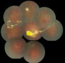
Coats' Disease with Cryotherapy 1065 viewsYoung male with Coat's Disease with Cryotherapy in the right eye. VA was 20/20, right eye. Left eye was completely normal. Patient will be followed up in 3-months.10/01/13 at 04:42CGUIER: Awesome montage
|
|

OU Stereo Pair Optic Disc Drusen1451 viewsText book example of OU optic discs with drusen in stereo.05/07/13 at 11:57photoscherf: 
|
|

RD with PVR1175 viewsRD with Proliferative Vitreoretinopathy 05/03/13 at 08:01kseal: Would be nice to see in stereo
|
|

OD Albinism with foveal hypoplasia1786 views28 year old female latina with vision of 20/40004/27/13 at 18:26calhoun16: Congrats
|
|

Optic Nerve Pit1037 viewsOptic Nerve Pit04/24/13 at 07:47photoscherf: 
|
|

Rubeosis Iridis1033 viewsPatient presents with Rubeosis Iridis in the right eye due to neovascular glaucoma. VA is 20/40 in the right eye. Will follow up in 3-months.04/09/13 at 08:34CGUIER: Yuck!
|
|

OD Albinism with foveal hypoplasia1786 views28 year old female latina with vision of 20/40003/05/13 at 13:03KClark: Great photo!
|
|

Macular Degeneration with Hemorrhage913 viewsPatient comes in for follow up exam for wet AMD. Fundus photography reveals retinal hemorrhage temporally to the macula. Patient was not treated. Wait to see if the blood will absorb on its own. 01/08/13 at 08:57photoscherf: 
|
|

Dislocated Intraocular Lens957 viewsDislocated Intraocular Lens 01/08/13 at 07:00photoscherf: 
|
|

Macular fold after PPV for RD2226 viewsA 53-year-old man with a macula-off RD underwent left eye pars plana vitrectomy with air-fluid exchange, laser retinopexy, and injection of 14% SF6 gas. There were 4 retinal breaks between 10 o’clock and 12 o’clock including one large tear. He was compliant with facedown positioning. On the 8th post-operative day, a retinal fold along the 4-10 o’clock meridian was seen coursing through his central macula. He underwent repeat PPV but multiple attempts to lift and flatten the retina were unsuccessful.11/15/12 at 12:18calhoun16: Awesome Photo, you deserve to win. Great Job.
|
|

Late414 viewsCorresponding FA for Cilioretinal artery occlusion with early venous stasis retinopathy 11/08/12 at 21:53tsaunders: hot picture, hot nerve, hot resident?
|
|

coats disease; exudative retinitis; retinal telangiectasis; leber multiple miliary aneurysm disease1484 viewsclassic coats11/06/12 at 20:11golenj: Simply excellent 
|
|

Macular fold after PPV for RD2226 viewsA 53-year-old man with a macula-off RD underwent left eye pars plana vitrectomy with air-fluid exchange, laser retinopexy, and injection of 14% SF6 gas. There were 4 retinal breaks between 10 o’clock and 12 o’clock including one large tear. He was compliant with facedown positioning. On the 8th post-operative day, a retinal fold along the 4-10 o’clock meridian was seen coursing through his central macula. He underwent repeat PPV but multiple attempts to lift and flatten the retina were unsuccessful.09/24/12 at 20:55sleva079: Great picture.
|
|

Macular fold after PPV for RD2226 viewsA 53-year-old man with a macula-off RD underwent left eye pars plana vitrectomy with air-fluid exchange, laser retinopexy, and injection of 14% SF6 gas. There were 4 retinal breaks between 10 o’clock and 12 o’clock including one large tear. He was compliant with facedown positioning. On the 8th post-operative day, a retinal fold along the 4-10 o’clock meridian was seen coursing through his central macula. He underwent repeat PPV but multiple attempts to lift and flatten the retina were unsuccessful.09/24/12 at 17:27Schendel: Exquisite image.
|
|

Melanoma1156 viewsYoung male with large Melanoma08/27/12 at 14:18sfoxman: great photo
|
|

Macular fold after PPV for RD2226 viewsA 53-year-old man with a macula-off RD underwent left eye pars plana vitrectomy with air-fluid exchange, laser retinopexy, and injection of 14% SF6 gas. There were 4 retinal breaks between 10 o’clock and 12 o’clock including one large tear. He was compliant with facedown positioning. On the 8th post-operative day, a retinal fold along the 4-10 o’clock meridian was seen coursing through his central macula. He underwent repeat PPV but multiple attempts to lift and flatten the retina were unsuccessful.08/27/12 at 08:58scohen125: Beautiful image useful for complications of retina...
|
|

58 year old man with a Subhyaloid Hemorrhage732 views08/27/12 at 08:52KClark: Great photo!
|
|

Melanoma1156 viewsYoung male with large Melanoma08/06/12 at 06:08bfoxman: Classic!
|
|

Melanoma1156 viewsYoung male with large Melanoma08/06/12 at 06:01nmahan: Very cool
|
|
|

Three years after Injury - Optic Atrophy - Vision is 1/200 in each eye913 views07/17/12 at 11:46scohen125: Used in: Teaching video titled 'Basic Ophthal...
|
|

Clinically Significant Diabetic Macular Edema both Eyes with Exudates1229 views56-year-old woman with diabetes for twelve years. She has noticed blurring of the vision in the right eye for about six months. OD is 20/70, OS is 20/25. 07/17/12 at 11:45scohen125: Used in: Teaching video titled 'Basic Ophthal...
|
|

PDR OS with NVE979 views57-year-old man has diabetic retinopathy in both eyes.
Diabetic for 14 years with HgB A1C often over 10.
VISUAL ACUITY: OD 20/30, OS 20/40. PDR OS BDR OD07/17/12 at 11:38scohen125: Used in: Teaching video titled 'Basic Ophthal...
|
|

Optic disc swelling1754 views56 year old woman07/17/12 at 11:38scohen125: Used in: Teaching video titled 'Basic Ophthal...
|
|

Occult Maculopathy - Thin Fovea on OCT and Normal Color VA, Photos, FA VA 20/80 OU734 views45-year-old man Normal FA05/23/12 at 02:32Pablo Carnota: What about an electrophysiological testing? In my ...
|
|

Melanoma1253 views40 Year old male05/22/12 at 11:53nmahan: very cool! 
|
|

CAVERNOUS HEMONGIOMA2287 views28 year old male w/ 20/200 vision at time of exam. Patient c/o poor vision since childhood. No significant medical history or family medical history. A problem was only noted when patient enlisted in the Army.05/22/12 at 11:52nmahan: nice!
|
|

Central Retinal Artery Occlusion1332 views60 year old male with LP vision due to extensive blood flow loss.05/03/12 at 07:08luebelu: great picture
|
|

CAVERNOUS HEMONGIOMA1731 views28 year old male. FA05/03/12 at 07:05luebelu: very sharp details
|
|

CAVERNOUS HEMONGIOMA2287 views28 year old male w/ 20/200 vision at time of exam. Patient c/o poor vision since childhood. No significant medical history or family medical history. A problem was only noted when patient enlisted in the Army.05/03/12 at 07:05luebelu: Great pic with good detail
|
|

Combined hamartoma of the retina and RPE2346 views16 year old female, diagnosed with a combined hamartoma of the retina and the retinal pigment epithelium.04/20/12 at 14:34emurphy: 
|
|

Wyburn-Mason Syndrome 1539 views28 year old female, diagnosed with Wyburn-Mason Syndrome at age 7. At time of exam, vision in the left eye was 4/200.04/20/12 at 12:07mblansing: fabulous photo!
|
|

Serous Choroidal Effusions2039 viewsSerous choroidal effusions following anterior segment surgery. Final visual outcome was 20/20. 04/19/12 at 16:26J.H: Great images. Better quality than a optoscan even....
|
|

Inferotemporal and Inferonasal BRAO1260 viewsBranch Retinal Artery Occlusions - Multiple04/17/12 at 06:37James L. Perron: James Perron
|
|
|

Total Retinal Detachment1220 viewsTotal Retinal Detachment04/11/12 at 11:33nmahan: very nice pic!
|
|

Wyburn Mason Syndrome3025 views28 year old female, visual acuity OS 20/20003/24/12 at 14:36vitrector@gmail.com: Awesome
|
|

Wyburn Mason Syndrome3025 views28 year old female, visual acuity OS 20/20003/03/12 at 20:00bfoxman: Cool!
|
|

Wyburn Mason Syndrome3025 views28 year old female, visual acuity OS 20/20002/28/12 at 19:45G.bronner: Great case
|
|

Wyburn Mason Syndrome3025 views28 year old female, visual acuity OS 20/20002/28/12 at 12:45: this is so cool.
|
|

Aplastic anemia1441 views29 y.o.m. with aplastic anemia OD VA 20/25. 02/27/12 at 12:21scohen125: Can you please note the patient's hemoglobin ...
|
|

Occult Metallic Intraocular Foreign Body - WINNER JANUARY 2011 - BEST IMAGE2913 viewsFundus photograph of a 28 year old male who presented with intermittent, painless, blurred vision of his left eye secondary to an occult metallic intraocular foreign body with old vitreous hemorrhage. He later admitted to feeling a mild gritty sensation of his affected eye while working with a circular saw the previous year. He was then misdiagnosed as having a subconjunctival hemorrhage from the emergency physician. He later underwent successful pars plana vitrectomy and foreign body removal.02/21/12 at 20:48joffeleo: good images. OCT case is not worthy of special men...
|
|

Wyburn Mason Syndrome3025 views28 year old female, visual acuity OS 20/20002/21/12 at 07:25: OMG the blood vessel looks like a eartworm!
|
|

Wyburn Mason Syndrome3025 views28 year old female, visual acuity OS 20/20002/21/12 at 07:21: Very cool
|
|

Wyburn Mason Syndrome3025 views28 year old female, visual acuity OS 20/20002/21/12 at 07:15: I hope this one wins
|
|

Wyburn Mason Syndrome3025 views28 year old female, visual acuity OS 20/20002/21/12 at 07:15: This is alesome.... 
|
|

Wyburn Mason Syndrome3025 views28 year old female, visual acuity OS 20/20002/21/12 at 07:04luebelu: Great Pic
|
|

Occult Metallic Intraocular Foreign Body - WINNER JANUARY 2011 - BEST IMAGE2913 viewsFundus photograph of a 28 year old male who presented with intermittent, painless, blurred vision of his left eye secondary to an occult metallic intraocular foreign body with old vitreous hemorrhage. He later admitted to feeling a mild gritty sensation of his affected eye while working with a circular saw the previous year. He was then misdiagnosed as having a subconjunctival hemorrhage from the emergency physician. He later underwent successful pars plana vitrectomy and foreign body removal.02/20/12 at 07:40Sac0067: Many images were noteworthy for their interesting ...
|
|

Wyburn Mason Syndrome3025 views28 year old female, visual acuity OS 20/20002/19/12 at 23:04cbtsai: Rare case, nice pic
|
|

Large mac hole OCT1747 views76 yof with large mac hole
VA: CF at 3ft
chance of spontaneous closure low; surgery option available02/19/12 at 09:56Barbara Parolini: NOT INTERESTING BECAUSE TOO COMMON
|
|

Wyburn Mason Syndrome3025 views28 year old female, visual acuity OS 20/20002/19/12 at 09:52Barbara Parolini: RARE CONDITION, WELL TAKEN
|
|

Acute Macula Neuroretinopathy2111 views39yr old male: Presents with Inferior Temporal Scotoma in his left eye, x 10 days with no change in shape or size, Visual acuity 20/25.
Most common sysptoms are described as sudden onset of one or more paracentral scotomas. {with the tip pointing toward the Fovea} without any other visual symptoms. Currently no treatment recommended.02/19/12 at 09:52Barbara Parolini: WELL TAKEN, GOOD CHOICE OF IMAGING, RARE CONDITION
|
|

Red Free Hemangioma left eye1429 views02/19/12 at 09:50Barbara Parolini: VERY SHARP WITH 3D EFFECT
|
|

Optic Nerve Tumor3213 views21 year old male: Presents with a history of Von Hippel-Lindau Syndrome, diagnosed in 2005 affecting his left Optic Nerve, Brain, Spine, Kidney and Pancreas. He has undergone Laser for Retinal Hemangioma measuring 2/3-4/5DD -vs_ 1/3DD when originally diagnosed. However his visual acuity remains good 20/25. Patient has also undergone Neurosurgery in 2005 and Spinal cord in 2006. Recent MRI of spinal cord hemangioma showed stable tumors. 02/19/12 at 09:48Barbara Parolini: INTRESTING SUBJECT AND WELL TAKEN PICTURE
|
|

Proliferative Diabetic Retinopathy with Macular Hole2537 viewsPatitent with diabetes diagnosed 12 years ago and low visual acuity. 02/19/12 at 09:46Barbara Parolini: well focused with 3d effect. less interesting subj...
|
|

Acute Macula Neuroretinopathy2111 views39yr old male: Presents with Inferior Temporal Scotoma in his left eye, x 10 days with no change in shape or size, Visual acuity 20/25.
Most common sysptoms are described as sudden onset of one or more paracentral scotomas. {with the tip pointing toward the Fovea} without any other visual symptoms. Currently no treatment recommended.01/31/12 at 14:15tomsteele: very rare
|
|

wyburn-mason sydrome1067 views28 year old female, visual acuity OS 20/20001/10/12 at 13:48luebelu: Lucinda Little, CCRC
|
|

Wyburn Mason Syndrome3025 views28 year old female, visual acuity OS 20/20001/10/12 at 13:47luebelu: Lucinda Little, CCRC Great Pic
|
|

Acute Macula Neuroretinopathy2111 views39yr old male: Presents with Inferior Temporal Scotoma in his left eye, x 10 days with no change in shape or size, Visual acuity 20/25.
Most common sysptoms are described as sudden onset of one or more paracentral scotomas. {with the tip pointing toward the Fovea} without any other visual symptoms. Currently no treatment recommended.01/06/12 at 12:27Char Harris: well done
|
|

Red Free Hemangioma left eye1429 views01/05/12 at 12:11tomsteele: Nice Image
|
|

Retinal Arterial Macroaneurysm - Recurrent Hemorrhage 6 months post laser1368 views6 months post laser: Her vision had improved, but then three weeks ago it worsened again. She is not on any blood thinners. She wasn’t doing any heavy lifting or straining.
VISUAL ACUITY: OD 3/20012/06/11 at 11:45ngpmedha: Would Anti-VEGF help
|
|

Retinal Pigment Epithelial Dysgenesis1545 views12/06/11 at 11:34ngpmedha: Was Vitrectomy cosidered to relieve foveal straie
|
|

706 views12/06/11 at 11:29ngpmedha: Is it trypan blue stain
|
|

Myopic CNVM - Wet - rx Avastin for 1 year717 views55-year-old woman OD is 20/40, OS is 20/20. There is a hyperpigmented disc diameter choroidal neovascular membrane with fluid touching the fovea.12/06/11 at 11:23ngpmedha: CNVM appears juxtafoveal,feel laser can be conside...
|
|

CNV secondary to Angioid Streaks, PXE798 views12/04/11 at 16:58scohen125: Beautiful photo - was this eye treated?
|
|

Retinal Pigment Epithelial Dysgenesis875 views10/09/11 at 11:18rlewis: Why is this not an eccentric disciform? It is unl...
|
|

Retinal Pigment Epithelial Dysgenesis1545 views09/25/11 at 18:10scohen: This looks like a combined hamartoma of the retina...
|
|

Low Tension Glaucoma1086 views59-year-old woman has a history of glaucoma dating back to 1990. She had trabeculectomy in the left eye in 1998 and then persisted to lose vision despite normal intraocular pressures from low-tension glaucoma in the left eye. She is now on Cosopt and Travatan in both eyes.
Vision OD is 20/20, OS is 20/16. IOP: OD 9, OS 6.
09/24/11 at 18:17scohen: Amazingly low intraocular pressure - has this pati...
|
|
|
|