Retina Gallery ~ Full Sized Retina Images

Library of Free, Non-Copyrighted Retina Images and Videos

Wähle Sprache:
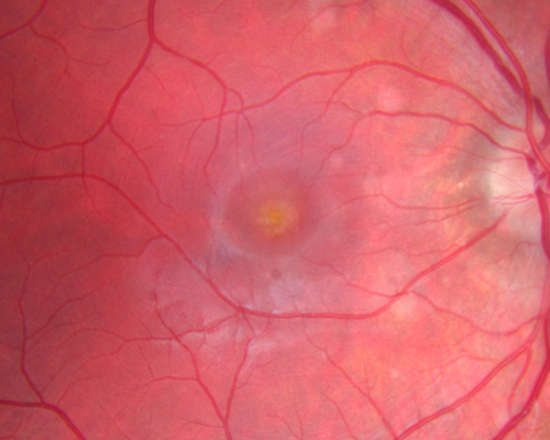
Multiple Evanescent White Dot Syndrome - Atypical Yellow Foveal Spot Right Eye
12-year-old had decreased vision in the left eye from reported amblyopia since around the year 2000.  His right eye was doing fine until  two days ago, when he had sudden severe vision loss.  He sees a blurred spot in the middle of the vision in the right eye wherever he looks. 
 OD 20/60 with eccentric movement, OS 20/200.   Pinhole 20/70.  IOP:  OD 14, OS 10.
Schlüsselwörter: mewds;multiple evanescent white dot syndrome;yellow spot;vitelliform

Multiple Evanescent White Dot Syndrome - Atypical Yellow Foveal Spot Right Eye

12-year-old had decreased vision in the left eye from reported amblyopia since around the year 2000. His right eye was doing fine until two days ago, when he had sudden severe vision loss. He sees a blurred spot in the middle of the vision in the right eye wherever he looks.
OD 20/60 with eccentric movement, OS 20/200. Pinhole 20/70. IOP: OD 14, OS 10.

plaque.png maculaod.png 49914_001.JPG 26640_001.JPG htnretinopathy_gadi_032013_08.jpg