Image search results - " BRVO"
|

Old Branch Retinal Vein Occlusion- Severe CME - Pucker - Diabetic Fundus Photo767 views61-year old man has diabetic retinopathy in the right eye. He also had a macular pucker and macular edema. I did a vitrectomy on September 5th. His vision was initially improving. His vision in the right eye seems to be a little more foggy.
VISUAL ACUITY: OD: 20/200
|
|

Old Branch Retinal Vein Occlusion- Severe CME - Pucker - Diabetic Map OCT720 views61-year old man has diabetic retinopathy in the right eye. He also had a macular pucker and macular edema. I did a vitrectomy on September 5th. His vision was initially improving. His vision in the right eye seems to be a little more foggy.
VISUAL ACUITY: OD: 20/200
|
|
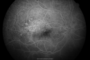
Old Branch Retinal Vein Occlusion- Severe CME - Pucker - Diabetic Early FA504 views61-year old man has diabetic retinopathy in the right eye. He also had a macular pucker and macular edema. I did a vitrectomy on September 5th. His vision was initially improving. His vision in the right eye seems to be a little more foggy.
VISUAL ACUITY: OD: 20/200
|
|

Old Branch Retinal Vein Occlusion- Severe CME - Pucker - Diabetic Early FA515 views61-year old man has diabetic retinopathy in the right eye. He also had a macular pucker and macular edema. I did a vitrectomy on September 5th. His vision was initially improving. His vision in the right eye seems to be a little more foggy.
VISUAL ACUITY: OD: 20/200
|
|

Old Branch Retinal Vein Occlusion- Severe CME - Pucker - Diabetic Late FA473 views61-year old man has diabetic retinopathy in the right eye. He also had a macular pucker and macular edema. I did a vitrectomy on September 5th. His vision was initially improving. His vision in the right eye seems to be a little more foggy.
VISUAL ACUITY: OD: 20/200
|
|

BRVO729 views42 year old female VA 20/25
Branch Retinal Vein Occlusion - Hot Spot on Vein at Occlusion Site
|
|

Cytomeglovirus1256 views25 year old male experiencing blurry vision OS for two days. Onset very rapid. VA 20/40 OS 20/30 OD. After HIV titer test, determined the pt. had HIV and Cytomeglovirus.
|
|

Cytomeglovirus494 views25 year old male experiencing blurry vision OS for two days. Onset very rapid. VA 20/40 OS 20/30 OD. After HIV titer test, determined the pt. had HIV and Cytomeglovirus.
|
|
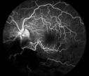
Cytomeglovirus559 views25 year old male experiencing blurry vision OS for two days. Onset very rapid. VA 20/40 OS 20/30 OD. After HIV titer test, determined the pt. had HIV and Cytomeglovirus.
|
|

Cytomeglovirus491 views25 year old male experiencing blurry vision OS for two days. Onset very rapid. VA 20/40 OS 20/30 OD. After HIV titer test, determined the pt. had HIV and Cytomeglovirus.
|
|

Cytomeglovirus435 views25 year old male experiencing blurry vision OS for two days. Onset very rapid. VA 20/40 OS 20/30 OD. After HIV titer test, determined the pt. had HIV and Cytomeglovirus.
|
|

Cytomeglovirus447 views25 year old male experiencing blurry vision OS for two days. Onset very rapid. VA 20/40 OS 20/30 OD. After HIV titer test, determined the pt. had HIV and Cytomeglovirus.
|
|
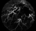
Cytomeglovirus381 views25 year old male experiencing blurry vision OS for two days. Onset very rapid. VA 20/40 OS 20/30 OD. After HIV titer test, determined the pt. had HIV and Cytomeglovirus.
|
|

Cytomeglovirus424 views25 year old male experiencing blurry vision OS for two days. Onset very rapid. VA 20/40 OS 20/30 OD. After HIV titer test, determined the pt. had HIV and Cytomeglovirus.
|
|

Cytomeglovirus402 views25 year old male experiencing blurry vision OS for two days. Onset very rapid. VA 20/40 OS 20/30 OD. After HIV titer test, determined the pt. had HIV and Cytomeglovirus.
|
|

Classic BRVO810 viewsClassic branch retinal vein occlusion
|
|

BRVO393 views50 yr old Female with a BRVO and ME in OS
|
|

Ischemic HRVO357 views63 y/ o African American female with advanced glaucoma and longstanding loss of vision OD presents with acute vision loss OS. Ophthalmic history is significant for uncomplicated phaco/IOL OS 2 months prior to event. Medical history is significant for poorly controlled HTN. Pt characterizes vision loss as blurring centrally OS.
|
|

BRVO with collaterals and cystoid macular edema1012 views66 Year Old Man with has had a branch retinal vein occlusion in the right eye with vision loss for the last few months. OD is 20/40, OS is 20/20. IOP: OD 18, OS 19. There is a superotemporal macular branch retinal vein occlusion with edema, hemorrhage, and exudate.
OCT SCAN: The right eye shows edema with a central foveal thickness of 354 microns FA: shows remodeling of the superotemporal vasculature. There are already venous-venous collaterals, suggesting the vein occlusion may be a little more than a few months old. There are multiple microaneurysms superotemporal to the fovea, and there is leakage in the late frames involving the fovea.
|
|

BRVO with collaterals and cystoid macular edema699 views66 Year Old Man with has had a branch retinal vein occlusion in the right eye with vision loss for the last few months. OD is 20/40, OS is 20/20. IOP: OD 18, OS 19. There is a superotemporal macular branch retinal vein occlusion with edema, hemorrhage, and exudate.
OCT SCAN: The right eye shows edema with a central foveal thickness of 354 microns FA: shows remodeling of the superotemporal vasculature. There are already venous-venous collaterals, suggesting the vein occlusion may be a little more than a few months old. There are multiple microaneurysms superotemporal to the fovea, and there is leakage in the late frames involving the fovea.
|
|

BRVO with collaterals and cystoid macular edema720 views66 Year Old Man with has had a branch retinal vein occlusion in the right eye with vision loss for the last few months. OD is 20/40, OS is 20/20. IOP: OD 18, OS 19. There is a superotemporal macular branch retinal vein occlusion with edema, hemorrhage, and exudate.
OCT SCAN: The right eye shows edema with a central foveal thickness of 354 microns FA: shows remodeling of the superotemporal vasculature. There are already venous-venous collaterals, suggesting the vein occlusion may be a little more than a few months old. There are multiple microaneurysms superotemporal to the fovea, and there is leakage in the late frames involving the fovea.
|
|
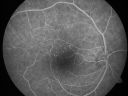
BRVO with collaterals and cystoid macular edema825 views66 Year Old Man with has had a branch retinal vein occlusion in the right eye with vision loss for the last few months. OD is 20/40, OS is 20/20. IOP: OD 18, OS 19. There is a superotemporal macular branch retinal vein occlusion with edema, hemorrhage, and exudate.
OCT SCAN: The right eye shows edema with a central foveal thickness of 354 microns FA: shows remodeling of the superotemporal vasculature. There are already venous-venous collaterals, suggesting the vein occlusion may be a little more than a few months old. There are multiple microaneurysms superotemporal to the fovea, and there is leakage in the late frames involving the fovea.
|
|

BRVO with collaterals and cystoid macular edema651 views66 Year Old Man with has had a branch retinal vein occlusion in the right eye with vision loss for the last few months. OD is 20/40, OS is 20/20. IOP: OD 18, OS 19. There is a superotemporal macular branch retinal vein occlusion with edema, hemorrhage, and exudate.
OCT SCAN: The right eye shows edema with a central foveal thickness of 354 microns FA: shows remodeling of the superotemporal vasculature. There are already venous-venous collaterals, suggesting the vein occlusion may be a little more than a few months old. There are multiple microaneurysms superotemporal to the fovea, and there is leakage in the late frames involving the fovea.
|
|
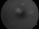
BRVO with collaterals and cystoid macular edema842 views66 Year Old Man with has had a branch retinal vein occlusion in the right eye with vision loss for the last few months. OD is 20/40, OS is 20/20. IOP: OD 18, OS 19. There is a superotemporal macular branch retinal vein occlusion with edema, hemorrhage, and exudate.
OCT SCAN: The right eye shows edema with a central foveal thickness of 354 microns FA: shows remodeling of the superotemporal vasculature. There are already venous-venous collaterals, suggesting the vein occlusion may be a little more than a few months old. There are multiple microaneurysms superotemporal to the fovea, and there is leakage in the late frames involving the fovea.
|
|

BRVO with collaterals and cystoid macular edema594 views66 Year Old Man with has had a branch retinal vein occlusion in the right eye with vision loss for the last few months. OD is 20/40, OS is 20/20. IOP: OD 18, OS 19. There is a superotemporal macular branch retinal vein occlusion with edema, hemorrhage, and exudate.
OCT SCAN: The right eye shows edema with a central foveal thickness of 354 microns FA: shows remodeling of the superotemporal vasculature. There are already venous-venous collaterals, suggesting the vein occlusion may be a little more than a few months old. There are multiple microaneurysms superotemporal to the fovea, and there is leakage in the late frames involving the fovea.
|
|

BRVO with collaterals and cystoid macular edema736 views66 Year Old Man with has had a branch retinal vein occlusion in the right eye with vision loss for the last few months. OD is 20/40, OS is 20/20. IOP: OD 18, OS 19. There is a superotemporal macular branch retinal vein occlusion with edema, hemorrhage, and exudate.
OCT SCAN: The right eye shows edema with a central foveal thickness of 354 microns FA: shows remodeling of the superotemporal vasculature. There are already venous-venous collaterals, suggesting the vein occlusion may be a little more than a few months old. There are multiple microaneurysms superotemporal to the fovea, and there is leakage in the late frames involving the fovea.
|
|
|
|
|
|


