Image search results - "Diabetic"
|

Old Branch Retinal Vein Occlusion- Severe CME - Pucker - Diabetic Fundus Photo843 views61-year old man has diabetic retinopathy in the right eye. He also had a macular pucker and macular edema. I did a vitrectomy on September 5th. His vision was initially improving. His vision in the right eye seems to be a little more foggy.
VISUAL ACUITY: OD: 20/200
|
|

Old Branch Retinal Vein Occlusion- Severe CME - Pucker - Diabetic Map OCT835 views61-year old man has diabetic retinopathy in the right eye. He also had a macular pucker and macular edema. I did a vitrectomy on September 5th. His vision was initially improving. His vision in the right eye seems to be a little more foggy.
VISUAL ACUITY: OD: 20/200
|
|
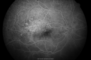
Old Branch Retinal Vein Occlusion- Severe CME - Pucker - Diabetic Early FA572 views61-year old man has diabetic retinopathy in the right eye. He also had a macular pucker and macular edema. I did a vitrectomy on September 5th. His vision was initially improving. His vision in the right eye seems to be a little more foggy.
VISUAL ACUITY: OD: 20/200
|
|

Old Branch Retinal Vein Occlusion- Severe CME - Pucker - Diabetic Early FA570 views61-year old man has diabetic retinopathy in the right eye. He also had a macular pucker and macular edema. I did a vitrectomy on September 5th. His vision was initially improving. His vision in the right eye seems to be a little more foggy.
VISUAL ACUITY: OD: 20/200
|
|

Old Branch Retinal Vein Occlusion- Severe CME - Pucker - Diabetic Late FA535 views61-year old man has diabetic retinopathy in the right eye. He also had a macular pucker and macular edema. I did a vitrectomy on September 5th. His vision was initially improving. His vision in the right eye seems to be a little more foggy.
VISUAL ACUITY: OD: 20/200
|
|

Retinal Vasculopathy associated with Facioscapulohumeral Muscular Dystrophy line os724 views59-year-old woman had a blood pressure spike to 170/90 and was on medications for that. She does have facioscapulohumeral muscular dystrophy, which runs in her family. She has a cousin with the same disorder.
VISUAL ACUITY: Her vision is 20/25 OU. IOP: OD 21, OS 18.
There is no posterior vitreous separation, telangiectatic vessels near the macula. There are patchy retinal hemorrhages. There is no edema or proliferation.
|
|
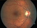
Retinal Vasculopathy associated with Facioscapulohumeral Muscular Dystrophy 820 views59-year-old woman had a blood pressure spike to 170/90 and was on medications for that. She does have facioscapulohumeral muscular dystrophy, which runs in her family. She has a cousin with the same disorder.
VISUAL ACUITY: Her vision is 20/25 OU. IOP: OD 21, OS 18.
There is no posterior vitreous separation, telangiectatic vessels near the macula. There are patchy retinal hemorrhages. There is no edema or proliferation.
59-year-old woman had a blood pressure spike to 170/90 and was on medications for tha
|
|

Retinal Vasculopathy associated with Facioscapulohumeral Muscular Dystrophy 788 views59-year-old woman had a blood pressure spike to 170/90 and was on medications for that. She does have facioscapulohumeral muscular dystrophy, which runs in her family. She has a cousin with the same disorder.
VISUAL ACUITY: Her vision is 20/25 OU. IOP: OD 21, OS 18.
There is no posterior vitreous separation, telangiectatic vessels near the macula. There are patchy retinal hemorrhages. There is no edema or proliferation.
|
|

Retinal Vasculopathy associated with Facioscapulohumeral Muscular Dystrophy 682 views59-year-old woman had a blood pressure spike to 170/90 and was on medications for that. She does have facioscapulohumeral muscular dystrophy, which runs in her family. She has a cousin with the same disorder.
VISUAL ACUITY: Her vision is 20/25 OU. IOP: OD 21, OS 18.
There is no posterior vitreous separation, telangiectatic vessels near the macula. There are patchy retinal hemorrhages. There is no edema or proliferation.
|
|

Retinal Vasculopathy associated with Facioscapulohumeral Muscular Dystrophy very early fa678 views59-year-old woman had a blood pressure spike to 170/90 and was on medications for that. She does have facioscapulohumeral muscular dystrophy, which runs in her family. She has a cousin with the same disorder.
VISUAL ACUITY: Her vision is 20/25 OU. IOP: OD 21, OS 18.
There is no posterior vitreous separation, telangiectatic vessels near the macula. There are patchy retinal hemorrhages. There is no edema or proliferation.
|
|

Retinal Vasculopathy associated with Facioscapulohumeral Muscular Dystrophy early fa827 views59-year-old woman had a blood pressure spike to 170/90 and was on medications for that. She does have facioscapulohumeral muscular dystrophy, which runs in her family. She has a cousin with the same disorder.
VISUAL ACUITY: Her vision is 20/25 OU. IOP: OD 21, OS 18.
There is no posterior vitreous separation, telangiectatic vessels near the macula. There are patchy retinal hemorrhages. There is no edema or proliferation.
|
|

Retinal Vasculopathy associated with Facioscapulohumeral Muscular Dystrophy early fa554 views59-year-old woman had a blood pressure spike to 170/90 and was on medications for that. She does have facioscapulohumeral muscular dystrophy, which runs in her family. She has a cousin with the same disorder.
VISUAL ACUITY: Her vision is 20/25 OU. IOP: OD 21, OS 18.
There is no posterior vitreous separation, telangiectatic vessels near the macula. There are patchy retinal hemorrhages. There is no edema or proliferation.
|
|

Retinal Vasculopathy associated with Facioscapulohumeral Muscular Dystrophy early fa540 views59-year-old woman had a blood pressure spike to 170/90 and was on medications for that. She does have facioscapulohumeral muscular dystrophy, which runs in her family. She has a cousin with the same disorder.
VISUAL ACUITY: Her vision is 20/25 OU. IOP: OD 21, OS 18.
There is no posterior vitreous separation, telangiectatic vessels near the macula. There are patchy retinal hemorrhages. There is no edema or proliferation.
|
|

Retinal Vasculopathy associated with Facioscapulohumeral Muscular Dystrophy early fa651 views59-year-old woman had a blood pressure spike to 170/90 and was on medications for that. She does have facioscapulohumeral muscular dystrophy, which runs in her family. She has a cousin with the same disorder.
VISUAL ACUITY: Her vision is 20/25 OU. IOP: OD 21, OS 18.
There is no posterior vitreous separation, telangiectatic vessels near the macula. There are patchy retinal hemorrhages. There is no edema or proliferation.
|
|

Retinal Vasculopathy associated with Facioscapulohumeral Muscular Dystrophy mid fa641 views59-year-old woman had a blood pressure spike to 170/90 and was on medications for that. She does have facioscapulohumeral muscular dystrophy, which runs in her family. She has a cousin with the same disorder.
VISUAL ACUITY: Her vision is 20/25 OU. IOP: OD 21, OS 18.
There is no posterior vitreous separation, telangiectatic vessels near the macula. There are patchy retinal hemorrhages. There is no edema or proliferation.
|
|
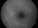
Retinal Vasculopathy associated with Facioscapulohumeral Muscular Dystrophy mid fa553 views59-year-old woman had a blood pressure spike to 170/90 and was on medications for that. She does have facioscapulohumeral muscular dystrophy, which runs in her family. She has a cousin with the same disorder.
VISUAL ACUITY: Her vision is 20/25 OU. IOP: OD 21, OS 18.
There is no posterior vitreous separation, telangiectatic vessels near the macula. There are patchy retinal hemorrhages. There is no edema or proliferation.
|
|

Retinal Vasculopathy associated with Facioscapulohumeral Muscular Dystrophy late fa656 views59-year-old woman had a blood pressure spike to 170/90 and was on medications for that. She does have facioscapulohumeral muscular dystrophy, which runs in her family. She has a cousin with the same disorder.
VISUAL ACUITY: Her vision is 20/25 OU. IOP: OD 21, OS 18.
There is no posterior vitreous separation, telangiectatic vessels near the macula. There are patchy retinal hemorrhages. There is no edema or proliferation.
|
|

Retinal Vasculopathy associated with Facioscapulohumeral Muscular Dystrophy late fa654 views59-year-old woman had a blood pressure spike to 170/90 and was on medications for that. She does have facioscapulohumeral muscular dystrophy, which runs in her family. She has a cousin with the same disorder.
VISUAL ACUITY: Her vision is 20/25 OU. IOP: OD 21, OS 18.
There is no posterior vitreous separation, telangiectatic vessels near the macula. There are patchy retinal hemorrhages. There is no edema or proliferation.
|
|

Retinal Vasculopathy associated with Facioscapulohumeral Muscular Dystrophy OCT right eye line scan775 views59-year-old woman had a blood pressure spike to 170/90 and was on medications for that. She does have facioscapulohumeral muscular dystrophy, which runs in her family. She has a cousin with the same disorder.
VISUAL ACUITY: Her vision is 20/25 OU. IOP: OD 21, OS 18.
There is no posterior vitreous separation, telangiectatic vessels near the macula. There are patchy retinal hemorrhages. There is no edema or proliferation.
|
|

Diabetic Patient with Macular Edema and Blood Pressure 200/95981 views49-year-old decreasing vision over the last year. OD is 20/80, OS 20/80. blood pressure which was 200/95.
|
|

Diabetic Patient with Macular Edema and Blood Pressure 200/951028 views49-year-old decreasing vision over the last year. OD is 20/80, OS 20/80. blood pressure which was 200/95.
|
|
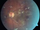
Diabetic Patient with Macular Edema and Blood Pressure 200/95754 views49-year-old decreasing vision over the last year. OD is 20/80, OS 20/80. blood pressure which was 200/95.
|
|

Diabetic Patient with Macular Edema and Blood Pressure 200/95612 views49-year-old decreasing vision over the last year. OD is 20/80, OS 20/80. blood pressure which was 200/95.
|
|

Diabetic Patient with Macular Edema and Blood Pressure 200/95587 views49-year-old decreasing vision over the last year. OD is 20/80, OS 20/80. blood pressure which was 200/95.
|
|

Diabetic Patient with Macular Edema and Blood Pressure 200/95603 views49-year-old decreasing vision over the last year. OD is 20/80, OS 20/80. blood pressure which was 200/95.
|
|
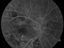
Diabetic Patient with Macular Edema and Blood Pressure 200/95579 views49-year-old decreasing vision over the last year. OD is 20/80, OS 20/80. blood pressure which was 200/95.
|
|

Diabetic Patient with Macular Edema and Blood Pressure 200/95610 views49-year-old decreasing vision over the last year. OD is 20/80, OS 20/80. blood pressure which was 200/95.
|
|

Diabetic Patient with Macular Edema and Blood Pressure 200/95593 views49-year-old decreasing vision over the last year. OD is 20/80, OS 20/80. blood pressure which was 200/95.
|
|

Diabetic Patient with Macular Edema and Blood Pressure 200/95606 views49-year-old decreasing vision over the last year. OD is 20/80, OS 20/80. blood pressure which was 200/95.
|
|

Diabetic Patient with Macular Edema and Blood Pressure 200/95643 views49-year-old decreasing vision over the last year. OD is 20/80, OS 20/80. blood pressure which was 200/95.
|
|

Diabetic Patient with Macular Edema and Blood Pressure 200/95597 views49-year-old decreasing vision over the last year. OD is 20/80, OS 20/80. blood pressure which was 200/95.
|
|

Diabetic Patient with Macular Edema and Blood Pressure 200/95694 views49-year-old decreasing vision over the last year. OD is 20/80, OS 20/80. blood pressure which was 200/95.
|
|

Diabetic Patient with Macular Edema and Blood Pressure 200/95731 views49-year-old decreasing vision over the last year. OD is 20/80, OS 20/80. blood pressure which was 200/95.
|
|

Diabetic Patient with Macular Edema and Blood Pressure 200/95748 views49-year-old decreasing vision over the last year. OD is 20/80, OS 20/80. blood pressure which was 200/95.
|
|

Diabetic Retinopathy - Non-Perfusion - Possible Central Retinal Vein Occlusion791 views68-year-old woman OD 20/70, OS 20/200 CYSTOID MACULAR EDEMA, POSSIBLE EITHER OCULAR ISCHEMIC SYNDROME IN THE RIGHT EYE OR OCCULT CENTRAL RETINAL VEIN OCCLUSION IN THE RIGHT EYE,S EVERE NON-PerfUSION WITH DIABETIC RETINOPATHY IN THE LEFT EYE.
|
|

Diabetic Retinopathy - Non-Perfusion - Possible Central Retinal Vein Occlusion771 views68-year-old woman OD 20/70, OS 20/200 CYSTOID MACULAR EDEMA, POSSIBLE EITHER OCULAR ISCHEMIC SYNDROME IN THE RIGHT EYE OR OCCULT CENTRAL RETINAL VEIN OCCLUSION IN THE RIGHT EYE,S EVERE NON-PerfUSION WITH DIABETIC RETINOPATHY IN THE LEFT EYE.
|
|
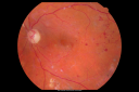
Diabetic Retinopathy - Non-Perfusion - Possible Central Retinal Vein Occlusion1030 views68-year-old woman OD 20/70, OS 20/200 CYSTOID MACULAR EDEMA, POSSIBLE EITHER OCULAR ISCHEMIC SYNDROME IN THE RIGHT EYE OR OCCULT CENTRAL RETINAL VEIN OCCLUSION IN THE RIGHT EYE,S EVERE NON-PerfUSION WITH DIABETIC RETINOPATHY IN THE LEFT EYE.
|
|
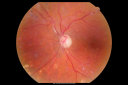
Diabetic Retinopathy - Non-Perfusion - Possible Central Retinal Vein Occlusion707 views68-year-old woman OD 20/70, OS 20/200 CYSTOID MACULAR EDEMA, POSSIBLE EITHER OCULAR ISCHEMIC SYNDROME IN THE RIGHT EYE OR OCCULT CENTRAL RETINAL VEIN OCCLUSION IN THE RIGHT EYE,S EVERE NON-PerfUSION WITH DIABETIC RETINOPATHY IN THE LEFT EYE.
|
|
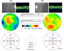
Diabetic Retinopathy - Non-Perfusion - Possible Central Retinal Vein Occlusion802 views68-year-old woman OD 20/70, OS 20/200 CYSTOID MACULAR EDEMA, POSSIBLE EITHER OCULAR ISCHEMIC SYNDROME IN THE RIGHT EYE OR OCCULT CENTRAL RETINAL VEIN OCCLUSION IN THE RIGHT EYE,S EVERE NON-PerfUSION WITH DIABETIC RETINOPATHY IN THE LEFT EYE.
|
|

Diabetic Retinopathy - Non-Perfusion - Possible Central Retinal Vein Occlusion574 views68-year-old woman OD 20/70, OS 20/200 CYSTOID MACULAR EDEMA, POSSIBLE EITHER OCULAR ISCHEMIC SYNDROME IN THE RIGHT EYE OR OCCULT CENTRAL RETINAL VEIN OCCLUSION IN THE RIGHT EYE,S EVERE NON-PerfUSION WITH DIABETIC RETINOPATHY IN THE LEFT EYE.
|
|
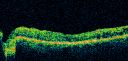
Diabetic Retinopathy - Non-Perfusion - Possible Central Retinal Vein Occlusion615 views68-year-old woman OD 20/70, OS 20/200 CYSTOID MACULAR EDEMA, POSSIBLE EITHER OCULAR ISCHEMIC SYNDROME IN THE RIGHT EYE OR OCCULT CENTRAL RETINAL VEIN OCCLUSION IN THE RIGHT EYE,S EVERE NON-PerfUSION WITH DIABETIC RETINOPATHY IN THE LEFT EYE.
|
|
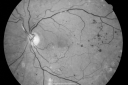
Diabetic Retinopathy - Non-Perfusion - Possible Central Retinal Vein Occlusion661 views68-year-old woman OD 20/70, OS 20/200 CYSTOID MACULAR EDEMA, POSSIBLE EITHER OCULAR ISCHEMIC SYNDROME IN THE RIGHT EYE OR OCCULT CENTRAL RETINAL VEIN OCCLUSION IN THE RIGHT EYE,S EVERE NON-PerfUSION WITH DIABETIC RETINOPATHY IN THE LEFT EYE.
|
|

Diabetic Retinopathy - Non-Perfusion - Possible Central Retinal Vein Occlusion645 views68-year-old woman OD 20/70, OS 20/200 CYSTOID MACULAR EDEMA, POSSIBLE EITHER OCULAR ISCHEMIC SYNDROME IN THE RIGHT EYE OR OCCULT CENTRAL RETINAL VEIN OCCLUSION IN THE RIGHT EYE,S EVERE NON-PerfUSION WITH DIABETIC RETINOPATHY IN THE LEFT EYE.
|
|
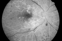
Diabetic Retinopathy - Non-Perfusion - Possible Central Retinal Vein Occlusion568 views68-year-old woman OD 20/70, OS 20/200 CYSTOID MACULAR EDEMA, POSSIBLE EITHER OCULAR ISCHEMIC SYNDROME IN THE RIGHT EYE OR OCCULT CENTRAL RETINAL VEIN OCCLUSION IN THE RIGHT EYE,S EVERE NON-PerfUSION WITH DIABETIC RETINOPATHY IN THE LEFT EYE.
|
|

Diabetic Retinopathy - Non-Perfusion - Possible Central Retinal Vein Occlusion619 views68-year-old woman OD 20/70, OS 20/200 CYSTOID MACULAR EDEMA, POSSIBLE EITHER OCULAR ISCHEMIC SYNDROME IN THE RIGHT EYE OR OCCULT CENTRAL RETINAL VEIN OCCLUSION IN THE RIGHT EYE,S EVERE NON-PerfUSION WITH DIABETIC RETINOPATHY IN THE LEFT EYE.
|
|

Diabetic Retinopathy - Non-Perfusion - Possible Central Retinal Vein Occlusion753 views68-year-old woman OD 20/70, OS 20/200 CYSTOID MACULAR EDEMA, POSSIBLE EITHER OCULAR ISCHEMIC SYNDROME IN THE RIGHT EYE OR OCCULT CENTRAL RETINAL VEIN OCCLUSION IN THE RIGHT EYE,S EVERE NON-PerfUSION WITH DIABETIC RETINOPATHY IN THE LEFT EYE.
|
|

Diabetic Retinopathy - Non-Perfusion - Possible Central Retinal Vein Occlusion647 views68-year-old woman OD 20/70, OS 20/200 CYSTOID MACULAR EDEMA, POSSIBLE EITHER OCULAR ISCHEMIC SYNDROME IN THE RIGHT EYE OR OCCULT CENTRAL RETINAL VEIN OCCLUSION IN THE RIGHT EYE,S EVERE NON-PerfUSION WITH DIABETIC RETINOPATHY IN THE LEFT EYE.
|
|

Diabetic Retinopathy - Non-Perfusion - Possible Central Retinal Vein Occlusion656 views68-year-old woman OD 20/70, OS 20/200 CYSTOID MACULAR EDEMA, POSSIBLE EITHER OCULAR ISCHEMIC SYNDROME IN THE RIGHT EYE OR OCCULT CENTRAL RETINAL VEIN OCCLUSION IN THE RIGHT EYE,S EVERE NON-PerfUSION WITH DIABETIC RETINOPATHY IN THE LEFT EYE.
|
|
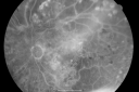
Diabetic Retinopathy - Non-Perfusion - Possible Central Retinal Vein Occlusion593 views68-year-old woman OD 20/70, OS 20/200 CYSTOID MACULAR EDEMA, POSSIBLE EITHER OCULAR ISCHEMIC SYNDROME IN THE RIGHT EYE OR OCCULT CENTRAL RETINAL VEIN OCCLUSION IN THE RIGHT EYE,S EVERE NON-PerfUSION WITH DIABETIC RETINOPATHY IN THE LEFT EYE.
|
|

Diabetic Retinopathy - Non-Perfusion - Possible Central Retinal Vein Occlusion588 views68-year-old woman OD 20/70, OS 20/200 CYSTOID MACULAR EDEMA, POSSIBLE EITHER OCULAR ISCHEMIC SYNDROME IN THE RIGHT EYE OR OCCULT CENTRAL RETINAL VEIN OCCLUSION IN THE RIGHT EYE,S EVERE NON-PerfUSION WITH DIABETIC RETINOPATHY IN THE LEFT EYE.
|
|

Diabetic Retinopathy - Non-Perfusion - Possible Central Retinal Vein Occlusion640 views68-year-old woman OD 20/70, OS 20/200 CYSTOID MACULAR EDEMA, POSSIBLE EITHER OCULAR ISCHEMIC SYNDROME IN THE RIGHT EYE OR OCCULT CENTRAL RETINAL VEIN OCCLUSION IN THE RIGHT EYE,S EVERE NON-PerfUSION WITH DIABETIC RETINOPATHY IN THE LEFT EYE.
|
|

Diabetic Macular Edema Left Eye - Post Laser1410 views82-year-old woman diabetic for many years, last eye exam 5 years ago with gradual vision loss. OD 20/60, OS 20/70. IOP: OD 16, OS 17.
|
|

Proliferative Diabetic Retinopathy both Eyes Neovascularization of the Disc851 views63-year-old woman has been diabetic for twenty one years who sees new floaters in the right eye. OD 20/40, OS 20/40.
|
|
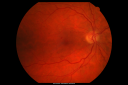
Proliferative Diabetic Retinopathy both Eyes Neovascularization of the Disc974 views63-year-old woman has been diabetic for twenty one years who sees new floaters in the right eye. OD 20/40, OS 20/40.
|
|

Proliferative Diabetic Retinopathy both Eyes Neovascularization of the Disc1635 views63-year-old woman has been diabetic for twenty one years who sees new floaters in the right eye. OD 20/40, OS 20/40.
|
|

Proliferative Diabetic Retinopathy both Eyes Neovascularization of the Disc803 views63-year-old woman has been diabetic for twenty one years who sees new floaters in the right eye. OD 20/40, OS 20/40.
|
|

Proliferative Diabetic Retinopathy both Eyes Neovascularization of the Disc769 views63-year-old woman has been diabetic for twenty one years who sees new floaters in the right eye. OD 20/40, OS 20/40.
|
|

Proliferative Diabetic Retinopathy both Eyes Neovascularization of the Disc789 views63-year-old woman has been diabetic for twenty one years who sees new floaters in the right eye. OD 20/40, OS 20/40.
|
|

Proliferative Diabetic Retinopathy both Eyes Neovascularization of the Disc1052 views63-year-old woman has been diabetic for twenty one years who sees new floaters in the right eye. OD 20/40, OS 20/40.
|
|

Proliferative Diabetic Retinopathy both Eyes Neovascularization of the Disc803 views63-year-old woman has been diabetic for twenty one years who sees new floaters in the right eye. OD 20/40, OS 20/40.
|
|

Proliferative Diabetic Retinopathy both Eyes Neovascularization of the Disc811 views63-year-old woman has been diabetic for twenty one years who sees new floaters in the right eye. OD 20/40, OS 20/40.
|
|

Proliferative Diabetic Retinopathy both Eyes Neovascularization of the Disc725 views63-year-old woman has been diabetic for twenty one years who sees new floaters in the right eye. OD 20/40, OS 20/40.
|
|

Proliferative Diabetic Retinopathy both Eyes Neovascularization of the Disc757 views63-year-old woman has been diabetic for twenty one years who sees new floaters in the right eye. OD 20/40, OS 20/40.
|
|

Proliferative Diabetic Retinopathy both Eyes Neovascularization of the Disc742 views63-year-old woman has been diabetic for twenty one years who sees new floaters in the right eye. OD 20/40, OS 20/40.
|
|

Proliferative Diabetic Retinopathy with Macular Hole3205 viewsPatitent with diabetes diagnosed 12 years ago and low visual acuity.
|
|

Proliferative Diabetic Retinopathy - Preretinal Hemorrhage1818 viewsPreretinal Hemorrhage secondary to Proliferative Diabetic Retinopathy (PDR)
James L. Perron, C.R.A.
|
|

524 views
|
|

480 views
|
|

536 views
|
|

BDR648 views
|
|

Capillary Pruning577 viewsCapillary drop out secondary to proliferative diabetic retinopathy
|
|

Capillary Drop Out of the FAZ697 viewsHigh magnification image of a damaged foveal avascular zone secondary to proliferative diabetic retinopathy. Seen here are multiple laser scars and capillary drop out in FAZ.
|
|

Severe Hemorrhage from NVD and NVE Due To PDR507 views60 year old female with Diabetic Retinopathy. VA 20/40 OD HM OS. History of Dot and Blot hemorrhages and cotton-wool spots associated with Diabetes. Severe Hemorrhaging due to NVD and NVE. Pt stated sight decreased very rapidly.
|
|

Severe Hemorrhage from NVD and NVE Due To PDR421 views60 year old female with Diabetic Retinopathy. VA 20/40 OD HM OS. History of Dot and Blot hemorrhages and cotton-wool spots associated with Diabetes. Severe Hemorrhaging due to NVD and NVE. Pt stated sight decreased very rapidly.
|
|

Juvenile Onset Diabetic - Proliferative Diabetic retinopathy with NVE and NVD4776 viewsProliferative Diabetic Retinopathy with Neovascularization of the Disc and Neovascularization Elsewhere.
James L. Perron, C.R.A.
|
|

Proliferative Diabetic Retinopathy with NVD, NVE, and Pre Retinal Hemorrhage620 viewsJuvenile Onset Diabetic with NVD, NVE and Pre Retinal Hemorrhage.
|
|

FAF Montage of Diabetic Retinopathy662 views70-year old male with severe Diabetic Retinopathy. VA at visit was 20/100 with no improvement with pinhole. Patient had recently undergone cataract surgery to try to regain some of his vison. Panoretinal Photocoagulation scars 360 degrees around the retina...
|
|

Proliferative Diabetic Retinopathy with Tractional Retinal Detachment1060 viewsProliferative Diabetic Retinopathy with Tractional Retinal Detachment
|
|

Nonprolific Diabetic Retinopathy with Circinate Ring1024 viewsNonprolific Diabetic Retinopathy with Circinate Ring
|
|

Diabetic Retinopathy with Hard Exudates591 views
|
|
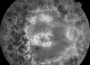
late phase FA1082 views50+ YO female presented with DM type 1;
PT presented with PDR s/p extensive PRP
FA showed CNV OD
VA stable at 20/150 for 1 year
|
|

early phase FA558 views50+ YO female presented with DM type 1;
PT presented with PDR s/p extensive PRP
FA showed CNV OD
VA stable at 20/150 for 1 year
|
|

early phase FA589 views50+ YO female presented with DM type 1;
PT presented with PDR s/p extensive PRP
FA showed CNV OD
VA stable at 20/150 for 1 year
|
|

Diabetic Tractional Detachment476 views
|
|
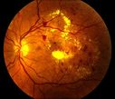
Nonproliferative Diabetic Retinopathy with Circinate Hard Exudates1522 views
|
|

Actively Bleeding NVE537 viewsThis male patient was referred for a diabetic examination. Upon dilated exam, he started to hemorrhage from a frond of NVE in the superior arcade. These six photographs follow the flow of the hemorrhage through the vitreous to the inferior arcade.
|
|
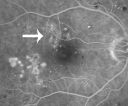
Focal Laser for diabetic macular edema1356 views66 year old female with diabetes for 45 years. VA 20/20 OU. Extrafoveal edema in the left eye treated with light focal laser.
|
|
|
|
|


