Image search results - "MA"
|

Macular Pucker875 viewsFundus photo with OCT superimposed, 20/100 macular pucker
|
|

Papilledema 10 year old child1594 views10-year-old child has uveitis in both eyes. She probably has tubulointerstitial nephritis-uveitis syndrome, and she apparently also independently has idiopathic elevated intracranial pressure. VISUAL ACUITY: Vision OU is 20/20.
|
|

a scan right eye 0 degree deviation 777 views10-year-old child has uveitis in both eyes. She probably has tubulointerstitial nephritis-uveitis syndrome, and she apparently also independently has idiopathic elevated intracranial pressure. VISUAL ACUITY: Vision OU is 20/20.
|
|
Papilledema 10 year old child710 views10-year-old child has uveitis in both eyes. She probably has tubulointerstitial nephritis-uveitis syndrome, and she apparently also independently has idiopathic elevated intracranial pressure. VISUAL ACUITY: Vision OU is 20/20.
|
|

Papilledema 10 year old child785 views10-year-old child has uveitis in both eyes. She probably has tubulointerstitial nephritis-uveitis syndrome, and she apparently also independently has idiopathic elevated intracranial pressure. VISUAL ACUITY: Vision OU is 20/20.
|
|

Papilledema 10 year old child740 views10-year-old child has uveitis in both eyes. She probably has tubulointerstitial nephritis-uveitis syndrome, and she apparently also independently has idiopathic elevated intracranial pressure. VISUAL ACUITY: Vision OU is 20/20.
|
|

Papilledema 10 year old child1086 views10-year-old child has uveitis in both eyes. She probably has tubulointerstitial nephritis-uveitis syndrome, and she apparently also independently has idiopathic elevated intracranial pressure. VISUAL ACUITY: Vision OU is 20/20.
|
|

Macular Pucker644 viewsThis pleasant 59-year-old man was seen in the office on 7/9/2010. He had new onset floaters about a year ago. Subsequent to that his vision was not quite right in the right eye and then he noticed, starting about three weeks ago, substantial change in the vision. In the morning he sees what he calls a small gray circle with a ring of fire around it and then subsequent to that the vision becomes more normal but not quite right. His vision in the left eye is fine. Vision OD is 20/50, OS is 20/16.
|
|

Macular Pucker555 views. This pleasant 59-year-old man had vitrectomy for a macular pucker in the right eye in July. His vision is gradually improving.
VISUAL ACUITY: Vision OD is 20/30. IOP: OD 22. There is 2+ nuclear sclerosis.
EXTENDED OPHTHALMOSCOPY:
OD: Vertical C/D ratio is 0.5. The macula is smooth and the retina is attached.
OCT SCAN: The OCT scan shows an average central foveal thickness of 349 microns, which compares favorably to the pre-op thickness of 482 microns.
|
|

Macular Pucker500 views. This pleasant 59-year-old man had vitrectomy for a macular pucker in the right eye in July. His vision is gradually improving.
VISUAL ACUITY: Vision OD is 20/30. IOP: OD 22. There is 2+ nuclear sclerosis.
EXTENDED OPHTHALMOSCOPY:
OD: Vertical C/D ratio is 0.5. The macula is smooth and the retina is attached.
OCT SCAN: The OCT scan shows an average central foveal thickness of 349 microns, which compares favorably to the pre-op thickness of 482 microns.
|
|

Macular Pucker567 viewsThis pleasant 59-year-old man was seen in the office on 7/9/2010. He had new onset floaters about a year ago. Subsequent to that his vision was not quite right in the right eye and then he noticed, starting about three weeks ago, substantial change in the vision. In the morning he sees what he calls a small gray circle with a ring of fire around it and then subsequent to that the vision becomes more normal but not quite right. His vision in the left eye is fine. Vision OD is 20/50, OS is 20/16.
|
|
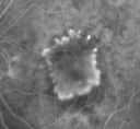
Subfoveal CNVM wet AMD early fa (contrast enhanced with photoshop)883 viewsShe has noticed decreased vision in the left eye. It is hard to say for how long, maybe a few months. She had her last eye exam about a year ago. She thoughts perhaps it was cataracts, but you saw a problem with the macula and suggested she come here for evaluation.
VISUAL ACUITY: Vision OD is 20/25, OS is 2/200
|
|

Myopic Macular Degeneration - Dry 1180 views55-year-old woman has myopic macular degeneration in both eyes with vision and an Amsler grid change starting a few days ago in the left eye.
VISUAL ACUITY: OD: 20/60. PH: 20/25. OS: 20/200. PH: 20/30.
|
|

Myopic Macular Degeneration - Dry 1070 views55-year-old woman has myopic macular degeneration in both eyes with vision and an Amsler grid change starting a few days ago in the left eye.
VISUAL ACUITY: OD: 20/60. PH: 20/25. OS: 20/200. PH: 20/30.
|
|

Myopic Macular Degeneration - Dry 1079 views55-year-old woman has myopic macular degeneration in both eyes with vision and an Amsler grid change starting a few days ago in the left eye.
VISUAL ACUITY: OD: 20/60. PH: 20/25. OS: 20/200. PH: 20/30.
|
|

Myopic Macular Degeneration - Dry 908 views55-year-old woman has myopic macular degeneration in both eyes with vision and an Amsler grid change starting a few days ago in the left eye.
VISUAL ACUITY: OD: 20/60. PH: 20/25. OS: 20/200. PH: 20/30.
|
|

Myopic Macular Degeneration - Dry 798 views55-year-old woman has myopic macular degeneration in both eyes with vision and an Amsler grid change starting a few days ago in the left eye.
VISUAL ACUITY: OD: 20/60. PH: 20/25. OS: 20/200. PH: 20/30.
|
|
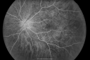
Myopic Macular Degeneration - Dry 806 views55-year-old woman has myopic macular degeneration in both eyes with vision and an Amsler grid change starting a few days ago in the left eye.
VISUAL ACUITY: OD: 20/60. PH: 20/25. OS: 20/200. PH: 20/30.
|
|

Myopic Macular Degeneration - Dry 715 views55-year-old woman has myopic macular degeneration in both eyes with vision and an Amsler grid change starting a few days ago in the left eye.
VISUAL ACUITY: OD: 20/60. PH: 20/25. OS: 20/200. PH: 20/30.
|
|

Myopic Macular Degeneration - Dry 780 views55-year-old woman has myopic macular degeneration in both eyes with vision and an Amsler grid change starting a few days ago in the left eye.
VISUAL ACUITY: OD: 20/60. PH: 20/25. OS: 20/200. PH: 20/30.
|
|
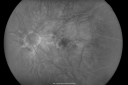
Myopic Macular Degeneration - Dry 696 views55-year-old woman has myopic macular degeneration in both eyes with vision and an Amsler grid change starting a few days ago in the left eye.
VISUAL ACUITY: OD: 20/60. PH: 20/25. OS: 20/200. PH: 20/30.
|
|

Myopic Macular Degeneration - Dry OCT OS1041 views55-year-old woman has myopic macular degeneration in both eyes with vision and an Amsler grid change starting a few days ago in the left eye.
VISUAL ACUITY: OD: 20/60. PH: 20/25. OS: 20/200. PH: 20/30.
|
|

Multifocal Choroiditis and Subretinal Fibrosis - 32 yo Female Macula OS 2 months post rx1189 views32-year-old woman vision loss in the right eye associated with macular scarring and multifocal choroiditis in 1999 with new vision loss in left eye: OD 20/400, OS 20/50.
2 months post-rx with posterior subtenons kenalog and intravitreal avastin - va os 20/30 and lesion has retracted and organized. It never subsequently grew over 2 years follow-up
|
|
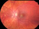
Multifocal Choroiditis and Subretinal Fibrosis - 32 yo Female Montage Initial Visit994 views32-year-old woman vision loss in the right eye associated with macular scarring and multifocal choroiditis in 1999 with new vision loss in left eye: OD 20/400, OS 20/50.
2 months post-rx with posterior subtenons kenalog and intravitreal avastin - va os 20/30 and lesion has retracted and organized. It never subsequently grew over 2 years follow-up
|
|

Choroidal Rupture and small Hemorrhage - Assault1393 views34 year old man. The patient reports he was assaulted by a couple of adolescents near downtown St. Petersburg about forty-eight hours ago. He reports the pain in the left eye has significantly improved, but his vision is a little fuzzy and he sees some floaters in the left eye. 20/125 OD, 20/20 OS. Pinhole improves in the right eye to 20/50. IOP: 15 OD, 13 OS.
SLIT LAMP EXAMINATION: Biomicroscopy reveals 3+ pupillary reaction OD, 2+ OS with no afferent pupillary defect. EXTENDED OPHTHALMOSCOPY: Extended funduscopy with Volk 90-diopter lens and fundus drawings of both eyes reveal a clear view with no posterior vitreous separation. C/D ratio is 0.2. There is no vitreous debris or hemorrhage. Large retinal vessels and macula are healthy. The retinal periphery, inspected with scleral depression for 360° shows no retinal tears, breaks, or detachments.
|
|

Choroidal Rupture and small Hemorrhage - Assault1038 views34 year old man. The patient reports he was assaulted by a couple of adolescents near downtown St. Petersburg about forty-eight hours ago. He reports the pain in the left eye has significantly improved, but his vision is a little fuzzy and he sees some floaters in the left eye. 20/125 OD, 20/20 OS. Pinhole improves in the right eye to 20/50. IOP: 15 OD, 13 OS.
SLIT LAMP EXAMINATION: Biomicroscopy reveals 3+ pupillary reaction OD, 2+ OS with no afferent pupillary defect. EXTENDED OPHTHALMOSCOPY: Extended funduscopy with Volk 90-diopter lens and fundus drawings of both eyes reveal a clear view with no posterior vitreous separation. C/D ratio is 0.2. There is no vitreous debris or hemorrhage. Large retinal vessels and macula are healthy. The retinal periphery, inspected with scleral depression for 360° shows no retinal tears, breaks, or detachments.
|
|

Choroidal Rupture and small Hemorrhage - Assault870 views34 year old man. The patient reports he was assaulted by a couple of adolescents near downtown St. Petersburg about forty-eight hours ago. He reports the pain in the left eye has significantly improved, but his vision is a little fuzzy and he sees some floaters in the left eye. 20/125 OD, 20/20 OS. Pinhole improves in the right eye to 20/50. IOP: 15 OD, 13 OS.
SLIT LAMP EXAMINATION: Biomicroscopy reveals 3+ pupillary reaction OD, 2+ OS with no afferent pupillary defect. EXTENDED OPHTHALMOSCOPY: Extended funduscopy with Volk 90-diopter lens and fundus drawings of both eyes reveal a clear view with no posterior vitreous separation. C/D ratio is 0.2. There is no vitreous debris or hemorrhage. Large retinal vessels and macula are healthy. The retinal periphery, inspected with scleral depression for 360° shows no retinal tears, breaks, or detachments.
|
|

Choroidal Rupture and small Hemorrhage - Assault880 views34 year old man. The patient reports he was assaulted by a couple of adolescents near downtown St. Petersburg about forty-eight hours ago. He reports the pain in the left eye has significantly improved, but his vision is a little fuzzy and he sees some floaters in the left eye. 20/125 OD, 20/20 OS. Pinhole improves in the right eye to 20/50. IOP: 15 OD, 13 OS.
SLIT LAMP EXAMINATION: Biomicroscopy reveals 3+ pupillary reaction OD, 2+ OS with no afferent pupillary defect. EXTENDED OPHTHALMOSCOPY: Extended funduscopy with Volk 90-diopter lens and fundus drawings of both eyes reveal a clear view with no posterior vitreous separation. C/D ratio is 0.2. There is no vitreous debris or hemorrhage. Large retinal vessels and macula are healthy. The retinal periphery, inspected with scleral depression for 360° shows no retinal tears, breaks, or detachments.
|
|

Choroidal Rupture and small Hemorrhage - Assault1099 views34 year old man. The patient reports he was assaulted by a couple of adolescents near downtown St. Petersburg about forty-eight hours ago. He reports the pain in the left eye has significantly improved, but his vision is a little fuzzy and he sees some floaters in the left eye. 20/125 OD, 20/20 OS. Pinhole improves in the right eye to 20/50. IOP: 15 OD, 13 OS.
SLIT LAMP EXAMINATION: Biomicroscopy reveals 3+ pupillary reaction OD, 2+ OS with no afferent pupillary defect. EXTENDED OPHTHALMOSCOPY: Extended funduscopy with Volk 90-diopter lens and fundus drawings of both eyes reveal a clear view with no posterior vitreous separation. C/D ratio is 0.2. There is no vitreous debris or hemorrhage. Large retinal vessels and macula are healthy. The retinal periphery, inspected with scleral depression for 360° shows no retinal tears, breaks, or detachments.
|
|

Choroidal Rupture and small Hemorrhage - Assault802 views34 year old man. The patient reports he was assaulted by a couple of adolescents near downtown St. Petersburg about forty-eight hours ago. He reports the pain in the left eye has significantly improved, but his vision is a little fuzzy and he sees some floaters in the left eye. 20/125 OD, 20/20 OS. Pinhole improves in the right eye to 20/50. IOP: 15 OD, 13 OS.
SLIT LAMP EXAMINATION: Biomicroscopy reveals 3+ pupillary reaction OD, 2+ OS with no afferent pupillary defect. EXTENDED OPHTHALMOSCOPY: Extended funduscopy with Volk 90-diopter lens and fundus drawings of both eyes reveal a clear view with no posterior vitreous separation. C/D ratio is 0.2. There is no vitreous debris or hemorrhage. Large retinal vessels and macula are healthy. The retinal periphery, inspected with scleral depression for 360° shows no retinal tears, breaks, or detachments.
|
|

Big Floater - From Spontaneously Peeled Vitreomacular Traction Macular Pucker760 views54-year-old woman has had a visually significant and debilitating vitreous opacity in the right eye, which has been there for 2 years. It is, if anything, getting worse. When she moves her eye around it lands right in her central vision most of the time.
VISUAL ACUITY: Her vision OD is 20/20
|
|

Big Floater - From Spontaneously Peeled Vitreomacular Traction Macular Pucker717 views54-year-old woman has had a visually significant and debilitating vitreous opacity in the right eye, which has been there for 2 years. It is, if anything, getting worse. When she moves her eye around it lands right in her central vision most of the time.
VISUAL ACUITY: Her vision OD is 20/20
|
|

Big Floater - From Spontaneously Peeled Vitreomacular Traction Macular Pucker711 views54-year-old woman has had a visually significant and debilitating vitreous opacity in the right eye, which has been there for 2 years. It is, if anything, getting worse. When she moves her eye around it lands right in her central vision most of the time.
VISUAL ACUITY: Her vision OD is 20/20
|
|

Myopic Degeneration - Staphyloma - Atrophy892 views53-year-old man was a -15.00 myope. He is aphakic. His left eye also had a retinal detachment repair and unfortunately the left eye does not see well. He is on Timolol in both eyes for glaucoma. VISUAL ACUITY: OD 20/50, OS: Faint light perception.
|
|

Myopic Degeneration - Staphyloma - Atrophy946 views53-year-old man was a -15.00 myope. He is aphakic. His left eye also had a retinal detachment repair and unfortunately the left eye does not see well. He is on Timolol in both eyes for glaucoma. VISUAL ACUITY: OD 20/50, OS: Faint light perception.
|
|

Myopic Degeneration - Staphyloma - Atrophy824 views53-year-old man was a -15.00 myope. He is aphakic. His left eye also had a retinal detachment repair and unfortunately the left eye does not see well. He is on Timolol in both eyes for glaucoma. VISUAL ACUITY: OD 20/50, OS: Faint light perception.
|
|

Retinoblastoma - Bilateral - Regressed - Chemotherapy, Thermal Therapy, Cryotherapy1054 views10-year-old child was seen in the office on August 6, 2008. She was treated with a combination of chemotherapy at 10 months old as well as thermal therapy and cryotherapy for bilateral retinoblastoma tumors. She reportedly had nine tumors in the left eye and five tumors in the right eye
|
|

Retinoblastoma - Bilateral - Regressed - Chemotherapy, Thermal Therapy, Cryotherapy901 views10-year-old child was seen in the office on August 6, 2008. She was treated with a combination of chemotherapy at 10 months old as well as thermal therapy and cryotherapy for bilateral retinoblastoma tumors. She reportedly had nine tumors in the left eye and five tumors in the right eye
|
|

Retinoblastoma - Bilateral - Regressed - Chemotherapy, Thermal Therapy, Cryotherapy807 views10-year-old child was seen in the office on August 6, 2008. She was treated with a combination of chemotherapy at 10 months old as well as thermal therapy and cryotherapy for bilateral retinoblastoma tumors. She reportedly had nine tumors in the left eye and five tumors in the right eye
|
|

Retinoblastoma - Bilateral - Regressed - Chemotherapy, Thermal Therapy, Cryotherapy739 views10-year-old child was seen in the office on August 6, 2008. She was treated with a combination of chemotherapy at 10 months old as well as thermal therapy and cryotherapy for bilateral retinoblastoma tumors. She reportedly had nine tumors in the left eye and five tumors in the right eye
|
|

Retinoblastoma - Bilateral - Regressed - Chemotherapy, Thermal Therapy, Cryotherapy880 views10-year-old child was seen in the office on August 6, 2008. She was treated with a combination of chemotherapy at 10 months old as well as thermal therapy and cryotherapy for bilateral retinoblastoma tumors. She reportedly had nine tumors in the left eye and five tumors in the right eye
|
|

Retinoblastoma - Bilateral - Regressed - Chemotherapy, Thermal Therapy, Cryotherapy851 views10-year-old child was seen in the office on August 6, 2008. She was treated with a combination of chemotherapy at 10 months old as well as thermal therapy and cryotherapy for bilateral retinoblastoma tumors. She reportedly had nine tumors in the left eye and five tumors in the right eye
|
|

Retinoblastoma - Bilateral - Regressed - Chemotherapy, Thermal Therapy, Cryotherapy792 views10-year-old child was seen in the office on August 6, 2008. She was treated with a combination of chemotherapy at 10 months old as well as thermal therapy and cryotherapy for bilateral retinoblastoma tumors. She reportedly had nine tumors in the left eye and five tumors in the right eye
|
|

Retinoblastoma - Bilateral - Regressed - Chemotherapy, Thermal Therapy, Cryotherapy735 views10-year-old child was seen in the office on August 6, 2008. She was treated with a combination of chemotherapy at 10 months old as well as thermal therapy and cryotherapy for bilateral retinoblastoma tumors. She reportedly had nine tumors in the left eye and five tumors in the right eye
|
|

Retinoblastoma - Bilateral - Regressed - Chemotherapy, Thermal Therapy, Cryotherapy738 views10-year-old child was seen in the office on August 6, 2008. She was treated with a combination of chemotherapy at 10 months old as well as thermal therapy and cryotherapy for bilateral retinoblastoma tumors. She reportedly had nine tumors in the left eye and five tumors in the right eye
|
|
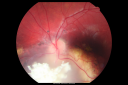
Retinoblastoma - Bilateral - Regressed - Chemotherapy, Thermal Therapy, Cryotherapy882 views10-year-old child was seen in the office on August 6, 2008. She was treated with a combination of chemotherapy at 10 months old as well as thermal therapy and cryotherapy for bilateral retinoblastoma tumors. She reportedly had nine tumors in the left eye and five tumors in the right eye
|
|

Retinoblastoma - Bilateral - Regressed - Chemotherapy, Thermal Therapy, Cryotherapy766 views10-year-old child was seen in the office on August 6, 2008. She was treated with a combination of chemotherapy at 10 months old as well as thermal therapy and cryotherapy for bilateral retinoblastoma tumors. She reportedly had nine tumors in the left eye and five tumors in the right eye
|
|

Retinoblastoma - Bilateral - Regressed - Chemotherapy, Thermal Therapy, Cryotherapy732 views10-year-old child was seen in the office on August 6, 2008. She was treated with a combination of chemotherapy at 10 months old as well as thermal therapy and cryotherapy for bilateral retinoblastoma tumors. She reportedly had nine tumors in the left eye and five tumors in the right eye
|
|

Retinoblastoma - Bilateral - Regressed - Chemotherapy, Thermal Therapy, Cryotherapy702 views10-year-old child was seen in the office on August 6, 2008. She was treated with a combination of chemotherapy at 10 months old as well as thermal therapy and cryotherapy for bilateral retinoblastoma tumors. She reportedly had nine tumors in the left eye and five tumors in the right eye
|
|

Retinoblastoma - Bilateral - Regressed - Chemotherapy, Thermal Therapy, Cryotherapy833 views10-year-old child was seen in the office on August 6, 2008. She was treated with a combination of chemotherapy at 10 months old as well as thermal therapy and cryotherapy for bilateral retinoblastoma tumors. She reportedly had nine tumors in the left eye and five tumors in the right eye
|
|
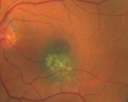
Macular Choroidal Nevus1723 views78-year-old woman has choroidal nevus in the macula of the left eye. You asked her to come in here for an evaluation and nevus was noted in the past about 7 years ago. She has not noticed any change in vision in that eye.
VISUAL ACUITY: OU 20/30.
|
|

Diabetic Patient with Macular Edema and Blood Pressure 200/95968 views49-year-old decreasing vision over the last year. OD is 20/80, OS 20/80. blood pressure which was 200/95.
|
|

Diabetic Patient with Macular Edema and Blood Pressure 200/951006 views49-year-old decreasing vision over the last year. OD is 20/80, OS 20/80. blood pressure which was 200/95.
|
|
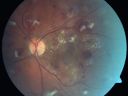
Diabetic Patient with Macular Edema and Blood Pressure 200/95743 views49-year-old decreasing vision over the last year. OD is 20/80, OS 20/80. blood pressure which was 200/95.
|
|

Diabetic Patient with Macular Edema and Blood Pressure 200/95597 views49-year-old decreasing vision over the last year. OD is 20/80, OS 20/80. blood pressure which was 200/95.
|
|

Diabetic Patient with Macular Edema and Blood Pressure 200/95564 views49-year-old decreasing vision over the last year. OD is 20/80, OS 20/80. blood pressure which was 200/95.
|
|

Diabetic Patient with Macular Edema and Blood Pressure 200/95591 views49-year-old decreasing vision over the last year. OD is 20/80, OS 20/80. blood pressure which was 200/95.
|
|
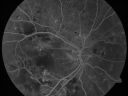
Diabetic Patient with Macular Edema and Blood Pressure 200/95565 views49-year-old decreasing vision over the last year. OD is 20/80, OS 20/80. blood pressure which was 200/95.
|
|

Diabetic Patient with Macular Edema and Blood Pressure 200/95597 views49-year-old decreasing vision over the last year. OD is 20/80, OS 20/80. blood pressure which was 200/95.
|
|

Diabetic Patient with Macular Edema and Blood Pressure 200/95581 views49-year-old decreasing vision over the last year. OD is 20/80, OS 20/80. blood pressure which was 200/95.
|
|

Diabetic Patient with Macular Edema and Blood Pressure 200/95598 views49-year-old decreasing vision over the last year. OD is 20/80, OS 20/80. blood pressure which was 200/95.
|
|

Diabetic Patient with Macular Edema and Blood Pressure 200/95636 views49-year-old decreasing vision over the last year. OD is 20/80, OS 20/80. blood pressure which was 200/95.
|
|

Diabetic Patient with Macular Edema and Blood Pressure 200/95587 views49-year-old decreasing vision over the last year. OD is 20/80, OS 20/80. blood pressure which was 200/95.
|
|

Diabetic Patient with Macular Edema and Blood Pressure 200/95684 views49-year-old decreasing vision over the last year. OD is 20/80, OS 20/80. blood pressure which was 200/95.
|
|

Diabetic Patient with Macular Edema and Blood Pressure 200/95721 views49-year-old decreasing vision over the last year. OD is 20/80, OS 20/80. blood pressure which was 200/95.
|
|

Diabetic Patient with Macular Edema and Blood Pressure 200/95739 views49-year-old decreasing vision over the last year. OD is 20/80, OS 20/80. blood pressure which was 200/95.
|
|

Wet AMD Fresh Submacular Hemorrhage - Displaced with Vitrectomy and Subretinal TPA1303 views85-year-old woman has wet age-related macular degeneration in her better eye and she was reading with it until two days ago when she had sudden severe vision loss in the eye. Her last injection with Avastin 2 weeks ago 20/160 (Pre-Surgical Displacement)
|
|

Wet AMD Fresh Submacular Hemorrhage - Displaced with Vitrectomy and Subretinal TPA657 views85-year-old woman has wet age-related macular degeneration in her better eye and she was reading with it until two days ago when she had sudden severe vision loss in the eye. Her last injection with Avastin 2 weeks ago 20/160 (Pre-Surgical Displacement)
|
|

Wet AMD Fresh Submacular Hemorrhage - Displaced with Vitrectomy and Subretinal TPA651 views85-year-old woman has wet age-related macular degeneration in her better eye and she was reading with it until two days ago when she had sudden severe vision loss in the eye. Her last injection with Avastin 2 weeks ago 20/160 (Pre-Surgical Displacement)
|
|

Wet AMD Fresh Submacular Hemorrhage - Displaced with Vitrectomy and Subretinal TPA635 views85-year-old woman has wet age-related macular degeneration in her better eye and she was reading with it until two days ago when she had sudden severe vision loss in the eye. Her last injection with Avastin 2 weeks ago 20/160 (Pre-Surgical Displacement)
|
|

Wet AMD Fresh Submacular Hemorrhage - Displaced with Vitrectomy and Subretinal TPA709 views85-year-old woman has wet age-related macular degeneration in her better eye and she was reading with it until two days ago when she had sudden severe vision loss in the eye. Her last injection with Avastin 2 weeks ago 20/160 (Pre-Surgical Displacement)
|
|

Wet AMD Fresh Submacular Hemorrhage - Displaced with Vitrectomy and Subretinal TPA1014 views85-year-old woman has wet age-related macular degeneration in her better eye and she was reading with it until two days ago when she had sudden severe vision loss in the eye. Post-surgical displacment vision was 20/120.
|
|

Wet AMD Fresh Submacular Hemorrhage - Displaced with Vitrectomy and Subretinal TPA728 views85-year-old woman has wet age-related macular degeneration in her better eye and she was reading with it until two days ago when she had sudden severe vision loss in the eye. Post-surgical displacment vision was 20/120.
|
|

X-Linked Juvenille Retinoschisis 10 Year Old Child treated with Trusopt924 views10-year-old child has x-linked juvenile retinoschisis VA 20/60, OS is 20/100. Macula and Vision improved with Trusopt drops.
|
|
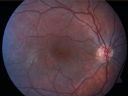
X-Linked Juvenille Retinoschisis 10 Year Old Child treated with Trusopt761 views10-year-old child has x-linked juvenile retinoschisis VA 20/60, OS is 20/100. Macula and Vision improved with Trusopt drops.
|
|

X-Linked Juvenille Retinoschisis 10 Year Old Child treated with Trusopt821 views10-year-old child has x-linked juvenile retinoschisis VA 20/60, OS is 20/100. Macula and Vision improved with Trusopt drops.
|
|

X-Linked Juvenille Retinoschisis 10 Year Old Child treated with Trusopt654 views10-year-old child has x-linked juvenile retinoschisis VA 20/60, OS is 20/100. Macula and Vision improved with Trusopt drops.
|
|

X-Linked Juvenille Retinoschisis 10 Year Old Child treated with Trusopt600 views10-year-old child has x-linked juvenile retinoschisis VA 20/60, OS is 20/100. Macula and Vision improved with Trusopt drops.
|
|
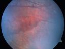
X-Linked Juvenille Retinoschisis 10 Year Old Child treated with Trusopt764 views10-year-old child has x-linked juvenile retinoschisis VA 20/60, OS is 20/100. Macula and Vision improved with Trusopt drops.
|
|

X-Linked Juvenille Retinoschisis 10 Year Old Child treated with Trusopt602 views10-year-old child has x-linked juvenile retinoschisis VA 20/60, OS is 20/100. Macula and Vision improved with Trusopt drops.
|
|
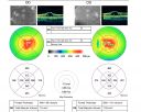
X-Linked Juvenille Retinoschisis 10 Year Old Child treated with Trusopt714 views10-year-old child has x-linked juvenile retinoschisis VA 20/60, OS is 20/100. Macula and Vision improved with Trusopt drops.
|
|

X-Linked Juvenille Retinoschisis 10 Year Old Child treated with Trusopt652 views10-year-old child has x-linked juvenile retinoschisis VA 20/60, OS is 20/100. Macula and Vision improved with Trusopt drops.
|
|

X-Linked Juvenille Retinoschisis 10 Year Old Child treated with Trusopt638 views10-year-old child has x-linked juvenile retinoschisis VA 20/60, OS is 20/100. Macula and Vision improved with Trusopt drops.
|
|

X-Linked Juvenille Retinoschisis 10 Year Old Child treated with Trusopt679 views10-year-old child has x-linked juvenile retinoschisis VA 20/60, OS is 20/100. Macula and Vision improved with Trusopt drops.
|
|

X-Linked Juvenille Retinoschisis 10 Year Old Child treated with Trusopt631 views10-year-old child has x-linked juvenile retinoschisis VA 20/60, OS is 20/100. Macula and Vision improved with Trusopt drops.
|
|

X-Linked Juvenille Retinoschisis 10 Year Old Child treated with Trusopt756 views10-year-old child has x-linked juvenile retinoschisis VA 20/60, OS is 20/100. Macula and Vision improved with Trusopt drops.
|
|

Disciform Macular Scar Both Eyes from Wet Macular Degeneration647 views86-year-old woman has disciform scars OD: 20/400; OS: 20/100
|
|

Group 2a Juxtafoveal Retinal Telangiectasis (type 2)703 views58-year-old man decreased vision right eye and not better since cataract surgery one year ago. OD 20/40, OS 20/40.
|
|

Group 2a Juxtafoveal Retinal Telangiectasis (type 2)595 views58-year-old man decreased vision right eye and not better since cataract surgery one year ago. OD 20/40, OS 20/40.
|
|

Group 2a Juxtafoveal Retinal Telangiectasis (type 2)581 views58-year-old man decreased vision right eye and not better since cataract surgery one year ago. OD 20/40, OS 20/40.
|
|

Group 2a Juxtafoveal Retinal Telangiectasis (type 2)594 views58-year-old man decreased vision right eye and not better since cataract surgery one year ago. OD 20/40, OS 20/40.
|
|

Group 2a Juxtafoveal Retinal Telangiectasis (type 2)610 views58-year-old man decreased vision right eye and not better since cataract surgery one year ago. OD 20/40, OS 20/40.
|
|

Group 2a Juxtafoveal Retinal Telangiectasis (type 2)534 views58-year-old man decreased vision right eye and not better since cataract surgery one year ago. OD 20/40, OS 20/40.
|
|

Group 2a Juxtafoveal Retinal Telangiectasis (type 2)532 views58-year-old man decreased vision right eye and not better since cataract surgery one year ago. OD 20/40, OS 20/40.
|
|

Group 2a Juxtafoveal Retinal Telangiectasis (type 2)568 views58-year-old man decreased vision right eye and not better since cataract surgery one year ago. OD 20/40, OS 20/40.
|
|

Group 2a Juxtafoveal Retinal Telangiectasis (type 2)587 views58-year-old man decreased vision right eye and not better since cataract surgery one year ago. OD 20/40, OS 20/40.
|
|

Group 2a Juxtafoveal Retinal Telangiectasis (type 2)623 views58-year-old man decreased vision right eye and not better since cataract surgery one year ago. OD 20/40, OS 20/40.
|
|

Group 2a Juxtafoveal Retinal Telangiectasis (type 2)669 views58-year-old man decreased vision right eye and not better since cataract surgery one year ago. OD 20/40, OS 20/40.
|
|

Plaquenil Toxicity - Bulls Eye Maculopathy1338 views70-year-old woman with systemic Lupus erythematosus and clotting problems. She was on the Plaquenil for about eight years and then off the Plaquenil for the last eight years because she developed macular toxicity. Although her vision was hazy, it was stable. Recent deceased vision left eye: OD 20/60, OS 20/100. IOP: OD 18, OS 19.
|
|

Plaquenil Toxicity - Bulls Eye Maculopathy1840 views70-year-old woman with systemic Lupus erythematosus and clotting problems. She was on the Plaquenil for about eight years and then off the Plaquenil for the last eight years because she developed macular toxicity. Although her vision was hazy, it was stable. Recent deceased vision left eye: OD 20/60, OS 20/100. IOP: OD 18, OS 19.
|
|
| 439 files on 5 page(s) |
1 |
 |
|
|


