Image search results - "Macular edema"
|

Diabetic Patient with Macular Edema and Blood Pressure 200/95881 views49-year-old decreasing vision over the last year. OD is 20/80, OS 20/80. blood pressure which was 200/95.
|
|

Diabetic Patient with Macular Edema and Blood Pressure 200/95903 views49-year-old decreasing vision over the last year. OD is 20/80, OS 20/80. blood pressure which was 200/95.
|
|
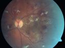
Diabetic Patient with Macular Edema and Blood Pressure 200/95662 views49-year-old decreasing vision over the last year. OD is 20/80, OS 20/80. blood pressure which was 200/95.
|
|

Diabetic Patient with Macular Edema and Blood Pressure 200/95539 views49-year-old decreasing vision over the last year. OD is 20/80, OS 20/80. blood pressure which was 200/95.
|
|

Diabetic Patient with Macular Edema and Blood Pressure 200/95526 views49-year-old decreasing vision over the last year. OD is 20/80, OS 20/80. blood pressure which was 200/95.
|
|

Diabetic Patient with Macular Edema and Blood Pressure 200/95538 views49-year-old decreasing vision over the last year. OD is 20/80, OS 20/80. blood pressure which was 200/95.
|
|
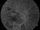
Diabetic Patient with Macular Edema and Blood Pressure 200/95514 views49-year-old decreasing vision over the last year. OD is 20/80, OS 20/80. blood pressure which was 200/95.
|
|

Diabetic Patient with Macular Edema and Blood Pressure 200/95534 views49-year-old decreasing vision over the last year. OD is 20/80, OS 20/80. blood pressure which was 200/95.
|
|

Diabetic Patient with Macular Edema and Blood Pressure 200/95523 views49-year-old decreasing vision over the last year. OD is 20/80, OS 20/80. blood pressure which was 200/95.
|
|

Diabetic Patient with Macular Edema and Blood Pressure 200/95551 views49-year-old decreasing vision over the last year. OD is 20/80, OS 20/80. blood pressure which was 200/95.
|
|

Diabetic Patient with Macular Edema and Blood Pressure 200/95567 views49-year-old decreasing vision over the last year. OD is 20/80, OS 20/80. blood pressure which was 200/95.
|
|

Diabetic Patient with Macular Edema and Blood Pressure 200/95544 views49-year-old decreasing vision over the last year. OD is 20/80, OS 20/80. blood pressure which was 200/95.
|
|

Diabetic Patient with Macular Edema and Blood Pressure 200/95634 views49-year-old decreasing vision over the last year. OD is 20/80, OS 20/80. blood pressure which was 200/95.
|
|

Diabetic Patient with Macular Edema and Blood Pressure 200/95645 views49-year-old decreasing vision over the last year. OD is 20/80, OS 20/80. blood pressure which was 200/95.
|
|

Diabetic Patient with Macular Edema and Blood Pressure 200/95671 views49-year-old decreasing vision over the last year. OD is 20/80, OS 20/80. blood pressure which was 200/95.
|
|

BRVO390 views50 yr old Female with a BRVO and ME in OS
|
|

Macroaneurysm FA636 viewsMacroaneurysm with Subretinal Hemorrhage and ME in OS of an Elderly Female
|
|

Macroaneurysm FP1091 viewsMacroaneurysm with Subretinal Hemorrhage and ME in OS of an Elderly Female
|
|
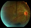
PDR w/ERM and severe CSDME669 views24 year old woman with uncontrolled blood sugar; progressively worsening vision OU - from level of 20/40 in August 2012 to 20/200:OD and 20/CF:OS in January 2013.
|
|

PDR, NVE - interesting shape489 views24 yr. old with uncontrolled blood sugar. Decreased vision from 20/40 O.U. in August of 2012, to 20/200: OD and 20/CF: OS in January 2013.
|
|

Pseudophakic Cystoid Macular Edema FA late frame1018 views
|
|
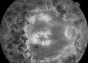
late phase FA985 views50+ YO female presented with DM type 1;
PT presented with PDR s/p extensive PRP
FA showed CNV OD
VA stable at 20/150 for 1 year
|
|

early phase FA498 views50+ YO female presented with DM type 1;
PT presented with PDR s/p extensive PRP
FA showed CNV OD
VA stable at 20/150 for 1 year
|
|

early phase FA544 views50+ YO female presented with DM type 1;
PT presented with PDR s/p extensive PRP
FA showed CNV OD
VA stable at 20/150 for 1 year
|
|

Central Retinal Venous Occlusion with Papilledema, Intraretinal Hemorrhage and Macular Edema847 views
|
|

Branch Retinal Vein Occlusion with worse macular edema from hypertension - FA shows Edema Right Eye522 views
|
|

branch retinal vein occlusion - fundus photo951 views70 year old woman with 20/80 vision from a branch retinal vein occlusion and macular edema. She has had laser treatment and intravitreal steroids. Her vision with further laser improved to 20/50.
|
|

Idiopathic Cystoid Macular Edema - CME - Responded to Subtenons Kenalog - Color Photo Showing Foveal Cysts899 views49-year-old man had a flu two weeks ago the right eye has been throbbing and his vision in the right eye has been foggy. OD 20/80, OS 20/40. IOP: 14 OU.
|
|

BRVO with collaterals and cystoid macular edema1010 views66 Year Old Man with has had a branch retinal vein occlusion in the right eye with vision loss for the last few months. OD is 20/40, OS is 20/20. IOP: OD 18, OS 19. There is a superotemporal macular branch retinal vein occlusion with edema, hemorrhage, and exudate.
OCT SCAN: The right eye shows edema with a central foveal thickness of 354 microns FA: shows remodeling of the superotemporal vasculature. There are already venous-venous collaterals, suggesting the vein occlusion may be a little more than a few months old. There are multiple microaneurysms superotemporal to the fovea, and there is leakage in the late frames involving the fovea.
|
|

branch retinal vein occlusion - oct map894 views70 year old woman with 20/80 vision from a branch retinal vein occlusion and macular edema. She has had laser treatment and intravitreal steroids. Her vision with further laser improved to 20/50.
|
|

Idiopathic Cystoid Macular Edema - CME - Responded to Subtenons Kenalog - Color Photo786 views49-year-old man had a flu two weeks ago the right eye has been throbbing and his vision in the right eye has been foggy. OD 20/80, OS 20/40. IOP: 14 OU.
|
|

BRVO with collaterals and cystoid macular edema699 views66 Year Old Man with has had a branch retinal vein occlusion in the right eye with vision loss for the last few months. OD is 20/40, OS is 20/20. IOP: OD 18, OS 19. There is a superotemporal macular branch retinal vein occlusion with edema, hemorrhage, and exudate.
OCT SCAN: The right eye shows edema with a central foveal thickness of 354 microns FA: shows remodeling of the superotemporal vasculature. There are already venous-venous collaterals, suggesting the vein occlusion may be a little more than a few months old. There are multiple microaneurysms superotemporal to the fovea, and there is leakage in the late frames involving the fovea.
|
|

brvo cme with small serous macular detachment 754 views70 year old woman with 20/80 vision from a branch retinal vein occlusion and macular edema. She has had laser treatment and intravitreal steroids. Her vision with further laser improved to 20/50.
|
|

Idiopathic Cystoid Macular Edema - CME - Pre Treatment Optical Coherence Tomography Map Showing Thickened Right Macula934 views49-year-old man had a flu two weeks ago the right eye has been throbbing and his vision in the right eye has been foggy. OD 20/80, OS 20/40. IOP: 14 OU.
|
|

BRVO with collaterals and cystoid macular edema717 views66 Year Old Man with has had a branch retinal vein occlusion in the right eye with vision loss for the last few months. OD is 20/40, OS is 20/20. IOP: OD 18, OS 19. There is a superotemporal macular branch retinal vein occlusion with edema, hemorrhage, and exudate.
OCT SCAN: The right eye shows edema with a central foveal thickness of 354 microns FA: shows remodeling of the superotemporal vasculature. There are already venous-venous collaterals, suggesting the vein occlusion may be a little more than a few months old. There are multiple microaneurysms superotemporal to the fovea, and there is leakage in the late frames involving the fovea.
|
|
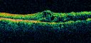
branch retinal vein occlusion - oct line scan768 views70 year old woman with 20/80 vision from a branch retinal vein occlusion and macular edema. She has had laser treatment and intravitreal steroids. Her vision with further laser improved to 20/50.
|
|

Idiopathic Cystoid Macular Edema - CME - Responded to Subtenons Kenalog - Pre Treatment Line Scan ShowingLarge Foveal Cysts694 views49-year-old man had a flu two weeks ago the right eye has been throbbing and his vision in the right eye has been foggy. OD 20/80, OS 20/40. IOP: 14 OU.
|
|
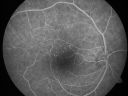
BRVO with collaterals and cystoid macular edema824 views66 Year Old Man with has had a branch retinal vein occlusion in the right eye with vision loss for the last few months. OD is 20/40, OS is 20/20. IOP: OD 18, OS 19. There is a superotemporal macular branch retinal vein occlusion with edema, hemorrhage, and exudate.
OCT SCAN: The right eye shows edema with a central foveal thickness of 354 microns FA: shows remodeling of the superotemporal vasculature. There are already venous-venous collaterals, suggesting the vein occlusion may be a little more than a few months old. There are multiple microaneurysms superotemporal to the fovea, and there is leakage in the late frames involving the fovea.
|
|

branch retinal vein occlusion - red free photo830 views70 year old woman with 20/80 vision from a branch retinal vein occlusion and macular edema. She has had laser treatment and intravitreal steroids. Her vision with further laser improved to 20/50.
|
|

Idiopathic Cystoid Macular Edema - CME - Responded to Subtenons Kenalog - Red Free Photos Shows Foveal Cysts624 views49-year-old man had a flu two weeks ago the right eye has been throbbing and his vision in the right eye has been foggy. OD 20/80, OS 20/40. IOP: 14 OU.
|
|

BRVO with collaterals and cystoid macular edema650 views66 Year Old Man with has had a branch retinal vein occlusion in the right eye with vision loss for the last few months. OD is 20/40, OS is 20/20. IOP: OD 18, OS 19. There is a superotemporal macular branch retinal vein occlusion with edema, hemorrhage, and exudate.
OCT SCAN: The right eye shows edema with a central foveal thickness of 354 microns FA: shows remodeling of the superotemporal vasculature. There are already venous-venous collaterals, suggesting the vein occlusion may be a little more than a few months old. There are multiple microaneurysms superotemporal to the fovea, and there is leakage in the late frames involving the fovea.
|
|

BRVO arrow showing obstruction point with collateral vessels - red free photo818 views70 year old woman with 20/80 vision from a branch retinal vein occlusion and macular edema. She has had laser treatment and intravitreal steroids. Her vision with further laser improved to 20/50.
|
|

Idiopathic Cystoid Macular Edema - CME - Responded to Subtenons Kenalog - Early Fluorescein Angiogram Showing Dilated Capillaries614 views49-year-old man had a flu two weeks ago the right eye has been throbbing and his vision in the right eye has been foggy. OD 20/80, OS 20/40. IOP: 14 OU.
|
|
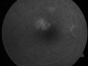
BRVO with collaterals and cystoid macular edema842 views66 Year Old Man with has had a branch retinal vein occlusion in the right eye with vision loss for the last few months. OD is 20/40, OS is 20/20. IOP: OD 18, OS 19. There is a superotemporal macular branch retinal vein occlusion with edema, hemorrhage, and exudate.
OCT SCAN: The right eye shows edema with a central foveal thickness of 354 microns FA: shows remodeling of the superotemporal vasculature. There are already venous-venous collaterals, suggesting the vein occlusion may be a little more than a few months old. There are multiple microaneurysms superotemporal to the fovea, and there is leakage in the late frames involving the fovea.
|
|

branch retinal vein occlusion - fluorescein angiogram575 views70 year old woman with 20/80 vision from a branch retinal vein occlusion and macular edema. She has had laser treatment and intravitreal steroids. Her vision with further laser improved to 20/50.
|
|

Idiopathic Cystoid Macular Edema - CME - Responded to Subtenons Kenalog - Early Fluorescein Showing Dilated Capillaries680 views49-year-old man had a flu two weeks ago the right eye has been throbbing and his vision in the right eye has been foggy. OD 20/80, OS 20/40. IOP: 14 OU.
|
|

BRVO with collaterals and cystoid macular edema594 views66 Year Old Man with has had a branch retinal vein occlusion in the right eye with vision loss for the last few months. OD is 20/40, OS is 20/20. IOP: OD 18, OS 19. There is a superotemporal macular branch retinal vein occlusion with edema, hemorrhage, and exudate.
OCT SCAN: The right eye shows edema with a central foveal thickness of 354 microns FA: shows remodeling of the superotemporal vasculature. There are already venous-venous collaterals, suggesting the vein occlusion may be a little more than a few months old. There are multiple microaneurysms superotemporal to the fovea, and there is leakage in the late frames involving the fovea.
|
|

branch retinal vein occlusion - fluorescein angiogram684 views70 year old woman with 20/80 vision from a branch retinal vein occlusion and macular edema. She has had laser treatment and intravitreal steroids. Her vision with further laser improved to 20/50.
|
|

Idiopathic Cystoid Macular Edema - Mid Phase Fluorescein Angiogram Showing Leakage of Dye into Center of Macula836 views49-year-old man had a flu two weeks ago the right eye has been throbbing and his vision in the right eye has been foggy. OD 20/80, OS 20/40. IOP: 14 OU.
|
|

BRVO with collaterals and cystoid macular edema736 views66 Year Old Man with has had a branch retinal vein occlusion in the right eye with vision loss for the last few months. OD is 20/40, OS is 20/20. IOP: OD 18, OS 19. There is a superotemporal macular branch retinal vein occlusion with edema, hemorrhage, and exudate.
OCT SCAN: The right eye shows edema with a central foveal thickness of 354 microns FA: shows remodeling of the superotemporal vasculature. There are already venous-venous collaterals, suggesting the vein occlusion may be a little more than a few months old. There are multiple microaneurysms superotemporal to the fovea, and there is leakage in the late frames involving the fovea.
|
|

branch retinal vein occlusion - fluorescein angiogram577 views70 year old woman with 20/80 vision from a branch retinal vein occlusion and macular edema. She has had laser treatment and intravitreal steroids. Her vision with further laser improved to 20/50.
|
|

Idiopathic Cystoid Macular Edema - CME - Responded to Subtenons Kenalog - Fluorescein Angiogram - Leakage of White Dye Into Center of Macula763 views49-year-old man had a flu two weeks ago the right eye has been throbbing and his vision in the right eye has been foggy. OD 20/80, OS 20/40. IOP: 14 OU.
|
|

branch retinal vein occlusion - fluorescein angiogram - arrow shows obstruction point with collateral vessels857 views70 year old woman with 20/80 vision from a branch retinal vein occlusion and macular edema. She has had laser treatment and intravitreal steroids. Her vision with further laser improved to 20/50.
|
|

Idiopathic Cystoid Macular Edema - CME - Responded to Subtenons Kenalog - Fellow (uninvolved) Eye Nearly Normal Dark Center FA761 views49-year-old man had a flu two weeks ago the right eye has been throbbing and his vision in the right eye has been foggy. OD 20/80, OS 20/40. IOP: 14 OU.
|
|

branch retinal vein occlusion - fluorescein angiogram631 views70 year old woman with 20/80 vision from a branch retinal vein occlusion and macular edema. She has had laser treatment and intravitreal steroids. Her vision with further laser improved to 20/50.
|
|
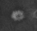
Idiopathic Cystoid Macular Edema - CME - Close up of Late Fluorescein Angiogram Showing Leakage into Foveal Cysts855 views49-year-old man had a flu two weeks ago the right eye has been throbbing and his vision in the right eye has been foggy. OD 20/80, OS 20/40. IOP: 14 OU.
|
|

branch retinal vein occlusion - fluorescein angiogram644 views70 year old woman with 20/80 vision from a branch retinal vein occlusion and macular edema. She has had laser treatment and intravitreal steroids. Her vision with further laser improved to 20/50.
|
|

branch retinal vein occlusion - fluorescein angiogram787 views70 year old woman with 20/80 vision from a branch retinal vein occlusion and macular edema. She has had laser treatment and intravitreal steroids. Her vision with further laser improved to 20/50.
|
|
|
|
|
|


