|
|

Neuroretinitis and Multifocal Retinitis449 views62 year old female with vision loss in the left eye to 20/160. Positive Bartonellas IgG. Vision recovered in 2 months. She was treated with Oral Erythromycin BID for 2 weeks.
|
|

Primary Toxoplasmosis449 views76 year old man with blurred vision left eye. He improved to 20/25 in 2 months with valtrex. No steroids were given
|
|

Lupus Choroidopathy - Multifocal PED449 views82 year old female with spot in her vision for over a year. PEDs have been stable with VA 20/20 OD; 20/40 OS
|
|

Neuroretinitis - Negative cat scratch serology twice - Possible Behcets449 views65 year old man with no direct cat exposure and vision loss from neuroretinitis. His work up was positive for HLA B51. He had negative cat scratch titers twice. Vision dropped from 20/60 - 20/200 and then improved to 20/40 over 2 months
|
|

Neuroretinitis - Negative cat scratch serology twice - Possible Behcets449 views65 year old man with no direct cat exposure and vision loss from neuroretinitis. His work up was positive for HLA B51. He had negative cat scratch titers twice. Vision dropped from 20/60 - 20/200 and then improved to 20/40 over 2 months
|
|

Retinitis Pigmentosa - RP1 Mutation449 views78 year old man: 20/32 OD and 20/50 OS - RP1 Gene is Bad -- > RP with Novel Amino Acid Change in GLY723Stop sequence of the RP1 gene - consistent with AD RP
|
|

Angioid Streaks - CNVM OS with vision loss449 views48 year old soccer player. Prior trauma OS (with soccer ball). Vision loss for 3 weeks to 20/60 - with Lucentis vision improved to 20/30
|
|

Microaneursyms and Macular Edema Not Diabetic449 views85 year old man has been tested about 2-3 times and is always negative for diabetes
|
|
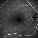
Microaneursyms and Macular Edema Not Diabetic449 views85 year old man has been tested about 2-3 times and is always negative for diabetes
|
|

Cat Scratch - Neuroretinitis OD AND retinitis OS - Bartonella henselae positive449 views27 year old female with exposure to cats and prior fever. Reports with recent vision loss right eye. She was treated with doxycycline and within 2 weeks the right eye looks better. Then she skipped all follow-up visits. The right eye presented 20/200 and the left 20/16
|
|

Acute Central Retinal Artery Occlusion449 views1 day of vision loss 79 year old man to HM. NVG developed 2 months later.
|
|

Nanophthalmous with Choroidal Folds449 views71 year old female with 20/20 vision and worsening cataracts. There is also a CHRPE lesion in the periphery
|
|

Moderate PDR449 views43 year old man 20/32 vision in both eyes. Being started on Anti-VEGF injections which will be followed by PRP.
|
|

Lung Cancer Metastasis to Right Eye449 views63 year old female with vision loss for 6 weeks. She had been scheduled for cataract surgery and then a tumor was found. She was ultimately diagnosed with lung cancer. Vision is 20/100
|
|

Papilledema - Benign Intracranial hypertension - Idiopathic intracranial hypertension449 views35 year old obese female. Normal vision and headache. Opening pressure was 30 cm water (normal 10-20). Nerve swelling responded to diamox.
|
|

Papilledema - Benign Intracranial hypertension - Idiopathic intracranial hypertension449 views35 year old obese female. Normal vision and headache. Opening pressure was 30 cm water (normal 10-20). Nerve swelling responded to diamox.
|
|

Heterochromia - Left Fundus - Possible Waardenburg Syndrome449 views49 year old female with normal vision. No hearing loss. Fundus in the left eye has large patchy hypopigmentation. OCT in that area shows decrease vascularity.
|
|
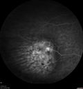
Melanosis Oculi and Choroidal Melanoma449 views88 year old female with 2.5 mm elevated choroidal melanoma and melanosis oculi. She also has geographic atrophy from AMD. The right eye is the better eye with 20/40 vision. After observation with growth, brachytherapy was done.
|
|

Punctate Inner Choroidopathy and Choroidal Neovascular Membrane449 views27 year old female with vision loss for 4 months. VA was 20/200. She had a CNVM which was extrafoveal with no subretinal fluid. Her vision improved over 6 months to 20/50 with Ranibizumab injections (she might have improved without treatment)
|
|

Acute Branch Retinal Vein Occlusion - 20/200449 views66 year old female with 3 weeks of vision loss in the left eye. VA 20/20 OD 20/200 OS
|
|

Choroidal Nevus with Drusen449 views82 year old female with flat nevus for 3 years.
|
|

Choroidal Neovascular Membrane following Prior Central Serous Chorioretinopathy - OCT-A449 views67 year old female with CSR in the left eye first 20 years ago. She was managed with out therapy and has had a few episodes since. Now ther eis distortion in the left eye for a few days. She had a steroid shot in her shoulder 5 months ago.
VA 20/20 OD, 20/50 OS- ICG and OCT-A confirm CNVM in the left eye
|
|

Acquired Toxoplasmosis Retinitis Para Foveal - Following Venison Consumption449 views56 year old man who became very sick after eating venison at the end of December 2019. Presented 6 weeks later with a scotoma. Initial photos show presentation with 20/63 vision. Vision dropped to 5/200. All tests (including PCR of anterior chamber tap) were negative except serum toxoplasmosis IgG and IgM. Vision improved to 20/32 but the scotoma remained on oral trimetheprim/sulfa
|
|

Cilioretinal Artery Occlusion - Nasal (not macular)449 views77 year old female There is a shadow in the top half of her temporal vision in the left eye that came on suddenly yesterday morning. It has not changed since then and the right eye is OK. She has had sinus headaches. She had no focal neurological changes. Â
VA OD: Dcc20/32
VA OS: Dcc20/16-1
IOP: TP: OD:18 OS:19Â
|
|

Stellate non-hereditary idiopathic foveomacular retinoschisis (SNIFR) and optic nerve drusen449 views84 year old man No visual complaints. Not diabetic, cataract surgery 8 years ago. Meds: Omeprazole, Tamsulosin (Flomax)
VA 20/32 OD, 20/20 OS
|
|

Branch retinal vein occlusion with macular edema449 views71 year old man Starting about 2 weeks ago the left eye was blurred. The right eye is seeing well.
VA OD: sc20/25+2 NccJ1+
VA OS: sc20/63+2 PH20/50-2 Ncc20/400
IOP: TP: OD:19 OS:16
|
|

Lipemia Retinalis - Lipid Obscuring Vitreous449 views31 year old man The patient at the time of imaging had triglycerides of >5680, cholesterol of 1136.
He also does have uncontrolled hypertension and insulin dependent type 2 diabetes, which may be the reason for vitreous extension of the lipids.
|
|

448 views
|
|

Proliferative Diabetic Retinopathy Both Eyes - Vitreous Hemorrahge Left Eye448 views67-year-old man has proliferative diabetic retinopathy in both eyes with vitreous hemorrhage in the left eye. He has had pan retinal laser in the left eye. The vitreous hemorrhage has been there for greater than two years and his vision is still hazy. He takes Lumigan in both eyes for his glaucoma.
VISUAL ACUITY: OD 20/40, OS 20/40
|
|

X-linked Juvenille Retinoschisis - Peripheral Retinal Vascular Anomalies - Vitreous Hemorrhage - 8 Year Old Boy448 views8-year-old child OD 20/40, OS 20/50
OD: Vertical C/D ratio is 0.2. There are foveal cysts. There is also a retinal elevation inferiorly and there are patchy peripheral retinal hemorrhages.
OS: Vertical C/D ratio is 0.2. There are foveal cysts. There is peripheral retinal issues with some hemorrhage in some areas of peripheral retinoschisis.
|
|

X-linked Juvenille Retinoschisis - Peripheral Retinal Vascular Anomalies - Vitreous Hemorrhage - 8 Year Old Boy448 views8-year-old child OD 20/40, OS 20/50
OD: Vertical C/D ratio is 0.2. There are foveal cysts. There is also a retinal elevation inferiorly and there are patchy peripheral retinal hemorrhages.
OS: Vertical C/D ratio is 0.2. There are foveal cysts. There is peripheral retinal issues with some hemorrhage in some areas of peripheral retinoschisis.
|
|

Hypertensive Retinopathy448 viewsPatient comes in complaining of spots in vision in both eyes. VA was 20/25 - right eye and 20/20- left eye. Fundus exam reveals little hemorrhages with cotton wool spots due to hypertension and anemia.
|
|
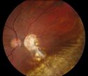
Reoccurring Retinal Detachment448 viewsPatient comes in for second opinion for RD in the right eye. Patient's VA was count fingers @ 2-ft in the right eye and 20/20 in the left eye. Patient is aphakic and has had 5- retinal surgeries in the past in the right eye including a scleral buckle. RD present with the macula off. Will consider surgery with silicone oil.
|
|

448 views
|
|

Lupus Retinopathy448 viewsFemale patient comes in for eval on Lupus Retinopathy. Has poor vision in the right eye. VA is hand motion in the right eye. Fundus photos show fibrosis along the temporal arcades and narrowing of the arteries. No macular edema found.
|
|

Gonioscopy, Mass in the Angle of Anterior Chamber448 viewsSlit Lamp and goinoscopy photos show a mass at 7-o'clock i the right eye. The mass extends beneath the iris behind the lens.
|
|

Wet AMD OD and Lamellar Macular hole OS448 viewsWet AMD in right and better eye failed extend to 2 months but was able to stop therapy once he started on systemic Avastin for cancer. The last image shows the macula dry 9 months without local therapy. The left eye has a lamellar macular hole.
|
|
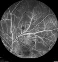
Hairy Cell Leukemia - Retinal Hemorrhage and twig Branch Vein Occlusion448 views79 year old man He has had hairy cell leukemia since 2002. He is in remission. His last blood tests were 9/2018. He just moved down here and needs a new leukemia doctor. His vision is fine.
VA OD: Dcc20/25
VA OS: Dcc20/25
IOP: TP: OD:12 OS:12
|
|

Hypertensive Retinopathy - BP 240 / 130 mmHg - Grade 4448 views71 year old woman who claims to be asymptomatic with decreased vision, macular edema, cotton wool spots, subretinal fluid and optic nerve edema
|
|

Multifocal Bilateral Central Serous Retinopathy in African American Male448 views67 year old african american male with : Blurred Vision OD. Duration of Problem: 4-6 weeks. VA 20/32 OD 20/20 OS
|
|

Multifocal Bilateral Central Serous Retinopathy in African American Male448 views67 year old african american male with : Blurred Vision OD. Duration of Problem: 4-6 weeks. VA 20/32 OD 20/20 OS
|
|

Central Serous Retinopathy - Subretinal Fibrin448 views66 year old female with 20 year history of single episode of CSR. Now with vision loss for a month but improving - VA 20/63 OD; 20/32 OS
|
|

Branch Retinal Arterial Occlusion Right Eye - Atheromata - Both Eyes448 views83 year old woman with no symptoms - VA 20/40 in both eyes. Right eye has ST BRAO with silver wiring. Left eye has superotemporal atheromata
|
|

Cystoid Macular Edema following Cataract surgery and vitrectomy for floaters448 views73 year old woman who had decreased vision 4 months following combined vitrectomy and cataract surgery. VA 20/63 improved in 1 month to 20/25 with ketorolac and prednisolone drops.
|
|

Persistent Subfoveal Fluid Following Retinal Detachment Repair448 viewsPatient had pneumatic retinopexy for macula off RD and subfoveal fluid persisted for 12 months but was gone at 18 months. IR images show pockets of fluid after the first week.
|
|

Vascular Remodeling Bilateral BRVO448 views85 year old female 20/25 OU with BRVO 7 years ago in both eyes. She has a change in vision. The FA shows extensive collaterallization and vascular remodeling in each macula.
|
|

448 views
|
|

Unilteral Acute Idiopathic Maculopathy - Initial Presentation448 views
|
|

Polypoidal Choroidal Vasculopathy Left Eye448 views74 year old man with vision loss to 20/100 over a few months. He improved to 20/20 with Lucentis. The color and ICG show the polyps.
|
|

Neuroretinitis - Negative cat scratch serology twice - Possible Behcets448 views65 year old man with no direct cat exposure and vision loss from neuroretinitis. His work up was positive for HLA B51. He had negative cat scratch titers twice. Vision dropped from 20/60 - 20/200 and then improved to 20/40 over 2 months
|
|

Macular Hole - Asymptomatic448 views71 year old female did not notice 20/200 macular hole. Post surgery vision improved to 20/30
|
|

Retinitis Pigmentosa - RP1 Mutation448 views78 year old man: 20/32 OD and 20/50 OS - RP1 Gene is Bad -- > RP with Novel Amino Acid Change in GLY723Stop sequence of the RP1 gene - consistent with AD RP
|
|

Retinitis Pigmentosa - RP1 Mutation448 views78 year old man: 20/32 OD and 20/50 OS - RP1 Gene is Bad -- > RP with Novel Amino Acid Change in GLY723Stop sequence of the RP1 gene - consistent with AD RP
|
|

Outer Retinal Tubulation448 views78 year old man chronic wet AMD - 20/200
|
|

pucker-prepeel-MC448 views
|
|

Pattern Dystrophy - Foveal Atrophy448 views73 year old female with vision loss - looks like macular hole
|
|

Unusual Intraretinal Deposits448 views71 year old diabetic man on dialysis with 20/20 vision and intraretinal deposits. Recent 150 lb weight loss, on dialysis, see album properties for full history
|
|
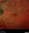
Vitreomacular Traction -> Macular Hole -> Aborted Macular hole448 viewsProgression of VMT in both eyes over time
|
|

Non-exudative wet AMD right eye (treatment naive quiescent)448 views82 year old man. ICG shows CNVM in the right eye. This stayed quiet for 3 years and then started leaking and responded well to Avastin.
|
|

Vitreous Hemorrhage with no evidence of PDR in the left eye448 views74 year old man with vision loss OS for about a week. The FA shows no PDR in the left eye but the right eye has very mild NVD. Diabetes for 40 years now on insulin.
|
|

Sickle Retinopathy - Chronic retinal detachment OS - Proliferation OD448 views40 year old with known SC disease. Failed to return 4 years ago for treatment and lost ision in the left eye. Then returned for one visit and refused further treatment.
|
|

Chronic CSC Both Eyes - Recent vision loss Right Eye Treated with PDT448 views50 year old man The right eye has had vision loss for a few weeks. He had previous episodes seen by me in 2012 OS and 2008 OD. This was 2018. VA OS has been poor for several years.
VA OD: Dcc20/100
VA OS: Dcc20/200-3
The patient had PDT to leaky area superotemporal to the fovea 1/2 dose and within 3 months was back up to 20/25. The last two OCT scans show fluid absorbing
|
|

Central Retinal Vein Occlusion - No macular edema - Pre-foveal opercula448 views62 year old female with 20/20 vision. She had been seeing a spot out of the left eye for 4 years and then noticed splotches in her vision. She never devleoped edema or NV
|
|

Central Retinal Vein Occlusion - No macular edema - Pre-foveal opercula448 views62 year old female with 20/20 vision. She had been seeing a spot out of the left eye for 4 years and then noticed splotches in her vision. She never devleoped edema or NV
|
|
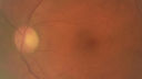
Late Onset Cone Dystrophy - Very Mild448 viewsOnly IR and OCT show very mild bull's eye. 67 year old female with difficulty seeing in bright light
|
|

Central Serous Chorioretinopathy - Moderate448 views49 year old man with episodes in 2011, 2017, and 2018 - now with some paracentral vision loss in the right eye and subretinal fluid.
|
|

Central Serous Chorioretinopathy - Moderate448 views49 year old man with episodes in 2011, 2017, and 2018 - now with some paracentral vision loss in the right eye and subretinal fluid.
|
|

Branch Retinal Artery Occlusion - No Plaque448 views74 year old man with 2 days of vision loss. Vision OS is 20/200 but improved to 20/25 in 1 month. He has dysfibrinogen anemia.
|
|

Macroaneurysm - Macular Hemorrhage - Branch Retinal Aterial Occlusion448 views62 year old African American female with chronic hypertension and vision loss for 2 weeks. VA is 20/400
|
|

Posterior Scleritis - positive T-sign448 views62 year ols man was sick about 6 weeks ago and then 2 weeks ago the the left eye got red. He was treated for conjunctivitis and the eye did not get better. Topical steroids and oral non-steroidal anti-inflammatory medications did not help.
VA OD: Dcc20/25 NccJ5
VA OS: Dcc5'/200
The scleritis cleared within 2 weeks of starting high dose (80 mg) oral steroids. Blood tests were all negative.
|
|

Multifocal Choroiditis and Subretinal Fibrosis with new Subretinal Neovascular Membrane448 views26 year old African American Girl Saw me at age 10 (16 years ago) with acute multifocal choroiditis and panuveitis which evolved into subretinal fibrosis. She was initially treated with 3 years of methotrexate. Vision ended up 20/20 in both eyes. Her eyes have been inactive since and she was lost to follow-up.
A week ago she noticed sudden decreased vision in the right eye. Within the past few weeks there are more floaters in the right eye.
VA OD: sc6’/200 PHNI
VA OS: sc20/16
|
|

Hyperpigmented Vasoproliferative Tumor on Buckle with New Serous Retinal Detachment 20 years post RD Repair448 views67 year old female RD repair OU 1999 then severe (20/400) macular pucker OD removed 3 months later (twin sister also had bilateral RD’s with PVR). Then dislocated IOL surgery right eye 9/2012. New vision loss in the right eye with serous retinal detachment. Argon laser to dark tumor led to resolution of subretinal fluid within a month.
|
|

Lung Cancer Metastasis to Right Eye448 views63 year old female with vision loss for 6 weeks. She had been scheduled for cataract surgery and then a tumor was found. She was ultimately diagnosed with lung cancer. Vision is 20/100
|
|
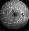
Chronic BRAO with Hemorrhages - Sclerosed Vessel448 views95 year old female with blurred vision in both eyes in for a checkup. History of perpapillary CNVM in the left eye 2 years prior not treated. History of stroke with multiple emboli 1 year ago.
Vision 20/32 OD, 20/40 OS
|
|

Macular Telangiectasis and Vision loss following vitrectomy with membrane peel448 views76 year old man with MacTel who had membrane peel in the right eye 10 years ago with permanent vision loss.
VA OD: sc20/200-4 and VA OS: sc20/25-1
|
|

Adult Pseudovitelliform Macular Dystrophy448 views89 year old female states dark smudge on ceiling during night time came and went. Decreased vision in the left eye
VA OD: Dcc20/32
VA OS: Dcc20/63
|
|

Chronic Endophthalmitis and Cystoid Macular Edema448 viewsVA 20/50 - Culture negative. Cleared with intravitreal antibiotics and decadron
|
|

Rapid macular atrophy with anti-VEGF therapy for myopic CNVM448 views64 year old female with 20/32 vision and distortion in the left and better eye. She was treated with monthly lucentis and her vision declined to 20/100. Medicine was stopped and she improved a little to 20/80.
|
|

Melanosis Oculi and Choroidal Melanoma448 views88 year old female with 2.5 mm elevated choroidal melanoma and melanosis oculi. She also has geographic atrophy from AMD. The right eye is the better eye with 20/40 vision. After observation with growth, brachytherapy was done.
|
|
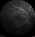
PDR and Vitreous Hemorrhage - High Risk Left Eye - Low Risk Right Eye448 views50 year old man with type I diabetes mellitus for 26 years. New Vitreous Hemorrhage in the left eye. Both eyes have NVE. Both also have foveal hypoplasia
|
|
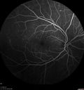
PDR and Vitreous Hemorrhage - High Risk Left Eye - Low Risk Right Eye448 views50 year old man with type I diabetes mellitus for 26 years. New Vitreous Hemorrhage in the left eye. Both eyes have NVE. Both also have foveal hypoplasia
|
|
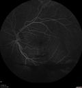
PDR and Vitreous Hemorrhage - High Risk Left Eye - Low Risk Right Eye448 views50 year old man with type I diabetes mellitus for 26 years. New Vitreous Hemorrhage in the left eye. Both eyes have NVE. Both also have foveal hypoplasia
|
|

Cystoid Macular Edema and Macular Pucker448 viewshas been particular bad for 3 years. She has a macular pucker and severe CME.
VA 20/160 left eye
|
|
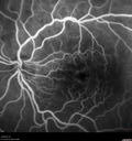
Cystoid Macular Edema and Macular Pucker448 viewshas been particular bad for 3 years. She has a macular pucker and severe CME.
VA 20/160 left eye
|
|

Normal Eye - Vision 20/16448 views61 year old female with multiple problems including chronic CME in the left eye.
|
|
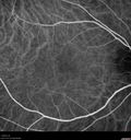
Normal Right eye448 views67 year old female with CSR in the left eye first 20 years ago. She was managed with out therapy and has had a few episodes since. Now ther eis distortion in the left eye for a few days. She had a steroid shot in her shoulder 5 months ago.
VA 20/20 OD, 20/50 OS
|
|

Eales Disease and fresh vitreous hemorrhage - 20 year old man448 views20 year old mane with fresh vitreous hemorrhage in the right eye. At age 15 he had a PPV and laser in the left eye and laser in the right eye. The vision is OD 20/80 PH 20/25, OS 20/25. The left eye has a mild cataract. He had prior testing for coagulopathies which was negative. Testing done for syphillis and TB was negative. Additional laser was done to prevent further bleeding in the right eye.
|
|

Myopic CNVM in the left eye. New Classic subfoveal lesion448 views64 year old female. She had retinal detachment repair in the right eye in 1996 (25 years ago). The left eye has new distortion for a week or two.
VA 20/40 OU.
Left eye has a very small subfoveal CNVM
|
|

Perifoveal Microaneurysm - Not Diabetic (also reticular pseudodrusen)448 views87 year old female with fluctuating macular edema in the right eye from a perifoveal MA. (no treatment). VA 20/40 OU
|
|

Perifoveal Microaneurysm - Not Diabetic (also reticular pseudodrusen)448 views87 year old female with fluctuating macular edema in the right eye from a perifoveal MA. (no treatment). VA 20/40 OU
|
|

Stellate non-hereditary idiopathic foveomacular retinoschisis (SNIFR) and optic nerve drusen448 views84 year old man No visual complaints. Not diabetic, cataract surgery 8 years ago. Meds: Omeprazole, Tamsulosin (Flomax)
VA 20/32 OD, 20/20 OS
|
|

Foveal Red Spot Syndrome Left Eye448 viewsShe noticed on covering the right eye that she had distortion off center in the left eye a few weeks ago. She had a temporal paracentral scotoma intermittently visible on the eye examination. She does notice that the left eye is not as good as the right eye now that it has been brought to her attention. She has every now and then a little twitch in the left eye. She has no pain on eye motion. Â
VA OD: sc20/20 NccJ1+
VA OS: sc20/20-1
|
|

Heavy Panretinal Photocoagulation for Proliferative Diabetic Retinopathy about 40 years ago448 views64 year old female - images from 2018 - heavy PRP about 30 years ago
VA 20/20 OD, 20/50 OS
|
|

Congenital Hypetrophy of the Retinal Pigment Epithelium (CHRPE)448 views63 year old female with normal vision and CHRPE lesion in the right eye.
|
|

448 views
|
|

Peripapillary Lesion Hemangioma or CNVM Early FA447 views67-year-old man with a history of a peripapillary lesion in the right eye.
|
|

Macular Pucker - Diabetic - Severe non-foveal Edema OS447 views74-year-old man has a macular pucker in the left eye and diabetic retinopathy.
VISUAL ACUITY: Vision OD is 20/25, OS is 20/40.
|
|

Basal Laminar Drusen - Cuticular Drusen - Equatorial Drusen - 70 Year Old Woman 447 views70 year old woman has had macular drusen since her thirties and she said her sister has a similar problem. Recently the vision in the left eye has substantially declined. OD 20/16, OS 10/200.
|
|

Retinal Pigment Epithelial Detachment with no Sub-Retinal Fluid...447 viewsA 38-year old male who comes in with blurred vision in the left eye. VA is 20/30. Notices a defect inferior of his central vision. Did an Fluorescence Angiogram to determine an RPE with no sub retinal fluid. Also OCT confirms. Patient was injected with Avastin.
|
|

BRVO447 views50 yr old Female with a BRVO and ME in OS
|
|
| 18117 files on 182 page(s) |
 |
|
|
|
|
113 | |
|
|
|
|


