|
|

Venous Macroaneurysm 6 year old CRVO - treated with focal laservaatamisi: 43593 year old female with CRVO in the right eye 6 years ago. The visual potential is 20/100 and has recently subjectively and objectively declined. VA is 20/200 and she has new onset macular edema from a venous macroaneurysm that formed at a collateral vessel. This responded nicely to a single laser spot.
|
|

Hyperpigmented Vasoproliferative Tumor on Buckle with New Serous Retinal Detachment 20 years post RD Repairvaatamisi: 43567 year old female RD repair OU 1999 then severe (20/400) macular pucker OD removed 3 months later (twin sister also had bilateral RD’s with PVR). Then dislocated IOL surgery right eye 9/2012. New vision loss in the right eye with serous retinal detachment. Argon laser to dark tumor led to resolution of subretinal fluid within a month.
|
|

Chronic Endophthalmitis and Cystoid Macular Edemavaatamisi: 435VA 20/50 - Culture negative. Cleared with intravitreal antibiotics and decadron
|
|

Punctate Inner Choroidopathy and Choroidal Neovascular Membranevaatamisi: 43527 year old female with vision loss for 4 months. VA was 20/200. She had a CNVM which was extrafoveal with no subretinal fluid. Her vision improved over 6 months to 20/50 with Ranibizumab injections (she might have improved without treatment)
|
|

Macular Telangiectasia - Mildvaatamisi: 43556 year old female with good vision and mild macular telangiectasia. VA 20/40 OD 20/16 OS
|
|

Choroidal folds right eye more than leftvaatamisi: 43568 year old man with prior laser corrective refractive surgery. VA OD 20/125 PH 20/32 OS 20/16 - Possible nanophthalmous
|
|
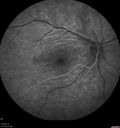
Choroidal folds right eye more than leftvaatamisi: 43568 year old man with prior laser corrective refractive surgery. VA OD 20/125 PH 20/32 OS 20/16 - Possible nanophthalmous
|
|

Sclerochoroidal Calcification and Peripheral Exudative Hemorrhagic Chorioretinopathyvaatamisi: 43567 year old man with asymptomatic peripheral subretinal hemorrhage associated with sclerochoroidal calcification.
VA 20/32 OU
|
|

Perifoveal Microaneurysm - Not Diabetic (also reticular pseudodrusen)vaatamisi: 43587 year old female with fluctuating macular edema in the right eye from a perifoveal MA. (no treatment). VA 20/40 OU
|
|

Perifoveal Microaneurysm - Not Diabetic (also reticular pseudodrusen)vaatamisi: 43587 year old female with fluctuating macular edema in the right eye from a perifoveal MA. (no treatment). VA 20/40 OU
|
|

Macular Telangiectasi - Stage V - CNVM OUvaatamisi: 43560 year old female who has been treated with injections for 5 years starting in the left eye. She had no treatment 6 months prior to this visit and her vision has been getting worse. Her vision is 20/100 OD, 20/32 OS. Vision improved with intravitreal Avastin to 20/30 OU.
|
|

Acquired Toxoplasmosis Retinitis Para Foveal - Following Venison Consumptionvaatamisi: 43556 year old man who became very sick after eating venison at the end of December 2019. Presented 6 weeks later with a scotoma. Initial photos show presentation with 20/63 vision. Vision dropped to 5/200. All tests (including PCR of anterior chamber tap) were negative except serum toxoplasmosis IgG and IgM. Vision improved to 20/32 but the scotoma remained on oral trimetheprim/sulfa
|
|

Enhanced S Cone Syndrome - Goldmann Favre - NR2E3 Mutationvaatamisi: 43582 year old man with poor vision for many years. VA HM OD, 5/200 OS. Diagnosed at age 12 with retinitis pigmentosa. Nystagmus.
|
|
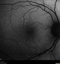
Mild Central Serous Chorioretinopathy - Flonase relatedvaatamisi: 43558 year old man with vision loss in the left eye for 8 days. This has never happened before. He started taking flonase about a month ago. He stopped the flonase and the vision improved in less than a month. Initial vision was 20/20 OD, 20/40 OS
|
|

Amelanotic Choroidal Nevus - Multimodal Imagingvaatamisi: 43561 year old man with amelanotic choroidal nevus. Vision Normal - Stable over 4 years.
|
|

Arteritic Anterior Ischmic Optic Neuropathy Left Eyevaatamisi: 43578 year old female with 5 day history of decreased vision in the left eye (sudden onset) She has been treated for GCA but her steroids were tapered down to 20 mg daily and she is not feeling well since the taper:
VA 20/20 OD, 20/25 OS ( APD OS)
|
|

Coloboma - Non Macular - Left Eyevaatamisi: 43571 year old female with cataracts. Left eye has inferior coloboma.
|
|
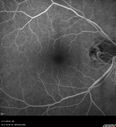
Branch retinal vein occlusion with macular edemavaatamisi: 43571 year old man Starting about 2 weeks ago the left eye was blurred. The right eye is seeing well.
VA OD: sc20/25+2 NccJ1+
VA OS: sc20/63+2 PH20/50-2 Ncc20/400
IOP: TP: OD:19 OS:16
|
|
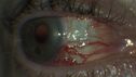
Elevated Episcleral Venous Pressure and Retinal Hemorrhages in both eyesvaatamisi: 43578 year old female with chronic red eye. The eyes are red all the time now. She is on Xeralto because of a history of blood clots in her lungs (2005 about). She has not had problems since. She has had floaters for many years. Ever since her eyes got funny she sees more floaters in both eyes. She had a brain scan over 6 months ago which was OK. She also had double vision when this first started which went away. She could not drive for a few months.
VA OD: Dcc20/32-1
VA OS: Dcc20/125
IOP: TP: OD:17 OS:16
Neuroimaging did not show a CC fistula
|
|

Central Serous Retinopathy 41 year old woman with Vision Loss for One Yearvaatamisi: 43441-year-old woman has chronic central serous retinopathy in the right eye with decreased vision in that eye for over a year. Her vision has been waxing and waning and recently is getting worse. Her vision in the left eye is fine.
VISUAL ACUITY: OD 20/40
|
|

Proliferative Diabetic Retinopathy - NVD regressed with PRP laservaatamisi: 43476 year old diabetic man - Presented one year ago with NVD in the left eye. This regressed with laser but then a year later worsened. Additional PRP was done and the NVD regressed again.
|
|
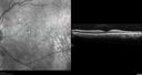
Normal Vision - Chronic Central Serous Chorioretinopathyvaatamisi: 43467 year old man - never had any symptoms - 20/16 vision in each eye. Lesions spare the fovea
|
|

Polypoidal Choroidal Vasculopathy Left Eyevaatamisi: 43474 year old man with vision loss to 20/100 over a few months. He improved to 20/20 with Lucentis. The color and ICG show the polyps.
|
|

Polypoidal Choroidal Vasculopathy Left Eyevaatamisi: 43474 year old man with vision loss to 20/100 over a few months. He improved to 20/20 with Lucentis. The color and ICG show the polyps.
|
|

Lupus Choroidopathy - Multifocal PEDvaatamisi: 43482 year old female with spot in her vision for over a year. PEDs have been stable with VA 20/20 OD; 20/40 OS
|
|

Neuroretinitis - Negative cat scratch serology twice - Possible Behcetsvaatamisi: 43465 year old man with no direct cat exposure and vision loss from neuroretinitis. His work up was positive for HLA B51. He had negative cat scratch titers twice. Vision dropped from 20/60 - 20/200 and then improved to 20/40 over 2 months
|
|

MacTel - Juxtafoveal Telangiectasisvaatamisi: 43456 year old man with a lot of difficulty adjusting to vision loss
|
|

Macroaneurysm - Resolved without treatmentvaatamisi: 43487 year old female with vision loss OD. Initial FA showed no leakage so no treatment was done and the fluid absorbed over 4 months. Initial VA 20/100, Final VA 20/80
|
|

Non-exudative wet AMD right eye (treatment naive quiescent)vaatamisi: 43482 year old man. ICG shows CNVM in the right eye. This stayed quiet for 3 years and then started leaking and responded well to Avastin.
|
|

Birdshot Chorioretintis and PAMMvaatamisi: 434Patient presented with paracentral acute middle maculopathy as the presenting symptom of birdshot. This case was published. RETINAL CASES & BRIEF REPORTS 10:151–153, 2016.
|
|

Central Retinal Vein Occlusion - No macular edema - Pre-foveal operculavaatamisi: 43462 year old female with 20/20 vision. She had been seeing a spot out of the left eye for 4 years and then noticed splotches in her vision. She never devleoped edema or NV
|
|

Nanophthalmous with Choroidal Foldsvaatamisi: 43471 year old female with 20/20 vision and worsening cataracts. There is also a CHRPE lesion in the periphery
|
|

Venous Macroaneurysm 6 year old CRVO - treated with focal laservaatamisi: 43493 year old female with CRVO in the right eye 6 years ago. The visual potential is 20/100 and has recently subjectively and objectively declined. VA is 20/200 and she has new onset macular edema from a venous macroaneurysm that formed at a collateral vessel. This responded nicely to a single laser spot.
|
|

Moderate PDRvaatamisi: 43443 year old man 20/32 vision in both eyes. Being started on Anti-VEGF injections which will be followed by PRP.
|
|

Hyperpigmented Vasoproliferative Tumor on Buckle with New Serous Retinal Detachment 20 years post RD Repairvaatamisi: 43467 year old female RD repair OU 1999 then severe (20/400) macular pucker OD removed 3 months later (twin sister also had bilateral RD’s with PVR). Then dislocated IOL surgery right eye 9/2012. New vision loss in the right eye with serous retinal detachment. Argon laser to dark tumor led to resolution of subretinal fluid within a month.
|
|
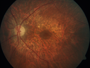
Concentric Geographic Atrophy - 76 year old manvaatamisi: 434Gradual vision loss, worse at night. VA 20/40 OU.
|
|

Choroidal Effusions following Glaucoma Surgery 9 months agovaatamisi: 43467 year old female with glaucoma surgery 9 month ago. Her IOP is 12. She has peripheral choroidal effusions. VA 20/100 ph 20/32
|
|
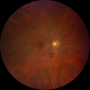
Retinitis Pigmentosa - Autosomal Recessive - 20/25 Visionvaatamisi: 43449 year old man 15 years ago he noticed decreased night vision.
VA OD: sc20/25 NscJ2
VA OS: sc20/25-2 NscJ2-1
IOP: TP: OD:17 OS:19
Genetic testing showed heterozygous for RPE65 - Probably not clinically important
|
|

Sclerochoroidal Calcification and Peripheral Exudative Hemorrhagic Chorioretinopathyvaatamisi: 43467 year old man with asymptomatic peripheral subretinal hemorrhage associated with sclerochoroidal calcification.
VA 20/32 OU
|
|

Eales Disease and fresh vitreous hemorrhage - 20 year old manvaatamisi: 43420 year old mane with fresh vitreous hemorrhage in the right eye. At age 15 he had a PPV and laser in the left eye and laser in the right eye. The vision is OD 20/80 PH 20/25, OS 20/25. The left eye has a mild cataract. He had prior testing for coagulopathies which was negative. Testing done for syphillis and TB was negative. Additional laser was done to prevent further bleeding in the right eye.
|
|

vaatamisi: 434
|
|

Severe Geographic Atrophy from Dry Age-Related Macular Degenerationvaatamisi: 43490 year old female with severe vision loss over the last few years.
VA 3/200 OD
1/200 OS
|
|

Choroidal Folds - Nanophthalmous - Axial length 20vaatamisi: 43467 year old female Decreased Vision OU with choriodal folds. Axial length is 20. There is a CHRPE lesion in the left eye.
VA 20/25 OU. Has already had cataract and glaucoma surgery.
|
|

Venous Anomaly - 80 year old female - normal visionvaatamisi: 434
|
|

Neuroretinitis Left Eyevaatamisi: 43429 year old female - The left eye blurred up and she had double vision back in 2/2019 and March. It has gradually improved so this past month the vision in the left eye is getting better. Her MRI scan of her orbits with and without contrast was normal. She had no pain in the eye with or without eye movement when the vision was blurred. She has a cat for over 5 years. She has no exposure to kittens or stray cats and her cat is an indoor cat.
VA OD: sc20/12
VA OS: sc20/16-1
All uveitis testing was negative (cat scratch, RPR, FTA-ABS, Sarcoid test, lyme test, TB testing. She later developed recurrent uveitis without optic nerve involvement.
|
|

Non-ischemic Central Retinal Vein Occlusion with Macular Edemavaatamisi: 43462 year old man The vision in the right eye started to decline about 10 days ago. Prior to that the two eyes were about the same.
VA OD: Dcc20/125 PH20/50-2
VA OS: Dcc20/20
IOP: TP: OD:21 OS:21
|
|

Branch retinal vein occlusion with macular edemavaatamisi: 43471 year old man Starting about 2 weeks ago the left eye was blurred. The right eye is seeing well.
VA OD: sc20/25+2 NccJ1+
VA OS: sc20/63+2 PH20/50-2 Ncc20/400
IOP: TP: OD:19 OS:16
|
|

hydroxychloroquine (Plaquenil) Toxicity - Taiwanese - 3 years post stoppingvaatamisi: 43459 year old female on 400 mg plaquenil for 22 years. 3 years ago, when stopping plaquenil, VA 20/50 both eyes. Plaquenil was stopped. The vision in the left eye continued to deteriorate to 3/200. 5'2" 127 lb (6.6 mg/kg body weight) -- Vision in these images is 20/80 OD, 5/200 OS - Vision loss now seems stable.
|
|

ABCA4 positive - Stargardt Diseasevaatamisi: 43424 year old female She has worn glasses since she was a kid. Since she became an adult her vision started to deteriorate more rapidly. This seems to have been happening since she entered the accounting department for the last two years. Her vision is worse in the light especially when she is driving. She can see green lights in the night but not in the day. She sees better on a dark background than on a light background.
Her mother and father both have inherited retinal diseases. His mother was diagnosed 23 years ago with Stargardts genetically confirmed in Boston. The father has Retinitis Pigmentosa. The mother has seven brothers and sisters and four have Stargardts and they are legally blind. Also one cousin has it.
PMHx benign, Meds: vitamins.
VA OD: Dcc20/160
VA OS: Dcc20/160
IOP: TP: OD:20 OS:21
|
|

Proliferative Diabetic Retinopathy Both Eyes - Vitreous Hemorrahge Left Eyevaatamisi: 43367-year-old man has proliferative diabetic retinopathy in both eyes with vitreous hemorrhage in the left eye. He has had pan retinal laser in the left eye. The vitreous hemorrhage has been there for greater than two years and his vision is still hazy. He takes Lumigan in both eyes for his glaucoma.
VISUAL ACUITY: OD 20/40, OS 20/40
|
|

Proliferative Diabetic Retinopathy Both Eyes - Vitreous Hemorrahge Left Eyevaatamisi: 43367-year-old man has proliferative diabetic retinopathy in both eyes with vitreous hemorrhage in the left eye. He has had pan retinal laser in the left eye. The vitreous hemorrhage has been there for greater than two years and his vision is still hazy. He takes Lumigan in both eyes for his glaucoma.
VISUAL ACUITY: OD 20/40, OS 20/40
|
|

Proliferative Diabetic Retinopathy Both Eyes - Vitreous Hemorrahge Left Eyevaatamisi: 43367-year-old man has proliferative diabetic retinopathy in both eyes with vitreous hemorrhage in the left eye. He has had pan retinal laser in the left eye. The vitreous hemorrhage has been there for greater than two years and his vision is still hazy. He takes Lumigan in both eyes for his glaucoma.
VISUAL ACUITY: OD 20/40, OS 20/40
|
|

Vitreomacular traction with traction over retinal vessels in non-diabetic - Fluorescein leakage in areas of traction - Progressing vision loss left eyevaatamisi: 43380-year-old woman had vitrectomy surgery done in the right eye in 2007. Vision has been poor since despite a good anatomical result. Recently her vision in the left eye has been declining. The vision has declined in the left eye. Vision OD is 20/160, OS is 20/80
|
|

X-linked Juvenille Retinoschisis - Peripheral Retinal Vascular Anomalies - Vitreous Hemorrhage - 8 Year Old Boyvaatamisi: 4338-year-old child OD 20/40, OS 20/50
OD: Vertical C/D ratio is 0.2. There are foveal cysts. There is also a retinal elevation inferiorly and there are patchy peripheral retinal hemorrhages.
OS: Vertical C/D ratio is 0.2. There are foveal cysts. There is peripheral retinal issues with some hemorrhage in some areas of peripheral retinoschisis.
|
|

Trans-Illumination Defect with Fuch's Dystrophy.vaatamisi: 433Patient comes in for eval on post cataract surgery. VA is 20/30 both eyes. Slit lamp exam shows Fuch's Dystrophy on the cornea on the right eye. IOL haptics are chaffing the iris therefore producing trans illumination defect in the right eye.
|
|

Wet AMD OD and Lamellar Macular hole OSvaatamisi: 433Wet AMD in right and better eye failed extend to 2 months but was able to stop therapy once he started on systemic Avastin for cancer. The last image shows the macula dry 9 months without local therapy. The left eye has a lamellar macular hole.
|
|

Multifocal Bilateral Central Serous Retinopathy in African American Malevaatamisi: 43367 year old african american male with : Blurred Vision OD. Duration of Problem: 4-6 weeks. VA 20/32 OD 20/20 OS
|
|

Multifocal Bilateral Central Serous Retinopathy in African American Malevaatamisi: 43367 year old african american male with : Blurred Vision OD. Duration of Problem: 4-6 weeks. VA 20/32 OD 20/20 OS
|
|

Proliferative Diabetic Retinopathy - NVD regressed with PRP laservaatamisi: 43376 year old diabetic man - Presented one year ago with NVD in the left eye. This regressed with laser but then a year later worsened. Additional PRP was done and the NVD regressed again.
|
|

Neuroretinitis - NOT cat scratch - 5 year oldvaatamisi: 433Presented with nerve swelling, serous RD and uveitis. Ultimately responded to steroids and methotrexate. ANA is positive. All other extensive testing negative including MRI worried about MS
|
|

Neuroretinitis - NOT cat scratch - 5 year oldvaatamisi: 433Presented with nerve swelling, serous RD and uveitis. Ultimately responded to steroids and methotrexate. ANA is positive. All other extensive testing negative including MRI worried about MS
|
|

Cystoid Macular Edema following Cataract surgery and vitrectomy for floatersvaatamisi: 43373 year old woman who had decreased vision 4 months following combined vitrectomy and cataract surgery. VA 20/63 improved in 1 month to 20/25 with ketorolac and prednisolone drops.
|
|

Persistent Subfoveal Fluid Following Retinal Detachment Repairvaatamisi: 433Patient had pneumatic retinopexy for macula off RD and subfoveal fluid persisted for 12 months but was gone at 18 months. IR images show pockets of fluid after the first week.
|
|

Lupus Choroidopathy - Multifocal PEDvaatamisi: 43382 year old female with spot in her vision for over a year. PEDs have been stable with VA 20/20 OD; 20/40 OS
|
|

Diabetic Macular Edema with Exudatesvaatamisi: 433Patient did not want injection therapy and did well with laser
|
|

vaatamisi: 433
|
|
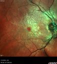
Vitreomacular Traction -> Macular Hole -> Aborted Macular holevaatamisi: 433Progression of VMT in both eyes over time
|
|

Microaneursyms and Macular Edema Not Diabeticvaatamisi: 43385 year old man has been tested about 2-3 times and is always negative for diabetes
|
|
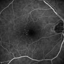
Microaneursyms and Macular Edema Not Diabeticvaatamisi: 43385 year old man has been tested about 2-3 times and is always negative for diabetes
|
|

Polypoidal Choroidal Vasculopathy - Right Eyevaatamisi: 43383 year old female presented in June 2013 with vision loss for 3 months. She had 2 injections elsewhere with no improvement (avastin). The first images show a peripapillary CNVM with polyps. I treated her once with PDT and she stayed dry with good vision for 5 years (2019). VA on presentation 20/80 OD. Improved to 20/40
|
|

Birdshot Chorioretintis and PAMMvaatamisi: 433Patient presented with paracentral acute middle maculopathy as the presenting symptom of birdshot. This case was published. RETINAL CASES & BRIEF REPORTS 10:151–153, 2016.
|
|
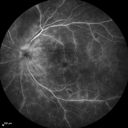
Birdshot Chorioretintis and PAMMvaatamisi: 433Patient presented with paracentral acute middle maculopathy as the presenting symptom of birdshot. This case was published. RETINAL CASES & BRIEF REPORTS 10:151–153, 2016.
|
|

Lung Cancer Metastasis to Right Eyevaatamisi: 43363 year old female with vision loss for 6 weeks. She had been scheduled for cataract surgery and then a tumor was found. She was ultimately diagnosed with lung cancer. Vision is 20/100
|
|
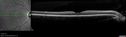
Punctate Inner Choroidopathy (PIC) Mildvaatamisi: 43324 year old female with high myopia. Noticing some distortion on grid. Vision 20/20 OU. Was followed frequently for several months with no progression and no CNVM.
|
|
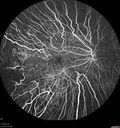
Choroideremia - Adultvaatamisi: 43369 year old man with no family history of choroideremia and gradual vision loss in both eyes. VVA 20/125 OD; 20/63 OS
|
|
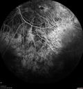
Choroideremia - Adultvaatamisi: 43369 year old man with no family history of choroideremia and gradual vision loss in both eyes. VVA 20/125 OD; 20/63 OS
|
|

Papilledema - Benign Intracranial hypertension - Idiopathic intracranial hypertensionvaatamisi: 43335 year old obese female. Normal vision and headache. Opening pressure was 30 cm water (normal 10-20). Nerve swelling responded to diamox.
|
|

PDR and DME and VHvaatamisi: 43357 year old diabetic man with vision loss in the left eye for several months. He has DME in the left eye and VH in the left eye and PDR in both eyes. He has been started in the left eye on Anti-VEGF therapy. VA on presentation was 20/25 OD and 20/200 OS
|
|

Concentric Geographic Atrophy - 76 year old manvaatamisi: 433Gradual vision loss, worse at night. VA 20/40 OU.
|
|
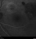
Polypiodal Choroidal Vasculopathy - Temporal Macula - Left eyevaatamisi: 43382 year old man with asymptomatic PCV in the left eye. Vision 20/50 OD, 20/32 OS did well with Lucentis
|
|
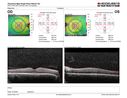
PDR and Vitreous Hemorrhage - High Risk Left Eye - Low Risk Right Eyevaatamisi: 43350 year old man with type I diabetes mellitus for 26 years. New Vitreous Hemorrhage in the left eye. Both eyes have NVE. Both also have foveal hypoplasia
|
|
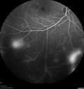
PDR and Vitreous Hemorrhage - High Risk Left Eye - Low Risk Right Eyevaatamisi: 43350 year old man with type I diabetes mellitus for 26 years. New Vitreous Hemorrhage in the left eye. Both eyes have NVE. Both also have foveal hypoplasia
|
|
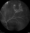
PDR and Vitreous Hemorrhage - High Risk Left Eye - Low Risk Right Eyevaatamisi: 43350 year old man with type I diabetes mellitus for 26 years. New Vitreous Hemorrhage in the left eye. Both eyes have NVE. Both also have foveal hypoplasia
|
|

Myopic CNVM in the left eye. New Classic subfoveal lesionvaatamisi: 43364 year old female. She had retinal detachment repair in the right eye in 1996 (25 years ago). The left eye has new distortion for a week or two.
VA 20/40 OU.
Left eye has a very small subfoveal CNVM
|
|

Myopic CNVM in the left eye. New Classic subfoveal lesionvaatamisi: 43364 year old female. She had retinal detachment repair in the right eye in 1996 (25 years ago). The left eye has new distortion for a week or two.
VA 20/40 OU.
Left eye has a very small subfoveal CNVM
|
|

Perifoveal Microaneurysm - Not Diabetic (also reticular pseudodrusen)vaatamisi: 43387 year old female with fluctuating macular edema in the right eye from a perifoveal MA. (no treatment). VA 20/40 OU
|
|
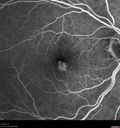
Perifoveal Microaneurysm - Not Diabetic (also reticular pseudodrusen)vaatamisi: 43387 year old female with fluctuating macular edema in the right eye from a perifoveal MA. (no treatment). VA 20/40 OU
|
|

Perifoveal Microaneurysm - Not Diabetic (also reticular pseudodrusen)vaatamisi: 43387 year old female with fluctuating macular edema in the right eye from a perifoveal MA. (no treatment). VA 20/40 OU
|
|

Enhanced S Cone Syndrome - Goldmann Favre - NR2E3 Mutationvaatamisi: 43382 year old man with poor vision for many years. VA HM OD, 5/200 OS. Diagnosed at age 12 with retinitis pigmentosa. Nystagmus.
|
|

Coloboma - Non Macular - Left Eyevaatamisi: 43371 year old female with cataracts. Left eye has inferior coloboma.
|
|

Coloboma - Non Macular - Left Eyevaatamisi: 43371 year old female with cataracts. Left eye has inferior coloboma.
|
|

Macular Telangiectasia (MacTel) and Pattern Dystrophyvaatamisi: 43371 year old man with diabetes for 5 years and blurred vision in the left eye. VA 20/25 OD, 20/50 OS. OCT OS shows MacTel
|
|

Branch Retinal Vein Occlusion - Both Eyes - Edema OD and NVE OS - Severe Non-perfusion Both Eyesvaatamisi: 43383 year old man with vision loss for 3 months not sure which eye. VA 20/100 OD and 20/50 OS. Tried avastin once without much improvement in the right eye and then lost to follow-up. FA in the left eye shows leaking NVE
|
|

Nonproliferative Diabetic Retinopathy and Good Visionvaatamisi: 43359 year old man Vision is good. Just referred following annual eye examination
PMHx: Type II DM, HTN, Cholesterol,
MEDS: Lantus, Metformin, lisinopril, atorvastatin, gabapentin
VA 20/20 OU
2+ NS OU
Observation
|
|

Vitreomacular traction with traction over retinal vessels in non-diabetic - Fluorescein leakage in areas of traction - Progressing vision loss left eyevaatamisi: 43280-year-old woman had vitrectomy surgery done in the right eye in 2007. Vision has been poor since despite a good anatomical result. Recently her vision in the left eye has been declining. The vision has declined in the left eye. Vision OD is 20/160, OS is 20/80
|
|

CVOvaatamisi: 432late FA (5min) optos photo of a 27 year old male with crvo os VA 20/20-
|
|

Total Dislocation of Intra Ocular Lens into the Vitreousvaatamisi: 43284-year old female comes in with blurred vision in the left eye. VA was 20/30, right eye and Count Fingers in the left eye. Fundus examination reveals dislocation of the IOL into the vitreous inferiorily @ 6-o'clock. Suggest surgery to fix the problem.
|
|

Macular Hole Photograph with superimposed OCTvaatamisi: 432
|
|

Red Free Macular Hole vaatamisi: 432Full Thickness Macular Hole
|
|

vaatamisi: 432
|
|
| 18117 faili 182 lehel |
 |
|
|
|
|
119 | |
|
|
|
|


