|
|

Central Serous Chorioretinopathy - Moderatevu 430 fois49 year old man with episodes in 2011, 2017, and 2018 - now with some paracentral vision loss in the right eye and subretinal fluid.
|
|

Macroaneurysm - Macular Hemorrhage - Branch Retinal Aterial Occlusionvu 430 fois62 year old African American female with chronic hypertension and vision loss for 2 weeks. VA is 20/400
|
|

Hyperpigmented Vasoproliferative Tumor on Buckle with New Serous Retinal Detachment 20 years post RD Repairvu 430 fois67 year old female RD repair OU 1999 then severe (20/400) macular pucker OD removed 3 months later (twin sister also had bilateral RD’s with PVR). Then dislocated IOL surgery right eye 9/2012. New vision loss in the right eye with serous retinal detachment. Argon laser to dark tumor led to resolution of subretinal fluid within a month.
|
|

Lung Cancer Metastasis to Right Eyevu 430 fois63 year old female with vision loss for 6 weeks. She had been scheduled for cataract surgery and then a tumor was found. She was ultimately diagnosed with lung cancer. Vision is 20/100
|
|

Lung Cancer Metastasis to Right Eyevu 430 fois63 year old female with vision loss for 6 weeks. She had been scheduled for cataract surgery and then a tumor was found. She was ultimately diagnosed with lung cancer. Vision is 20/100
|
|
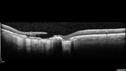
Macular Scars - Laser (1980s) Ocular Histoplasmosisvu 430 fois70 year old man who had laser at Wills, Wilmer and the Mayo clinic before better therapies were available for ocular histoplasmosis related CNVM
|
|

Rapid macular atrophy with anti-VEGF therapy for myopic CNVMvu 430 fois64 year old female with 20/32 vision and distortion in the left and better eye. She was treated with monthly lucentis and her vision declined to 20/100. Medicine was stopped and she improved a little to 20/80.
|
|
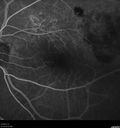
Polypiodal Choroidal Vasculopathy - Temporal Macula - Left eyevu 430 fois82 year old man with asymptomatic PCV in the left eye. Vision 20/50 OD, 20/32 OS did well with Lucentis
|
|

Peripheral Exudative Hemorrhagic Chorioretinopathyvu 430 fois93 year old female with shadow in side vision in the left eye. Referred for retinal detachment - Exudative lesion subsided in about 6 months without treatment.
Vision 20/63 OD and 20/100 OS (from dry AMD)
|
|
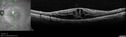
Cystoid Macular Edema and Macular Puckervu 430 foishas been particular bad for 3 years. She has a macular pucker and severe CME.
VA 20/160 left eye
|
|

Choroidal Neovascular Membrane following Prior Central Serous Chorioretinopathy - OCT-Avu 430 fois67 year old female with CSR in the left eye first 20 years ago. She was managed with out therapy and has had a few episodes since. Now ther eis distortion in the left eye for a few days. She had a steroid shot in her shoulder 5 months ago.
VA 20/20 OD, 20/50 OS- ICG and OCT-A confirm CNVM in the left eye
|
|

Perifoveal Microaneurysm - Not Diabetic (also reticular pseudodrusen)vu 430 fois87 year old female with fluctuating macular edema in the right eye from a perifoveal MA. (no treatment). VA 20/40 OU
|
|

Branch Retinal Vein Occlusion with Edema - Failed avastin (bevacizumab) and did well with Eylea (Aflibercept)vu 430 fois67 year old female The vision in the left eye was good until about 4 months ago. Prior to that it was OK. The vision in the right eye is fine. She is not sure if she has had strokes from studies taken in December. She was, at that time, sent to the ER for high blood pressure. Â
VA OD: Dcc20/40 OS: 20/100
Four treatments with Avastin she was still 20/100 with edema. With two Eylea treatments she improved to 20/32 and has remained dry and with good vision
|
|

Congenital Hypetrophy of the Retinal Pigment Epithelium (CHRPE)vu 430 fois63 year old female with normal vision and CHRPE lesion in the right eye.
|
|

Mild Central Serous Chorioretinopathy - Flonase relatedvu 430 fois58 year old man with vision loss in the left eye for 8 days. This has never happened before. He started taking flonase about a month ago. He stopped the flonase and the vision improved in less than a month. Initial vision was 20/20 OD, 20/40 OS
|
|

Neuroretinitis Left Eyevu 430 fois29 year old female - The left eye blurred up and she had double vision back in 2/2019 and March. It has gradually improved so this past month the vision in the left eye is getting better. Her MRI scan of her orbits with and without contrast was normal. She had no pain in the eye with or without eye movement when the vision was blurred. She has a cat for over 5 years. She has no exposure to kittens or stray cats and her cat is an indoor cat.
VA OD: sc20/12
VA OS: sc20/16-1
All uveitis testing was negative (cat scratch, RPR, FTA-ABS, Sarcoid test, lyme test, TB testing. She later developed recurrent uveitis without optic nerve involvement.
|
|

Non-ischemic Central Retinal Vein Occlusion with Macular Edemavu 430 fois62 year old man The vision in the right eye started to decline about 10 days ago. Prior to that the two eyes were about the same.
VA OD: Dcc20/125 PH20/50-2
VA OS: Dcc20/20
IOP: TP: OD:21 OS:21
|
|

vu 430 fois
|
|

Asymptomatic progressing retinal detachment 27 year old manvu 430 foisThis detachment was followed for one month, progressed, and was repaired with a scleral buckle.
|
|

Proliferative Diabetic Retinopathy both eyes Type I Diabetic for 25 yearsvu 429 fois47-year-old woman with diabetes for 25 years and decreased vision for 3 months. OD 20/40, OS 20/40
|
|

Polypoidal Choroidal Vasculopathy Right Eye - Disciform Scar Left Eyevu 429 fois84-year-old woman has polypoidal choroidal vasculopathy in both eyes with macular degeneration. She has macular scars in both eyes and she remains with remarkably good vision in the right eye despite the scarring.
VISUAL ACUITY: OD 20/30, OS 1/200
Avastin Rx OD
|
|

vu 429 fois75 year old male seen for ARMD. Pt developed massive hemorrhage with lipid and macular edema.
|
|

Total Dislocation of Intra Ocular Lens into the Vitreousvu 429 fois84-year old female comes in with blurred vision in the left eye. VA was 20/30, right eye and Count Fingers in the left eye. Fundus examination reveals dislocation of the IOL into the vitreous inferiorily @ 6-o'clock. Suggest surgery to fix the problem.
|
|

Peripheral Retinal Degenerationvu 429 foisPatient comes in with double vision. VA was 20/20 in both eyes. Fundus exam shows Retinal Degenerative changes in both eyes. Offer to correct double vision with temporary Fresnel prism
|
|
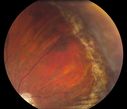
Reoccurring Retinal Detachmentvu 429 foisPatient comes in for second opinion for RD in the right eye. Patient's VA was count fingers @ 2-ft in the right eye and 20/20 in the left eye. Patient is aphakic and has had 5- retinal surgeries in the past in the right eye including a scleral buckle. RD present with the macula off. Will consider surgery with silicone oil.
|
|

vu 429 fois
|
|

Pseudoexfoliation with Cortical Cataract Changesvu 429 foisPatient comes in for eval on cataract surgery for the right eye. Slit lamp exam shows pseudoexfoliation (XFS) scattered on the lens in the right eye. Patient will be followed for glaucoma suspect and to schedule cataract surgery in the future...
|
|

Severe Pseudoexfoliation Ring, Left Eyevu 429 foisPatient comes in for blurred vision. VA is 20/30 both eyes. Mostly at near which was corrected with refraction. Slit lamp photos show severe pseudoexfoliation in the left eye. Patient will be followed up for glaucoma eval in a few weeks.
|
|
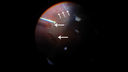
vu 429 fois
|
|

Dry AMD - Confluent Drusen Sparing Center of Macula which has Atrophyvu 429 fois65 year old woman, VA 20/40 OD; 20/80 OS. The center of the macula has few or no drusen with predominantly non-geographic atrophy
|
|

Neuroretinitis - Negative cat scratch serology twice - Possible Behcetsvu 429 fois65 year old man with no direct cat exposure and vision loss from neuroretinitis. His work up was positive for HLA B51. He had negative cat scratch titers twice. Vision dropped from 20/60 - 20/200 and then improved to 20/40 over 2 months
|
|

Outer Retinal Tubulationvu 429 fois78 year old man chronic wet AMD - 20/200
|
|

MacTel - Juxtafoveal Telangiectasisvu 429 fois56 year old man with a lot of difficulty adjusting to vision loss
|
|

vu 429 fois
|
|
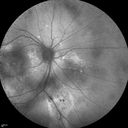
Chronic Central Serous Chorioretinopathyvu 429 fois77 year old man was seen at uveitis clinic and tested positive to Anti-retinal antibody against 35 kDa and anti-optic nerve antibody. He did not have EDI OCT, FAF, or ICG. All testing was consistent with Chronic CSR. His immunosuppresives were stopped and he did fine for 3 years.
|
|

Macular Telangiectasis - Very Mildvu 429 fois52 year old female with normal vision and suspicious OCT referred. Multimodal imaging shows very mild mactel (loss of macular pigment, ring on multicolor image and very mild OCT findings)
|
|

Macular Telangiectasis - Very Mildvu 429 fois52 year old female with normal vision and suspicious OCT referred. Multimodal imaging shows very mild mactel (loss of macular pigment, ring on multicolor image and very mild OCT findings)
|
|

Multifocal Choroiditis - Pneumocystis - aspergillis - cryptococcusvu 429 fois31 year old male with anaplastic astrocytoma on chemotherapy with mild vision loss in the right eye. LP did not reveal organism. He was placed on a trial of anti-fungal medications and lost to follow-up
|
|

Macroaneurysm - Macular Hemorrhage - Branch Retinal Aterial Occlusionvu 429 fois62 year old African American female with chronic hypertension and vision loss for 2 weeks. VA is 20/400
|
|

Posterior Scleritis - positive T-signvu 429 fois62 year ols man was sick about 6 weeks ago and then 2 weeks ago the the left eye got red. He was treated for conjunctivitis and the eye did not get better. Topical steroids and oral non-steroidal anti-inflammatory medications did not help.
VA OD: Dcc20/25 NccJ5
VA OS: Dcc5'/200
The scleritis cleared within 2 weeks of starting high dose (80 mg) oral steroids. Blood tests were all negative.
|
|

Multifocal Choroiditis and Subretinal Fibrosis with new Subretinal Neovascular Membranevu 429 fois26 year old African American Girl Saw me at age 10 (16 years ago) with acute multifocal choroiditis and panuveitis which evolved into subretinal fibrosis. She was initially treated with 3 years of methotrexate. Vision ended up 20/20 in both eyes. Her eyes have been inactive since and she was lost to follow-up.
A week ago she noticed sudden decreased vision in the right eye. Within the past few weeks there are more floaters in the right eye.
VA OD: sc6’/200 PHNI
VA OS: sc20/16
|
|

Central Retinal Vein Occlusion with Macular Edemavu 429 fois74 year old female The vision in the left eye has gotten worse over the last 3 weeks.
VA OD: Dcc20/25
VA OS: DccCF 3ft
IOP: TP: OD:16 OS:14
Vision improved to 20/40 in 3 months with monthly Aflibercept
|
|

PDR and DME and VHvu 429 fois57 year old diabetic man with vision loss in the left eye for several months. He has DME in the left eye and VH in the left eye and PDR in both eyes. He has been started in the left eye on Anti-VEGF therapy. VA on presentation was 20/25 OD and 20/200 OS
|
|

Concentric Geographic Atrophy - 76 year old manvu 429 foisGradual vision loss, worse at night. VA 20/40 OU.
|
|
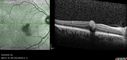
Valsalva Retinopathy - Macular Hemorrhage - Permanent Vision loss - Intraretinal Bloodvu 429 fois65 year old man with sudden vision loss in the right eye. Initial vision 20/200. VA improved to 20/50 in one year. Had carotid stenosis
|
|
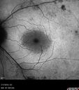
Retinitis Pigmentosa - Autosomal Recessive - 20/25 Visionvu 429 fois49 year old man 15 years ago he noticed decreased night vision.
VA OD: sc20/25 NscJ2
VA OS: sc20/25-2 NscJ2-1
IOP: TP: OD:17 OS:19
Genetic testing showed heterozygous for RPE65 - Probably not clinically important
|
|
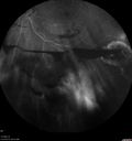
PDR and Vitreous Hemorrhage - High Risk Left Eye - Low Risk Right Eyevu 429 fois50 year old man with type I diabetes mellitus for 26 years. New Vitreous Hemorrhage in the left eye. Both eyes have NVE. Both also have foveal hypoplasia
|
|
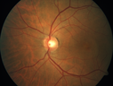
Choroidal Neovascular Membrane following Prior Central Serous Chorioretinopathy - OCT-Avu 429 fois67 year old female with CSR in the left eye first 20 years ago. She was managed with out therapy and has had a few episodes since. Now ther eis distortion in the left eye for a few days. She had a steroid shot in her shoulder 5 months ago.
VA 20/20 OD, 20/50 OS- ICG and OCT-A confirm CNVM in the left eye
|
|

Choroidal Neovascular Membrane following Prior Central Serous Chorioretinopathy - OCT-Avu 429 fois67 year old female with CSR in the left eye first 20 years ago. She was managed with out therapy and has had a few episodes since. Now ther eis distortion in the left eye for a few days. She had a steroid shot in her shoulder 5 months ago.
VA 20/20 OD, 20/50 OS- ICG and OCT-A confirm CNVM in the left eye
|
|

Choroidal Neovascular Membrane following Prior Central Serous Chorioretinopathy - OCT-Avu 429 fois67 year old female with CSR in the left eye first 20 years ago. She was managed with out therapy and has had a few episodes since. Now ther eis distortion in the left eye for a few days. She had a steroid shot in her shoulder 5 months ago.
VA 20/20 OD, 20/50 OS- ICG and OCT-A confirm CNVM in the left eye
|
|

vu 429 fois
|
|
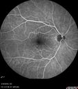
Cilioretinal Artery Occlusion - Nasal (not macular)vu 429 fois77 year old female There is a shadow in the top half of her temporal vision in the left eye that came on suddenly yesterday morning. It has not changed since then and the right eye is OK. She has had sinus headaches. She had no focal neurological changes. Â
VA OD: Dcc20/32
VA OS: Dcc20/16-1
IOP: TP: OD:18 OS:19Â
|
|

Hydroxychloroquine retinopathy - Plaquenil - Moderate - Myelinated nerve fiber layer vu 429 fois51 year old female on with trouble driving at night for a year. No eye examination for several years. 2009 (11 years ago) Plaquenil 300 then up to 400 a few years ago now down to 300 - 5'2" 135 lbs
VA 20/20 OD, 20/25 OS
|
|
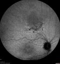
Amelanotic Choroidal Nevus - Multimodal Imagingvu 429 fois61 year old man with amelanotic choroidal nevus. Vision Normal - Stable over 4 years.
|
|

Intermediate Dry Age-Related Macular Degeneration - Soft Drusenvu 429 fois70-year-old Left eye worse than the right. Both eyes see distortion in the amsler grid for a few months
HTN, High Lipids, Osteoporosis
Meds: Lutein once daily, Calcium, Fish oil, Red Yeast Rice, CoQ10, Magnesium, MVI, Xanax
VA 20/16 OU
2+NS cataract
|
|
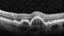
Focal Hyperpigmentation in Dry Age-Related Macular Degenerationvu 429 fois74 year old female with 20/32 vision right eye and 20/25 vision in the left eye. Images show focal hyper-pigmentation which is a risk factor for progression to wet AMD
|
|

Amelanotic Choroidal Nevusvu 429 fois60 year old man. Nevus is stable over 6 years. Images are Photo, FAF, OCT, FA, ICG, and B-Scan
|
|

Vitreomacular traction with traction over retinal vessels in non-diabetic - Fluorescein leakage in areas of traction - Progressing vision loss left eyevu 428 fois80-year-old woman had vitrectomy surgery done in the right eye in 2007. Vision has been poor since despite a good anatomical result. Recently her vision in the left eye has been declining. The vision has declined in the left eye. Vision OD is 20/160, OS is 20/80
|
|

vu 428 fois38-year old male presents with decreased vision in the left eye. VA was 20/20-right eye and 20/70-left eye with pinhole no improvement. Fundus exam shows exudates near the macula and also exudates with small hemorrhages, superior nasally in the left eye.
|
|

Vitreomacular traction - recent vision loss Right Eye vu 428 fois
|
|

Severe Pseudoexfoliation Ring, Left Eyevu 428 foisPatient comes in for blurred vision. VA is 20/30 both eyes. Mostly at near which was corrected with refraction. Slit lamp photos show severe pseudoexfoliation in the left eye. Patient will be followed up for glaucoma eval in a few weeks.
|
|

Wet AMD OD and Lamellar Macular hole OSvu 428 foisWet AMD in right and better eye failed extend to 2 months but was able to stop therapy once he started on systemic Avastin for cancer. The last image shows the macula dry 9 months without local therapy. The left eye has a lamellar macular hole.
|
|

Syphilitic Optic Neuritis - possible Gummavu 428 fois82 year old woman with vision loss in the left eye 20/32 and positive FTA-ABS (negative RPR). - this may be a gumma
|
|
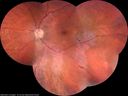
Normal Vision - Chronic Central Serous Chorioretinopathyvu 428 fois67 year old man - never had any symptoms - 20/16 vision in each eye. Lesions spare the fovea
|
|

Polypoidal Choroidal Vasculopathy Left Eyevu 428 fois74 year old man with vision loss to 20/100 over a few months. He improved to 20/20 with Lucentis. The color and ICG show the polyps.
|
|

Retinitis Pigmentosa - RP1 Mutationvu 428 fois78 year old man: 20/32 OD and 20/50 OS - RP1 Gene is Bad -- > RP with Novel Amino Acid Change in GLY723Stop sequence of the RP1 gene - consistent with AD RP
|
|

vu 428 fois
|
|

Photic Maculopathy Following Cataract Surgeryvu 428 foisInitial images are from 1 week and subsequent images from 1 month post surgery.
|
|
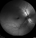
White Retinal Artiole Left eye - Inferotemporalvu 428 fois31 year old female with migraines and headaches for the last 12-13 years. Sometimes she gets the visual symptoms with the migraine. When she gets the migraines the pain is on the left side of her head. She gets the problem a few times a month, sometimes more. They usually last 5-6 hours. She has not had a permanent vision change. When she gets a vision change there are spotty dots of blue neon lights in her vision. With her glasses her two eyes are about the same. VA 20/16 in Each Eye
|
|

Vitreous Hemorrhage with no evidence of PDR in the left eyevu 428 fois74 year old man with vision loss OS for about a week. The FA shows no PDR in the left eye but the right eye has very mild NVD. Diabetes for 40 years now on insulin.
|
|
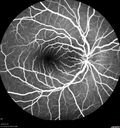
Cat Scratch - Neuroretinitis OD AND retinitis OS - Bartonella henselae positivevu 428 fois27 year old female with exposure to cats and prior fever. Reports with recent vision loss right eye. She was treated with doxycycline and within 2 weeks the right eye looks better. Then she skipped all follow-up visits. The right eye presented 20/200 and the left 20/16
|
|

Central Retinal Vein Occlusion - No macular edema - Pre-foveal operculavu 428 fois62 year old female with 20/20 vision. She had been seeing a spot out of the left eye for 4 years and then noticed splotches in her vision. She never devleoped edema or NV
|
|

Outer retinal holes and cataract from YAG laser vitreolysisvu 428 fois44 year old man with 7 year history of multiple lasers for floaters. FAF and OCT show outer retinal holes and lens has focal defects
|
|

Late Onset Cone Dystrophy - Very Mildvu 428 foisOnly IR and OCT show very mild bull's eye. 67 year old female with difficulty seeing in bright light
|
|

Central Serous Chorioretinopathy - Moderatevu 428 fois49 year old man with episodes in 2011, 2017, and 2018 - now with some paracentral vision loss in the right eye and subretinal fluid.
|
|

Multifocal Choroiditis and Subretinal Fibrosis with new Subretinal Neovascular Membranevu 428 fois26 year old African American Girl Saw me at age 10 (16 years ago) with acute multifocal choroiditis and panuveitis which evolved into subretinal fibrosis. She was initially treated with 3 years of methotrexate. Vision ended up 20/20 in both eyes. Her eyes have been inactive since and she was lost to follow-up.
A week ago she noticed sudden decreased vision in the right eye. Within the past few weeks there are more floaters in the right eye.
VA OD: sc6’/200 PHNI
VA OS: sc20/16
|
|

Hyperpigmented Vasoproliferative Tumor on Buckle with New Serous Retinal Detachment 20 years post RD Repairvu 428 fois67 year old female RD repair OU 1999 then severe (20/400) macular pucker OD removed 3 months later (twin sister also had bilateral RD’s with PVR). Then dislocated IOL surgery right eye 9/2012. New vision loss in the right eye with serous retinal detachment. Argon laser to dark tumor led to resolution of subretinal fluid within a month.
|
|
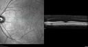
Lung Cancer Metastasis to Right Eyevu 428 fois63 year old female with vision loss for 6 weeks. She had been scheduled for cataract surgery and then a tumor was found. She was ultimately diagnosed with lung cancer. Vision is 20/100
|
|

Macular Telangiectasis - Mild - Retinal Crystalsvu 428 fois58 year old man with long term mild vision loss. Considering Cataract surgery. VA 20/50 OD and 20/63 OS - Moderate cataract are present
|
|
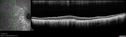
Choroideremia - Adultvu 428 fois69 year old man with no family history of choroideremia and gradual vision loss in both eyes. VVA 20/125 OD; 20/63 OS
|
|

Choroideremia - Adultvu 428 fois69 year old man with no family history of choroideremia and gradual vision loss in both eyes. VVA 20/125 OD; 20/63 OS
|
|

Chronic Endophthalmitis and Cystoid Macular Edemavu 428 foisVA 20/50 - Culture negative. Cleared with intravitreal antibiotics and decadron
|
|
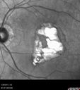
Macular Scars - Laser (1980s) Ocular Histoplasmosisvu 428 fois70 year old man who had laser at Wills, Wilmer and the Mayo clinic before better therapies were available for ocular histoplasmosis related CNVM
|
|

Recurrent Toxoplasmosis - Resolves with no treatmentvu 428 fois36 year old man with recent vision loss. He has a sulfa allergy. The eye was NOT treated and the uveitis resolved in about 6 weeks.
|
|

Acute Bilateral Central Serous Chorioretinopathy - Female - Smokestackvu 428 fois39 year old female She had no prior epsisodes of vision loss and then a week ago she noticed decreased vision in the right eye. She has not had any health problems and she is not pregnant. Phendimetrazine started 3 months ago. She is not on steroids. VA OD: sc20/63-1 NscJ16 VA OS: sc20/125-1 Dcc20/80 NscJ16 IOP: TP: OD:14 OS:14 Time:11:23 AM
|
|

Angioid Streaks Scar and new CNVMvu 428 fois49 y/o WF with decreased vision x 1 month. Her OD is 20/400 and her OS is seeing 20/70. No cutaneous lesions or Ehlers-Danlos. The FP and FA show a predominant classic CNV (with fibrosis) OD and an Occult CNV OS with what appears to be a RAP lesion.
|
|

Punctate Inner Choroidopathy and Choroidal Neovascular Membranevu 428 fois27 year old female with vision loss for 4 months. VA was 20/200. She had a CNVM which was extrafoveal with no subretinal fluid. Her vision improved over 6 months to 20/50 with Ranibizumab injections (she might have improved without treatment)
|
|
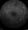
Eales Disease and fresh vitreous hemorrhage - 20 year old manvu 428 fois20 year old mane with fresh vitreous hemorrhage in the right eye. At age 15 he had a PPV and laser in the left eye and laser in the right eye. The vision is OD 20/80 PH 20/25, OS 20/25. The left eye has a mild cataract. He had prior testing for coagulopathies which was negative. Testing done for syphillis and TB was negative. Additional laser was done to prevent further bleeding in the right eye.
|
|

Acquired Toxoplasmosis Retinitis Para Foveal - Following Venison Consumptionvu 428 fois56 year old man who became very sick after eating venison at the end of December 2019. Presented 6 weeks later with a scotoma. Initial photos show presentation with 20/63 vision. Vision dropped to 5/200. All tests (including PCR of anterior chamber tap) were negative except serum toxoplasmosis IgG and IgM. Vision improved to 20/32 but the scotoma remained on oral trimetheprim/sulfa
|
|
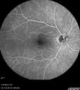
Cilioretinal Artery Occlusion - Nasal (not macular)vu 428 fois77 year old female There is a shadow in the top half of her temporal vision in the left eye that came on suddenly yesterday morning. It has not changed since then and the right eye is OK. She has had sinus headaches. She had no focal neurological changes. Â
VA OD: Dcc20/32
VA OS: Dcc20/16-1
IOP: TP: OD:18 OS:19Â
|
|

Heavy Panretinal Photocoagulation for Proliferative Diabetic Retinopathy about 40 years agovu 428 fois64 year old female - images from 2018 - heavy PRP about 30 years ago
VA 20/20 OD, 20/50 OS
|
|

Toxoplasmosis Macular Scar - Inactivevu 428 fois49 year old man Toxoplasmosis was diagnosed in his 20s. No past eye surgeries OU. He had floaters a few weeks ago with dots and a long line in the vision. Since then they have gone away.
VA OD: Dcc20/20 NccJ1+
VA OS: Dcc20/16 NccJ1+
|
|

Hydroxychloroquine retinopathy - Plaquenil - Moderatevu 428 fois51 year old female on with trouble driving at night for a year. No eye examination for several years. 2009 (11 years ago) Plaquenil 300 then up to 400 a few years ago now down to 300 - 5'2" 135 lbs
VA 20/20 OD, 20/25 OS
|
|

Amelanotic Choroidal Nevus - Multimodal Imagingvu 428 fois61 year old man with amelanotic choroidal nevus. Vision Normal - Stable over 4 years.
|
|

Macular Telangiectasia (MacTel) and Pattern Dystrophyvu 428 fois71 year old man with diabetes for 5 years and blurred vision in the left eye. VA 20/25 OD, 20/50 OS. OCT OS shows MacTel
|
|

Branch Retinal Vein Occlusion - Both Eyes - Edema OD and NVE OS - Severe Non-perfusion Both Eyesvu 428 fois83 year old man with vision loss for 3 months not sure which eye. VA 20/100 OD and 20/50 OS. Tried avastin once without much improvement in the right eye and then lost to follow-up. FA in the left eye shows leaking NVE
|
|

Age-related macular degeneration - Geographic Atrophy - Intermittent CME left eye from CRVO, Anemia, Diabetesvu 428 fois79 year old female - 3 years post CRVO in the left eye (20/32 VA) with intermittent CME. Left eye is the better eye. Vision is stable since 2 years ago
PMHx: Type II DM x 20 years, Chronic Anemia, Hyperlipidemia
VA 20/200 OD, 20/50 OS (usually 20/40)
|
|

Macular Pre-Retinal Hemorrhage vu 427 foisPatient complains of loss of vision in his left eye. Patient is diabetic. VA was 20/20, right eye and 20/150, left eye with no improvement pinhole. Fundus exam reveals very large pre retinal hemorrhage in the left eye. Embolism located inferior, nasally just off the optic nerve in the left eye. Patient underwent PRP for treatment of the hemorrhage.
|
|

CRVO, Left Eyevu 427 foisPatient comes in with central retinal vein occlusion in the left eye. VA was count fingers in the left eye. Fundus photo shows scattered hemorrhages with cotton wool spots. Patient was treated with Avastin in the left eye.
|
|

vu 427 fois
|
|
| 18117 photos sur 182 page(s) |
 |
|
|
|
|
121 | |
|
|
|
|


