|
|

Myopic CNVM in the left eye. New Classic subfoveal lesion404 views64 year old female. She had retinal detachment repair in the right eye in 1996 (25 years ago). The left eye has new distortion for a week or two.
VA 20/40 OU.
Left eye has a very small subfoveal CNVM
|
|
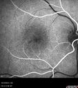
Macular Telangiectasi - Stage V - CNVM OU404 views60 year old female who has been treated with injections for 5 years starting in the left eye. She had no treatment 6 months prior to this visit and her vision has been getting worse. Her vision is 20/100 OD, 20/32 OS. Vision improved with intravitreal Avastin to 20/30 OU.
|
|

404 views
|
|

Acquired Toxoplasmosis Retinitis Para Foveal - Following Venison Consumption404 views56 year old man who became very sick after eating venison at the end of December 2019. Presented 6 weeks later with a scotoma. Initial photos show presentation with 20/63 vision. Vision dropped to 5/200. All tests (including PCR of anterior chamber tap) were negative except serum toxoplasmosis IgG and IgM. Vision improved to 20/32 but the scotoma remained on oral trimetheprim/sulfa
|
|

Acquired Toxoplasmosis Retinitis Para Foveal - Following Venison Consumption404 views56 year old man who became very sick after eating venison at the end of December 2019. Presented 6 weeks later with a scotoma. Initial photos show presentation with 20/63 vision. Vision dropped to 5/200. All tests (including PCR of anterior chamber tap) were negative except serum toxoplasmosis IgG and IgM. Vision improved to 20/32 but the scotoma remained on oral trimetheprim/sulfa
|
|

Acquired Toxoplasmosis Retinitis Para Foveal - Following Venison Consumption404 views56 year old man who became very sick after eating venison at the end of December 2019. Presented 6 weeks later with a scotoma. Initial photos show presentation with 20/63 vision. Vision dropped to 5/200. All tests (including PCR of anterior chamber tap) were negative except serum toxoplasmosis IgG and IgM. Vision improved to 20/32 but the scotoma remained on oral trimetheprim/sulfa
|
|

Enhanced S Cone Syndrome - Goldmann Favre - NR2E3 Mutation404 views82 year old man with poor vision for many years. VA HM OD, 5/200 OS. Diagnosed at age 12 with retinitis pigmentosa. Nystagmus.
|
|

Branch Retinal Vein Occlusion with Edema - Failed avastin (bevacizumab) and did well with Eylea (Aflibercept)404 views67 year old female The vision in the left eye was good until about 4 months ago. Prior to that it was OK. The vision in the right eye is fine. She is not sure if she has had strokes from studies taken in December. She was, at that time, sent to the ER for high blood pressure. Â
VA OD: Dcc20/40 OS: 20/100
Four treatments with Avastin she was still 20/100 with edema. With two Eylea treatments she improved to 20/32 and has remained dry and with good vision
|
|
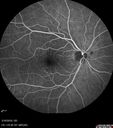
Cilioretinal Artery Occlusion - Nasal (not macular)404 views77 year old female There is a shadow in the top half of her temporal vision in the left eye that came on suddenly yesterday morning. It has not changed since then and the right eye is OK. She has had sinus headaches. She had no focal neurological changes. Â
VA OD: Dcc20/32
VA OS: Dcc20/16-1
IOP: TP: OD:18 OS:19Â
|
|

Cilioretinal Artery Occlusion - Good vision404 views67 year old female - 5 days ago she was getting ready for bed and closed one eye (she had worked for an optometrist for 20 years) and she noticed a spot in the vision in the right eye and came in. She has no focal neurological problems. She once in a while gets some weakness. She has a neurologist and there are some brain lesions. Father had stroke at age 52.
VA OD: Dcc20/20 NccJ1
VA OS: Dcc20/16-2 NccJ1-2
|
|

Neuroretinitis Left Eye404 views29 year old female - The left eye blurred up and she had double vision back in 2/2019 and March. It has gradually improved so this past month the vision in the left eye is getting better. Her MRI scan of her orbits with and without contrast was normal. She had no pain in the eye with or without eye movement when the vision was blurred. She has a cat for over 5 years. She has no exposure to kittens or stray cats and her cat is an indoor cat.
VA OD: sc20/12
VA OS: sc20/16-1
All uveitis testing was negative (cat scratch, RPR, FTA-ABS, Sarcoid test, lyme test, TB testing. She later developed recurrent uveitis without optic nerve involvement.
|
|

Branch retinal vein occlusion with macular edema404 views71 year old man Starting about 2 weeks ago the left eye was blurred. The right eye is seeing well.
VA OD: sc20/25+2 NccJ1+
VA OS: sc20/63+2 PH20/50-2 Ncc20/400
IOP: TP: OD:19 OS:16
|
|

Branch Retinal Vein Occlusion - Both Eyes - Edema OD and NVE OS - Severe Non-perfusion Both Eyes404 views83 year old man with vision loss for 3 months not sure which eye. VA 20/100 OD and 20/50 OS. Tried avastin once without much improvement in the right eye and then lost to follow-up. FA in the left eye shows leaking NVE
|
|

Bilateral Acute Central Serous Chorioretinopathy - Evidence of old disease OS404 views64 year old man noticed in the right eye that is a black spot 4 days ago. Also vertical lines in his kitchen were a little wavy. He is not on any steroid medications. He has vertigo.
VA OD: Dcc20/25-2 NccJ1+-3
VA OS: Dcc20/32-1 PHNI NccJ5-1
IOP: TP: OD:12 OS:15
|
|

Peripheral reticular pigment degeneration - pavingstone degeneration - age-related macular degeneration404 views60 year old healthy man with intermediate AMD and peripheral pigment degeneration and pavinstone degeneration. Vision is 20/20 OU and the patient is asymptomatic. No significant history of AMD and he is a non-smoker.
|
|

Acute Syphilitic Posterior Placoid Chorioretinitis404 views40 year old man About 10 days ago the patient woke up with a dark circle in the middle of the left eye that blocks his vision. He thought it might be from fatigue. The month of March was very stressful. After a few days the spot in the left eye got darker and he could not drive at night. During the last week while not going to work and sleeping more his vision is better but not normal. He has never had vision loss in the past. Â
PMHx: Benign, Meds: Multivitamin
VA OD: sc20/16 NscJ1+VA OS: sc20/32+2 NscJ4
IOP: TP: OD:12 OS:12
|
|
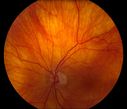
Total Dislocation of Intra Ocular Lens into the Vitreous403 views84-year old female comes in with blurred vision in the left eye. VA was 20/30, right eye and Count Fingers in the left eye. Fundus examination reveals dislocation of the IOL into the vitreous inferiorily @ 6-o'clock. Suggest surgery to fix the problem.
|
|

Reoccurring Retinal Detachment403 viewsPatient comes in for second opinion for RD in the right eye. Patient's VA was count fingers @ 2-ft in the right eye and 20/20 in the left eye. Patient is aphakic and has had 5- retinal surgeries in the past in the right eye including a scleral buckle. RD present with the macula off. Will consider surgery with silicone oil.
|
|

Polypoidal Choroidal Vasculopathy Left Eye403 views74 year old man with vision loss to 20/100 over a few months. He improved to 20/20 with Lucentis. The color and ICG show the polyps.
|
|

Polypoidal Choroidal Vasculopathy Left Eye403 views74 year old man with vision loss to 20/100 over a few months. He improved to 20/20 with Lucentis. The color and ICG show the polyps.
|
|

Angioid Streaks - CNVM OS with vision loss403 views48 year old soccer player. Prior trauma OS (with soccer ball). Vision loss for 3 weeks to 20/60 - with Lucentis vision improved to 20/30
|
|

Angioid Streaks - CNVM OS with vision loss403 views48 year old soccer player. Prior trauma OS (with soccer ball). Vision loss for 3 weeks to 20/60 - with Lucentis vision improved to 20/30
|
|

Chronic Central Serous Chorioretinopathy403 views77 year old man was seen at uveitis clinic and tested positive to Anti-retinal antibody against 35 kDa and anti-optic nerve antibody. He did not have EDI OCT, FAF, or ICG. All testing was consistent with Chronic CSR. His immunosuppresives were stopped and he did fine for 3 years.
|
|

403 views
|
|

Hyperpigmented Vasoproliferative Tumor on Buckle with New Serous Retinal Detachment 20 years post RD Repair403 views67 year old female RD repair OU 1999 then severe (20/400) macular pucker OD removed 3 months later (twin sister also had bilateral RD’s with PVR). Then dislocated IOL surgery right eye 9/2012. New vision loss in the right eye with serous retinal detachment. Argon laser to dark tumor led to resolution of subretinal fluid within a month.
|
|

Lung Cancer Metastasis to Right Eye403 views63 year old female with vision loss for 6 weeks. She had been scheduled for cataract surgery and then a tumor was found. She was ultimately diagnosed with lung cancer. Vision is 20/100
|
|

Giant Cell Arteritis - Anterior Ischemic Optic Neuropathy - Left Eye403 views78 year old female with vision loss on steroid taper for GCA. Superior pole of optic nerve left eye is swollen
|
|

Acute Bilateral Central Serous Chorioretinopathy - Female - Smokestack403 views39 year old female She had no prior epsisodes of vision loss and then a week ago she noticed decreased vision in the right eye. She has not had any health problems and she is not pregnant. Phendimetrazine started 3 months ago. She is not on steroids. VA OD: sc20/63-1 NscJ16 VA OS: sc20/125-1 Dcc20/80 NscJ16 IOP: TP: OD:14 OS:14 Time:11:23 AM
|
|

PDR and DME and VH403 views57 year old diabetic man with vision loss in the left eye for several months. He has DME in the left eye and VH in the left eye and PDR in both eyes. He has been started in the left eye on Anti-VEGF therapy. VA on presentation was 20/25 OD and 20/200 OS
|
|

Concentric Geographic Atrophy - 76 year old man403 viewsGradual vision loss, worse at night. VA 20/40 OU.
|
|

Macular Telangiectasia - Mild403 views56 year old female with good vision and mild macular telangiectasia. VA 20/40 OD 20/16 OS
|
|

Polypiodal Choroidal Vasculopathy - Temporal Macula - Left eye403 views82 year old man with asymptomatic PCV in the left eye. Vision 20/50 OD, 20/32 OS did well with Lucentis
|
|

Acute Branch Retinal Vein Occlusion - 20/200403 views66 year old female with 3 weeks of vision loss in the left eye. VA 20/20 OD 20/200 OS
|
|
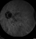
Peripheral Exudative Hemorrhagic Chorioretinopathy403 views93 year old female with shadow in side vision in the left eye. Referred for retinal detachment - Exudative lesion subsided in about 6 months without treatment.
Vision 20/63 OD and 20/100 OS (from dry AMD)
|
|

Choroidal folds right eye more than left403 views68 year old man with prior laser corrective refractive surgery. VA OD 20/125 PH 20/32 OS 20/16 - Possible nanophthalmous
|
|

403 views
|
|
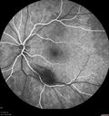
Acquired Toxoplasmosis Retinitis Para Foveal - Following Venison Consumption403 views56 year old man who became very sick after eating venison at the end of December 2019. Presented 6 weeks later with a scotoma. Initial photos show presentation with 20/63 vision. Vision dropped to 5/200. All tests (including PCR of anterior chamber tap) were negative except serum toxoplasmosis IgG and IgM. Vision improved to 20/32 but the scotoma remained on oral trimetheprim/sulfa
|
|

Acquired Toxoplasmosis Retinitis Para Foveal - Following Venison Consumption403 views56 year old man who became very sick after eating venison at the end of December 2019. Presented 6 weeks later with a scotoma. Initial photos show presentation with 20/63 vision. Vision dropped to 5/200. All tests (including PCR of anterior chamber tap) were negative except serum toxoplasmosis IgG and IgM. Vision improved to 20/32 but the scotoma remained on oral trimetheprim/sulfa
|
|

Enhanced S Cone Syndrome - Goldmann Favre - NR2E3 Mutation403 views82 year old man with poor vision for many years. VA HM OD, 5/200 OS. Diagnosed at age 12 with retinitis pigmentosa. Nystagmus.
|
|

Severe Geographic Atrophy from Dry Age-Related Macular Degeneration403 views90 year old female with severe vision loss over the last few years.
VA 3/200 OD
1/200 OS
|
|

Toxoplasmosis Macular Scar - Inactive403 views49 year old man Toxoplasmosis was diagnosed in his 20s. No past eye surgeries OU. He had floaters a few weeks ago with dots and a long line in the vision. Since then they have gone away.
VA OD: Dcc20/20 NccJ1+
VA OS: Dcc20/16 NccJ1+
|
|
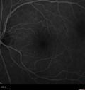
Mild Central Serous Chorioretinopathy - Flonase related403 views58 year old man with vision loss in the left eye for 8 days. This has never happened before. He started taking flonase about a month ago. He stopped the flonase and the vision improved in less than a month. Initial vision was 20/20 OD, 20/40 OS
|
|

Mild Central Serous Chorioretinopathy - Flonase related403 views58 year old man with vision loss in the left eye for 8 days. This has never happened before. He started taking flonase about a month ago. He stopped the flonase and the vision improved in less than a month. Initial vision was 20/20 OD, 20/40 OS
|
|

Venous Anomaly - 80 year old female - normal vision403 views
|
|

Amelanotic Choroidal Nevus - Multimodal Imaging403 views61 year old man with amelanotic choroidal nevus. Vision Normal - Stable over 4 years.
|
|

Lamellar Macular Hole - Evoluation over 5 years403 views65 year old man with Sturge weber and a choroidal hemangioma in the left eye. (See other images under sturge weber on this website). Has ongoing problem with his left eye. Now with Blurred vision in the right eye gradually worse the last few weeks. He has had a vitrectomy in the right eye 4 years ago for floaters. Laser for tears and cryo in the right eye 5 years ago. He is on Dorzolamide and Lumigan for glaucoma.
VA 20/32 OD, 20/40 OS
IOP 23 OD, 19 OS
|
|

Lamellar Macular Hole - Evoluation over 5 years403 views65 year old man with Sturge weber and a choroidal hemangioma in the left eye. (See other images under sturge weber on this website). Has ongoing problem with his left eye. Now with Blurred vision in the right eye gradually worse the last few weeks. He has had a vitrectomy in the right eye 4 years ago for floaters. Laser for tears and cryo in the right eye 5 years ago. He is on Dorzolamide and Lumigan for glaucoma.
VA 20/32 OD, 20/40 OS
IOP 23 OD, 19 OS
|
|

Tilted Disc Syndrome403 views56 year old female. Recent motor vehicle accident. Vision is 20/50 OD, 20/25 OS.
|
|

Branch retinal vein occlusion with macular edema403 views71 year old man Starting about 2 weeks ago the left eye was blurred. The right eye is seeing well.
VA OD: sc20/25+2 NccJ1+
VA OS: sc20/63+2 PH20/50-2 Ncc20/400
IOP: TP: OD:19 OS:16
|
|

Sclerochoroidal Calcification - Large Lesion403 views77 year old white male retired physician. The patient was noticing a change in vision and saw you for a checkup. He used to see better from a distance without glasses and now he takes his glasses off to read. There is a mass in the right eye. Â
VA OD: Dcc20/50-1 PH20/40-1 NccJ1
VA OS: Dcc20/32+1 NccJ1-1
IOP: TP: OD:14 OS:15
|
|

Large and Extensive Macular Drusen403 views64 year old female with 20/25 vision taking vitamins in for a checkup.
|
|
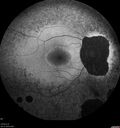
Retinitis Pigmentosa - X-linked Recessive403 views60 year old man The vision in the right eye is blurry recently. He has trouble with night vision for about 3 years. His brother has eye disease in Michigan.
VA OD: Dcc20/32-1 PHNI NccJ1-1
VA OS: Dcc20/20 NccJ1+-1
IOP: TP: OD:36 OS:18
He has pseudoexfoliative glaucoma in the right eye.
|
|

Branch Retinal Vein Occlusion - Both Eyes - Edema OD and NVE OS - Severe Non-perfusion Both Eyes403 views83 year old man with vision loss for 3 months not sure which eye. VA 20/100 OD and 20/50 OS. Tried avastin once without much improvement in the right eye and then lost to follow-up. FA in the left eye shows leaking NVE
|
|

Bilateral Acute Central Serous Chorioretinopathy - Evidence of old disease OS403 views64 year old man noticed in the right eye that is a black spot 4 days ago. Also vertical lines in his kitchen were a little wavy. He is not on any steroid medications. He has vertigo.
VA OD: Dcc20/25-2 NccJ1+-3
VA OS: Dcc20/32-1 PHNI NccJ5-1
IOP: TP: OD:12 OS:15
|
|
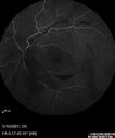
Branch Retinal Vein Occlusion with Neovascularization Elsewhere and Vitreous Hemorrhage403 views57 year old man New floaters in the left eye about a month ago. Now they are getting better. PMHx A-Fib on Pradaxa
VA 20/32 OD, 20/40 OS
1+ NS OU
BP 137/77, 200LBs, 5’8â€
PRP laser was done
|
|

Peripheral reticular pigment degeneration - pavingstone degeneration - age-related macular degeneration403 views60 year old healthy man with intermediate AMD and peripheral pigment degeneration and pavinstone degeneration. Vision is 20/20 OU and the patient is asymptomatic. No significant history of AMD and he is a non-smoker.
|
|
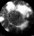
Severe Proliferative Diabetic Retinopathy and Good Vision403 views49 year old man with vision loss in the right eye for 2 weeks. VA 20/40 OD, 20/63 OS. DM x 15 years and HTN.
|
|
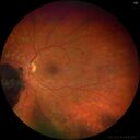
Pregnant - 25 weeks - new onset PDR and DME both eyes - Treated with PRP and ozurdex403 views29 year old female. Patient is 25 weeks pregnant and having flare up of retinopathy in both eyes. She states it is worse in the left. Patient has trouble with distant vision. At most recent visit, 1 years ago, VA was 20/20 OU. Two years ago anterior uveitis x 1 episode with normal labs (HLAB27, FTA, ACE, Lyme). 2017-2020 – Lucentis for DME left eye only.
PMHx: Type I DM since teenager, hyperlipidemia
BP was normal
|
|

Nonproliferative Diabetic Retinopathy and Good Vision403 views59 year old man Vision is good. Just referred following annual eye examination
PMHx: Type II DM, HTN, Cholesterol,
MEDS: Lantus, Metformin, lisinopril, atorvastatin, gabapentin
VA 20/20 OU
2+ NS OU
Observation
|
|
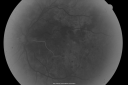
PDR OS with NVE402 views57-year-old man has diabetic retinopathy in both eyes.
Diabetic for 14 years with HgB A1C often over 10.
VISUAL ACUITY: OD 20/30, OS 20/40. PDR OS BDR OD
|
|

Central Retinal Vein Occlusion with Macular Edema and Retinal Hemorrhages 402 viewsFemale patient of 86-years of age presents a central retinal vein occlusion with macular edema in the right eye. Patient's VA was 20/70 in the right eye. Proceeded with laser treatment to the right eye.
|
|
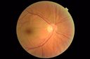
PAMM post CRAO402 views
|
|

Polypoidal Choroidal Vasculopathy Left Eye402 views74 year old man with vision loss to 20/100 over a few months. He improved to 20/20 with Lucentis. The color and ICG show the polyps.
|
|

Chronic Central Serous Chorioretinopathy402 views77 year old man was seen at uveitis clinic and tested positive to Anti-retinal antibody against 35 kDa and anti-optic nerve antibody. He did not have EDI OCT, FAF, or ICG. All testing was consistent with Chronic CSR. His immunosuppresives were stopped and he did fine for 3 years.
|
|

Chronic Central Serous Chorioretinopathy402 views77 year old man was seen at uveitis clinic and tested positive to Anti-retinal antibody against 35 kDa and anti-optic nerve antibody. He did not have EDI OCT, FAF, or ICG. All testing was consistent with Chronic CSR. His immunosuppresives were stopped and he did fine for 3 years.
|
|

Chronic CSC Both Eyes - Recent vision loss Right Eye Treated with PDT402 views50 year old man The right eye has had vision loss for a few weeks. He had previous episodes seen by me in 2012 OS and 2008 OD. This was 2018. VA OS has been poor for several years.
VA OD: Dcc20/100
VA OS: Dcc20/200-3
The patient had PDT to leaky area superotemporal to the fovea 1/2 dose and within 3 months was back up to 20/25. The last two OCT scans show fluid absorbing
|
|

Retinitis Pigmentosa with Cystoid Macular Edema responsive to topical carbonic anhydrase inhibitors402 views67 year old female She thought it was time to have her glasses changed.She has no family history of reitnal disease and has never been diagnosed with a problem. She started having night vision trouble the last year. Two sisters and a brother with normal vision.
VA OD: Dcc20/40-2 NccJ3
VA OS: Dcc20/20 NccJ1
IOP: TP: OD:9
|
|

Rapid macular atrophy with anti-VEGF therapy for myopic CNVM402 views64 year old female with 20/32 vision and distortion in the left and better eye. She was treated with monthly lucentis and her vision declined to 20/100. Medicine was stopped and she improved a little to 20/80.
|
|

PDR and DME and VH402 views57 year old diabetic man with vision loss in the left eye for several months. He has DME in the left eye and VH in the left eye and PDR in both eyes. He has been started in the left eye on Anti-VEGF therapy. VA on presentation was 20/25 OD and 20/200 OS
|
|

Valsalva Retinopathy - Macular Hemorrhage - Permanent Vision loss - Intraretinal Blood402 views65 year old man with sudden vision loss in the right eye. Initial vision 20/200. VA improved to 20/50 in one year. Had carotid stenosis
|
|
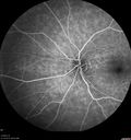
Choroidal folds right eye more than left402 views68 year old man with prior laser corrective refractive surgery. VA OD 20/125 PH 20/32 OS 20/16 - Possible nanophthalmous
|
|
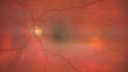
Cystoid Macular Edema and Macular Pucker402 viewshas been particular bad for 3 years. She has a macular pucker and severe CME.
VA 20/160 left eye
|
|

Myopic CNVM in the left eye. New Classic subfoveal lesion402 views64 year old female. She had retinal detachment repair in the right eye in 1996 (25 years ago). The left eye has new distortion for a week or two.
VA 20/40 OU.
Left eye has a very small subfoveal CNVM
|
|

Acquired Toxoplasmosis Retinitis Para Foveal - Following Venison Consumption402 views56 year old man who became very sick after eating venison at the end of December 2019. Presented 6 weeks later with a scotoma. Initial photos show presentation with 20/63 vision. Vision dropped to 5/200. All tests (including PCR of anterior chamber tap) were negative except serum toxoplasmosis IgG and IgM. Vision improved to 20/32 but the scotoma remained on oral trimetheprim/sulfa
|
|
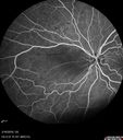
Cilioretinal Artery Occlusion - Nasal (not macular)402 views77 year old female There is a shadow in the top half of her temporal vision in the left eye that came on suddenly yesterday morning. It has not changed since then and the right eye is OK. She has had sinus headaches. She had no focal neurological changes. Â
VA OD: Dcc20/32
VA OS: Dcc20/16-1
IOP: TP: OD:18 OS:19Â
|
|
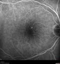
Mild Central Serous Chorioretinopathy - Flonase related402 views58 year old man with vision loss in the left eye for 8 days. This has never happened before. He started taking flonase about a month ago. He stopped the flonase and the vision improved in less than a month. Initial vision was 20/20 OD, 20/40 OS
|
|

Hydroxychloroquine retinopathy - Plaquenil - Moderate402 views51 year old female on with trouble driving at night for a year. No eye examination for several years. 2009 (11 years ago) Plaquenil 300 then up to 400 a few years ago now down to 300 - 5'2" 135 lbs
VA 20/20 OD, 20/25 OS
|
|

Lamellar Macular Hole - Evoluation over 5 years402 views65 year old man with Sturge weber and a choroidal hemangioma in the left eye. (See other images under sturge weber on this website). Has ongoing problem with his left eye. Now with Blurred vision in the right eye gradually worse the last few weeks. He has had a vitrectomy in the right eye 4 years ago for floaters. Laser for tears and cryo in the right eye 5 years ago. He is on Dorzolamide and Lumigan for glaucoma.
VA 20/32 OD, 20/40 OS
IOP 23 OD, 19 OS
|
|

Proliferative Diabetic Retinopathy and vitreous hemorrhage left eye402 views44 year old man with type II diabetes for 14 years and no eye examinations in the last 5 years. VA 20/40 right eye and 20/60 left eye. Vision loss has been for about a week in the left eye.
|
|

Amelanotic Choroidal Nevus402 views60 year old man. Nevus is stable over 6 years. Images are Photo, FAF, OCT, FA, ICG, and B-Scan
|
|

Idiopathic Iris Cyst402 views85 year old man History of wet AMD OD x 7 years under treatment with Lucentis. VA 20/40 OD, 20/32 OS
PMHx High cholesterol, GERD, COPD, A-Fib,
MEDS: coumadin and ASA and others
|
|

ABCA4 positive - Stargardt Disease402 views24 year old female She has worn glasses since she was a kid. Since she became an adult her vision started to deteriorate more rapidly. This seems to have been happening since she entered the accounting department for the last two years. Her vision is worse in the light especially when she is driving. She can see green lights in the night but not in the day. She sees better on a dark background than on a light background.
Her mother and father both have inherited retinal diseases. His mother was diagnosed 23 years ago with Stargardts genetically confirmed in Boston. The father has Retinitis Pigmentosa. The mother has seven brothers and sisters and four have Stargardts and they are legally blind. Also one cousin has it.
PMHx benign, Meds: vitamins.
VA OD: Dcc20/160
VA OS: Dcc20/160
IOP: TP: OD:20 OS:21
|
|

Diabetic Papillitis401 viewsPatient is about to undergo surgery for CNS Aneurysm. Patient woke up with little spots which never cleared up. Patients VA was 20/30-OD and 20/40-OS. Both eyes appeared to have disc edema with hemorrhages in the right eye. Ordered a CT of the brain to make sure the aneurysm didn't ruptured.
|
|

New wet AMD OS with subretinal hemorrhage and recent vision loss, Scar OD401 views
|
|

Neuroretinitis - Negative cat scratch serology twice - Possible Behcets401 views65 year old man with no direct cat exposure and vision loss from neuroretinitis. His work up was positive for HLA B51. He had negative cat scratch titers twice. Vision dropped from 20/60 - 20/200 and then improved to 20/40 over 2 months
|
|

MacTel - Juxtafoveal Telangiectasis401 views56 year old man with a lot of difficulty adjusting to vision loss
|
|

Macular Telangiectasis - Very Mild401 views52 year old female with normal vision and suspicious OCT referred. Multimodal imaging shows very mild mactel (loss of macular pigment, ring on multicolor image and very mild OCT findings)
|
|

Spontaneously Re-attached Asymptomatic Chronic Retinal Detachment401 views47 year old female The vision has been gradually worsening for a few years. The two eyes have always been different. She has no knowledge of having had a retinal detachment - She now has mild CME.
VA OD: Dcc20/32 NscJ2
VA OS: Dcc20/20 NscJ1
|
|

Hyperpigmented Vasoproliferative Tumor on Buckle with New Serous Retinal Detachment 20 years post RD Repair401 views67 year old female RD repair OU 1999 then severe (20/400) macular pucker OD removed 3 months later (twin sister also had bilateral RD’s with PVR). Then dislocated IOL surgery right eye 9/2012. New vision loss in the right eye with serous retinal detachment. Argon laser to dark tumor led to resolution of subretinal fluid within a month.
|
|

Rapid macular atrophy with anti-VEGF therapy for myopic CNVM401 views64 year old female with 20/32 vision and distortion in the left and better eye. She was treated with monthly lucentis and her vision declined to 20/100. Medicine was stopped and she improved a little to 20/80.
|
|

Angioid Streaks Scar and new CNVM401 views49 y/o WF with decreased vision x 1 month. Her OD is 20/400 and her OS is seeing 20/70. No cutaneous lesions or Ehlers-Danlos. The FP and FA show a predominant classic CNV (with fibrosis) OD and an Occult CNV OS with what appears to be a RAP lesion.
|
|

Punctate Inner Choroidopathy and Choroidal Neovascular Membrane401 views27 year old female with vision loss for 4 months. VA was 20/200. She had a CNVM which was extrafoveal with no subretinal fluid. Her vision improved over 6 months to 20/50 with Ranibizumab injections (she might have improved without treatment)
|
|

Concentric Geographic Atrophy - 76 year old man401 viewsGradual vision loss, worse at night. VA 20/40 OU.
|
|
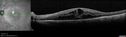
Cystoid Macular Edema and Macular Pucker401 viewshas been particular bad for 3 years. She has a macular pucker and severe CME.
VA 20/160 left eye
|
|
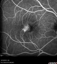
Macular Telangiectasi - Stage V - CNVM OU401 views60 year old female who has been treated with injections for 5 years starting in the left eye. She had no treatment 6 months prior to this visit and her vision has been getting worse. Her vision is 20/100 OD, 20/32 OS. Vision improved with intravitreal Avastin to 20/30 OU.
|
|

Acquired Toxoplasmosis Retinitis Para Foveal - Following Venison Consumption401 views56 year old man who became very sick after eating venison at the end of December 2019. Presented 6 weeks later with a scotoma. Initial photos show presentation with 20/63 vision. Vision dropped to 5/200. All tests (including PCR of anterior chamber tap) were negative except serum toxoplasmosis IgG and IgM. Vision improved to 20/32 but the scotoma remained on oral trimetheprim/sulfa
|
|

Acquired Toxoplasmosis Retinitis Para Foveal - Following Venison Consumption401 views56 year old man who became very sick after eating venison at the end of December 2019. Presented 6 weeks later with a scotoma. Initial photos show presentation with 20/63 vision. Vision dropped to 5/200. All tests (including PCR of anterior chamber tap) were negative except serum toxoplasmosis IgG and IgM. Vision improved to 20/32 but the scotoma remained on oral trimetheprim/sulfa
|
|
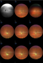
Acquired Toxoplasmosis Retinitis Para Foveal - Following Venison Consumption401 views56 year old man who became very sick after eating venison at the end of December 2019. Presented 6 weeks later with a scotoma. Initial photos show presentation with 20/63 vision. Vision dropped to 5/200. All tests (including PCR of anterior chamber tap) were negative except serum toxoplasmosis IgG and IgM. Vision improved to 20/32 but the scotoma remained on oral trimetheprim/sulfa
|
|

Lamellar Macular Hole - Evoluation over 5 years401 views65 year old man with Sturge weber and a choroidal hemangioma in the left eye. (See other images under sturge weber on this website). Has ongoing problem with his left eye. Now with Blurred vision in the right eye gradually worse the last few weeks. He has had a vitrectomy in the right eye 4 years ago for floaters. Laser for tears and cryo in the right eye 5 years ago. He is on Dorzolamide and Lumigan for glaucoma.
VA 20/32 OD, 20/40 OS
IOP 23 OD, 19 OS
|
|

Macular Telangiectasia (MacTel) and Pattern Dystrophy401 views71 year old man with diabetes for 5 years and blurred vision in the left eye. VA 20/25 OD, 20/50 OS. OCT OS shows MacTel
|
|
| 18117 files on 182 page(s) |
 |
|
|
|
|
129 | |
|
|
|
|


