|
|
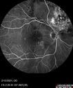
Branch Retinal Vein Occlusion with Venous Macroaneurysm (collateral)vu 372 fois66 year old female Did not notice vision loss in the right eye. The right eye has been a little worse than the left but now is much worse. Picked up during annual eye examination. VA had been 20/20 in 5 years ago.
History of brain and spinal cord radiation 1986 for T-cell lymphoma. Now with breast cancer, ablation for tachycardia, HTN
VA OD 6/200, OS 20/32
|
|

Age-related macular degeneration - Geographic Atrophy - Intermittent CME left eye from CRVO, Anemia, Diabetesvu 372 fois79 year old female - 3 years post CRVO in the left eye (20/32 VA) with intermittent CME. Left eye is the better eye. Vision is stable since 2 years ago
PMHx: Type II DM x 20 years, Chronic Anemia, Hyperlipidemia
VA 20/200 OD, 20/50 OS (usually 20/40)
|
|

Central Serous Chorioretinopathy in a childvu 372 fois12-year-old African American Female A few weeks ago she felt like something was covering the right eye and she couldn't see well. She was told that she had a swollen optic nerve. She had a recent diagnosis of sinus infection. She also recently had onset menarche on August 7th. Eye problem started about a week before menarche. She's been getting some headaches, partly because of sinus infection. They started about a month ago and now are getting better. She is on zytrhomax for the sinus problem. Patient is hypothyroid and is on thyroid supplementation and that has been stable. She has no eye pain.
MEDS: Synthroid, Flonase
PMHx - Thyroid Disease
VA OD: Dcc20/400 PH20/125 NccJ10
VA OS: Dcc20/20 NccJ1+
IOP: TP: OD:14 OS:21
Macular detachments resolved in 1 month with no treatment and vision returned to 20/20 in one month and 20/16 in 2 months
|
|

Bilateral Diffuse Melanocytic Proliferation (BDUMP)vu 372 fois67 year old man
The patient started noticing dimming of the vision in November of 2020. He had rapidly worsening cataracts. He had the cataract surgery done in both eyes in April of 2021. The vision did not improve with the cataract surgery. On June 1 of 2021 he had immunotherapy for his renal cell carcinoma and then developed severe vision loss. The patient noticed that one of the commercials for nivolumab (checkpoint inhibitor targeting programmed death receptor-1) said it may cause vision loss. He recalled that he started the nivolumab in October of 2020 and had vision loss starting then.  A retinal evaluation showed subretinal fluid and vision loss.  The peripheral vision and central vision were pretty good. The vision did seem to get better off the nivolumab. Then when the patient returned to Florida in September and now the vision is getting worse. The patient did not have any treatments in either eye. The patient is now off the oncology treatment. He was then started on a TKI cabometex for the cancer which was dosed in mid June through mid July just for a month. It was stopped because of a skin rash and reactions. Â
VA OD: Dcc20/200-2 PHNI Ncc20/800
VA OS: Dcc20/63 PHNI NccJ7 He has been off the checkpoint inhibitor for a year.
|
|

Central retinal artery occlusion and paracentral acute middle maculopathy and embolivu 372 fois86 year old female with sudden vision loss a week ago and imaging shows PAMM lesions and emboli. Her vision is 20/200 in the left eye
|
|

Central retinal artery occlusion and paracentral acute middle maculopathy and embolivu 372 fois86 year old female with sudden vision loss a week ago and imaging shows PAMM lesions and emboli. Her vision is 20/200 in the left eye
|
|

Choroidal Hemangioma Requiring PDT repeatedlyvu 372 fois64 year old man. Diagnosed with hemangioma in 2015. PDT in 2016. Images are from 2020 when fluid started to reaccumulate. Needed PDT repeated two more times.
PDT treatments: [1] 2/19/2015 -Â Pavan (IOPÂ problem after PDT)Â AND [2] 11/2/21 PDT (7 mm temporal to fovea) - SMCÂ AND [3] 7/22/22 PDT 7.5 mm Temporal to the fovea
|
|

hydroxychloroquine (Plaquenil) toxicityvu 371 fois
|
|
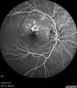
Punctate Inner Choroidopathy and Choroidal Neovascular Membranevu 371 fois27 year old female with vision loss for 4 months. VA was 20/200. She had a CNVM which was extrafoveal with no subretinal fluid. Her vision improved over 6 months to 20/50 with Ranibizumab injections (she might have improved without treatment)
|
|
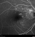
Polypiodal Choroidal Vasculopathy - Temporal Macula - Left eyevu 371 fois82 year old man with asymptomatic PCV in the left eye. Vision 20/50 OD, 20/32 OS did well with Lucentis
|
|

Acquired Toxoplasmosis Retinitis Para Foveal - Following Venison Consumptionvu 371 fois56 year old man who became very sick after eating venison at the end of December 2019. Presented 6 weeks later with a scotoma. Initial photos show presentation with 20/63 vision. Vision dropped to 5/200. All tests (including PCR of anterior chamber tap) were negative except serum toxoplasmosis IgG and IgM. Vision improved to 20/32 but the scotoma remained on oral trimetheprim/sulfa
|
|

Acquired Toxoplasmosis Retinitis Para Foveal - Following Venison Consumptionvu 371 fois56 year old man who became very sick after eating venison at the end of December 2019. Presented 6 weeks later with a scotoma. Initial photos show presentation with 20/63 vision. Vision dropped to 5/200. All tests (including PCR of anterior chamber tap) were negative except serum toxoplasmosis IgG and IgM. Vision improved to 20/32 but the scotoma remained on oral trimetheprim/sulfa
|
|

hydroxychloroquine (Plaquenil) Toxicity - Taiwanese - 3 years post stoppingvu 371 fois59 year old female on 400 mg plaquenil for 22 years. 3 years ago, when stopping plaquenil, VA 20/50 both eyes. Plaquenil was stopped. The vision in the left eye continued to deteriorate to 3/200. 5'2" 127 lb (6.6 mg/kg body weight) -- Vision in these images is 20/80 OD, 5/200 OS - Vision loss now seems stable.
|
|

Retinitis Pigmentosa - X-linked Recessivevu 371 fois60 year old man The vision in the right eye is blurry recently. He has trouble with night vision for about 3 years. His brother has eye disease in Michigan.
VA OD: Dcc20/32-1 PHNI NccJ1-1
VA OS: Dcc20/20 NccJ1+-1
IOP: TP: OD:36 OS:18
He has pseudoexfoliative glaucoma in the right eye.
|
|
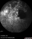
Branch Retinal Vein Occlusion with Neovascularization Elsewhere and Vitreous Hemorrhagevu 371 fois57 year old man New floaters in the left eye about a month ago. Now they are getting better. PMHx A-Fib on Pradaxa
VA 20/32 OD, 20/40 OS
1+ NS OU
BP 137/77, 200LBs, 5’8â€
PRP laser was done
|
|

Branch Retinal Vein Occlusion with NVE and vitreous hemorrhage and non-perfusionvu 371 fois50 year old man The right eye was fine and then there was a sudden change in the vision. When he is outside looking at something bright there are thousands of dots with squiggly lines through them.Â
PMHx: Type II DM (1 year), HTN, AnemiaÂ
VA OD: sc20/40 PH20/32 NscJ7
VA OS: sc20/160 PH20/25 NscJ2
Myelinate RNFL in the left eye
|
|

Central Retinal Artery Occlusion - Fluorescein Angiogram Videovu 371 fois63 year old man Sudden severe vision loss in the left eye for 3 days. Six months following macula off retinal detachment repair, Pneumatic then PPV, then PPV buckle. VA was 20/63 on 4/2021 (3 months post RD repair).
PMHx – HTN. Meds: HCT, Meloxicam
VA OD 20/25, OS HM
|
|
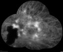
Pregnant - 25 weeks - new onset PDR and DME both eyes - Treated with PRP and ozurdexvu 371 fois29 year old female. Patient is 25 weeks pregnant and having flare up of retinopathy in both eyes. She states it is worse in the left. Patient has trouble with distant vision. At most recent visit, 1 years ago, VA was 20/20 OU. Two years ago anterior uveitis x 1 episode with normal labs (HLAB27, FTA, ACE, Lyme). 2017-2020 – Lucentis for DME left eye only.
PMHx: Type I DM since teenager, hyperlipidemia
BP was normal
|
|

Ocular Lymphoma - Initially presented like auto-immune retinopathyvu 371 fois43 year old man: initial presentation - The vision in the left eye is blurry and opaque for about 2 weeks. In the central of the vision, when he covers the right eye, in the center of the left eye there is a film with a light in the middle. This has never happened before. He has not been sick for the last few months. A few weeks ago he saw waves on the floor when looking down but that has gone away. Since the vision loss started the vision has not changed much. The central vision is not getting better. Â
PMHx – Borderline HTN
VA OD: Dcc20/20
VA OS: Dcc20/40-1 IOP: TP: OD:15 OS:19
1+ NS
No uveitis
Labs normal. One positive antiretinal antibody - Arrestin
VA improved over 2 months to normal. Then the patient had 2 more episodes of self-limited vision loss (no treatment). Then 2 years later he presented with a diagnosis of CNS lymphoma and later developed subretinal deposits (last images)
|
|

Central Retinal Vein Occlusion following Cataract Surgeryvu 371 fois79 year old man with decreased vision The patient had cataract surgery in the right eye 9 days ago and in the left eye 21 days ago and presents with vision loss in the right eye.
Meds: HCTZ, Garlic, Tumeric
VA 20/60 OD, 20/25 OS
|
|
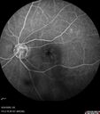
Central Retinal Vein Occlusion following Cataract Surgeryvu 371 fois79 year old man with decreased vision The patient had cataract surgery in the right eye 9 days ago and in the left eye 21 days ago and presents with vision loss in the right eye.
Meds: HCTZ, Garlic, Tumeric
VA 20/60 OD, 20/25 OS
|
|
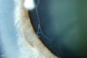
Pseudoexfoliation Material Peeling.vu 370 foisPatient comes in for regular eye exam. Last exam was 8-years ago. Complains of decrease in VA. VA was 20/30, right eye and 20/40, left eye. Slit lamp photography shows pseudoexfoliation peeling of the lens within 360-degrees. Pseudoexfoliation ring is present centrally. IOP was normal. Will revisit in 3-months for follow up.
|
|

Acute Bilateral Central Serous Chorioretinopathy - Female - Smokestackvu 370 fois39 year old female She had no prior epsisodes of vision loss and then a week ago she noticed decreased vision in the right eye. She has not had any health problems and she is not pregnant. Phendimetrazine started 3 months ago. She is not on steroids. VA OD: sc20/63-1 NscJ16 VA OS: sc20/125-1 Dcc20/80 NscJ16 IOP: TP: OD:14 OS:14 Time:11:23 AM
|
|

Perifoveal Microaneurysm - Not Diabetic (also reticular pseudodrusen)vu 370 fois87 year old female with fluctuating macular edema in the right eye from a perifoveal MA. (no treatment). VA 20/40 OU
|
|

Proliferative Diabetic Retinopathy and vitreous hemorrhage left eyevu 370 fois44 year old man with type II diabetes for 14 years and no eye examinations in the last 5 years. VA 20/40 right eye and 20/60 left eye. Vision loss has been for about a week in the left eye.
|
|

hydroxychloroquine (Plaquenil) Toxicity - Taiwanese - 3 years post stoppingvu 370 fois59 year old female on 400 mg plaquenil for 22 years. 3 years ago, when stopping plaquenil, VA 20/50 both eyes. Plaquenil was stopped. The vision in the left eye continued to deteriorate to 3/200. 5'2" 127 lb (6.6 mg/kg body weight) -- Vision in these images is 20/80 OD, 5/200 OS - Vision loss now seems stable.
|
|

Recurrent Toxoplasmosis Right eye then Left eye 8 years latervu 370 fois33 year old brazilian man. Initially presented with uveitis in the right eye in 2011. Then in the left eye in 2019. Since has had two more episodes.
|
|

Bilateral Acute Central Serous Chorioretinopathy - Evidence of old disease OSvu 370 fois64 year old man noticed in the right eye that is a black spot 4 days ago. Also vertical lines in his kitchen were a little wavy. He is not on any steroid medications. He has vertigo.
VA OD: Dcc20/25-2 NccJ1+-3
VA OS: Dcc20/32-1 PHNI NccJ5-1
IOP: TP: OD:12 OS:15
|
|

vu 370 fois
|
|
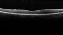
Elevated Episcleral Venous Pressure and Retinal Hemorrhages in both eyesvu 370 fois78 year old female with chronic red eye. The eyes are red all the time now. She is on Xeralto because of a history of blood clots in her lungs (2005 about). She has not had problems since. She has had floaters for many years. Ever since her eyes got funny she sees more floaters in both eyes. She had a brain scan over 6 months ago which was OK. She also had double vision when this first started which went away. She could not drive for a few months.
VA OD: Dcc20/32-1
VA OS: Dcc20/125
IOP: TP: OD:17 OS:16
Neuroimaging did not show a CC fistula
|
|
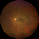
Intermediate Dry Age-Related Macular Degeneration - Soft Drusenvu 370 fois70-year-old Left eye worse than the right. Both eyes see distortion in the amsler grid for a few months
HTN, High Lipids, Osteoporosis
Meds: Lutein once daily, Calcium, Fish oil, Red Yeast Rice, CoQ10, Magnesium, MVI, Xanax
VA 20/16 OU
2+NS cataract
|
|

Amelanotic Choroidal Nevusvu 370 fois60 year old man. Nevus is stable over 6 years. Images are Photo, FAF, OCT, FA, ICG, and B-Scan
|
|
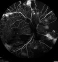
Severe Proliferative Diabetic Retinopathy and Good Visionvu 370 fois49 year old man with vision loss in the right eye for 2 weeks. VA 20/40 OD, 20/63 OS. DM x 15 years and HTN.
|
|
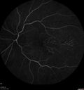
Dry AMD geographic atrophy and cystoid macular edemavu 370 fois79 year old vision is in for a checkup with no visual complaints in the left eye. This is her better eye. VA 20/200 OD, 20/50 OS. 3 years ago she had a CRVO in the left eye. She is also diabetic for 20 years, has carotid insufficiency and anemia. Left eye shows CME. This was not treated and the vision improved to 20/40 over the next year although mild edema persisted.
|
|
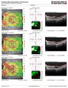
Dry AMD geographic atrophy and cystoid macular edemavu 370 fois79 year old vision is in for a checkup with no visual complaints in the left eye. This is her better eye. VA 20/200 OD, 20/50 OS. 3 years ago she had a CRVO in the left eye. She is also diabetic for 20 years, has carotid insufficiency and anemia. Left eye shows CME. This was not treated and the vision improved to 20/40 over the next year although mild edema persisted.
|
|

Bilateral Choroidal Tumors - Metastatic Breast Cancervu 370 fois51 year old female. First 3 sets of images show lesions growing despite systemic chemotherapy. She eventually had external beam radiation x 15 sessions and the tumors shrunk in the last two sets of images
|
|
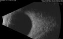
Bilateral Choroidal Tumors - Metastatic Breast Cancervu 370 fois51 year old female. First 3 sets of images show lesions growing despite systemic chemotherapy. She eventually had external beam radiation x 15 sessions and the tumors shrunk in the last two sets of images
|
|

Normal Fluorescein Angiogram 03vu 370 fois
|
|
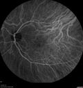
Normal Indocyanine Green Angiogram 03vu 370 fois
|
|
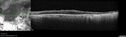
Pan Retinal Laser from original DRS study in the 1970s - 50 years latervu 370 fois67 year old female with diabetes since age 8. She had heavy PRP laser to both eyes in the 1970s by Lloyd M. Aiello, M.D, the father of PRP laser. Her vision is poor now, 1/200 OD and 20/200 OS.
|
|

Age-related macular degeneration - Geographic Atrophy - Intermittent CME left eye from CRVO, Anemia, Diabetesvu 370 fois79 year old female - 3 years post CRVO in the left eye (20/32 VA) with intermittent CME. Left eye is the better eye. Vision is stable since 2 years ago
PMHx: Type II DM x 20 years, Chronic Anemia, Hyperlipidemia
VA 20/200 OD, 20/50 OS (usually 20/40)
|
|
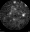
Pregnant - 25 weeks - new onset PDR and DME both eyes - Treated with PRP and ozurdexvu 370 fois29 year old female. Patient is 25 weeks pregnant and having flare up of retinopathy in both eyes. She states it is worse in the left. Patient has trouble with distant vision. At most recent visit, 1 years ago, VA was 20/20 OU. Two years ago anterior uveitis x 1 episode with normal labs (HLAB27, FTA, ACE, Lyme). 2017-2020 – Lucentis for DME left eye only.
PMHx: Type I DM since teenager, hyperlipidemia
BP was normal
|
|
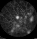
Pregnant - 25 weeks - new onset PDR and DME both eyes - Treated with PRP and ozurdexvu 370 fois29 year old female. Patient is 25 weeks pregnant and having flare up of retinopathy in both eyes. She states it is worse in the left. Patient has trouble with distant vision. At most recent visit, 1 years ago, VA was 20/20 OU. Two years ago anterior uveitis x 1 episode with normal labs (HLAB27, FTA, ACE, Lyme). 2017-2020 – Lucentis for DME left eye only.
PMHx: Type I DM since teenager, hyperlipidemia
BP was normal
|
|

Ocular Lymphoma - Initially presented like auto-immune retinopathyvu 370 fois43 year old man: initial presentation - The vision in the left eye is blurry and opaque for about 2 weeks. In the central of the vision, when he covers the right eye, in the center of the left eye there is a film with a light in the middle. This has never happened before. He has not been sick for the last few months. A few weeks ago he saw waves on the floor when looking down but that has gone away. Since the vision loss started the vision has not changed much. The central vision is not getting better. Â
PMHx – Borderline HTN
VA OD: Dcc20/20
VA OS: Dcc20/40-1 IOP: TP: OD:15 OS:19
1+ NS
No uveitis
Labs normal. One positive antiretinal antibody - Arrestin
VA improved over 2 months to normal. Then the patient had 2 more episodes of self-limited vision loss (no treatment). Then 2 years later he presented with a diagnosis of CNS lymphoma and later developed subretinal deposits (last images)
|
|

Bilateral Diffuse Melanocytic Proliferation (BDUMP)vu 370 fois67 year old man
Vision is better after 14 plasma exchanges, OD 20/160, OS 20/32
|
|

Central Retinal Vein Occlusion following Cataract Surgeryvu 370 fois79 year old man with decreased vision The patient had cataract surgery in the right eye 9 days ago and in the left eye 21 days ago and presents with vision loss in the right eye.
Meds: HCTZ, Garlic, Tumeric
VA 20/60 OD, 20/25 OS
|
|

Central Serous Retinopathyvu 369 foisPatient comes in with blurred vision. Patient also had a lung transplant. VA was 20/150 with pinhole correction to 20/60 in the right eye. Fundus exam in the right eye shows a great amount of sub retinal fluid over the macula. Will monitor to see if it goes away on its own, if not then will consider laser treatment.
|
|

Trans-Illumination Defect with Fuch's Dystrophy.vu 369 foisPatient comes in for eval on post cataract surgery. VA is 20/30 both eyes. Slit lamp exam shows Fuch's Dystrophy on the cornea on the right eye. IOL haptics are chaffing the iris therefore producing trans illumination defect in the right eye.
|
|

Spontaneously Re-attached Asymptomatic Chronic Retinal Detachmentvu 369 fois47 year old female The vision has been gradually worsening for a few years. The two eyes have always been different. She has no knowledge of having had a retinal detachment - She now has mild CME.
VA OD: Dcc20/32 NscJ2
VA OS: Dcc20/20 NscJ1
|
|

Valsalva Retinopathy - Macular Hemorrhage - Permanent Vision loss - Intraretinal Bloodvu 369 fois65 year old man with sudden vision loss in the right eye. Initial vision 20/200. VA improved to 20/50 in one year. Had carotid stenosis
|
|
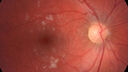
Cotton Wool Spots - Severe Anemia - Acute Lymphocytic Leukemiavu 369 fois23 year old just diagnosed with anemia and acute lymphocytic leukemia
: If he covers each eye neither eye is lacking. When he looks to the left he gets double vision when he looks to the right his vision is fine. This started about 2 weeks ago. Since it started it has been variable. He is on a bunch of medications for leukemia and he might be having high sugars. He noticed the vision change after his first blood transfusion in the hospital. He is currently very anemic.
VA OD: sc20/20 NscJ1+
VA OS: sc20/20 NscJ1+
|
|

Bilateral Acute Central Serous Chorioretinopathy - Evidence of old disease OSvu 369 fois64 year old man noticed in the right eye that is a black spot 4 days ago. Also vertical lines in his kitchen were a little wavy. He is not on any steroid medications. He has vertigo.
VA OD: Dcc20/25-2 NccJ1+-3
VA OS: Dcc20/32-1 PHNI NccJ5-1
IOP: TP: OD:12 OS:15
|
|
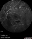
Branch Retinal Vein Occlusion with Neovascularization Elsewhere and Vitreous Hemorrhagevu 369 fois57 year old man New floaters in the left eye about a month ago. Now they are getting better. PMHx A-Fib on Pradaxa
VA 20/32 OD, 20/40 OS
1+ NS OU
BP 137/77, 200LBs, 5’8â€
PRP laser was done
|
|
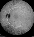
Punctate Inner Choroidopathy and Choroidal Neovascular Membrane (CNVM)vu 369 fois24 year old female She was doing fine until 4 days ago when the right eye fogged up on her. The left eye is seeing fine. In the end of January (3 months ago) she went to the doctor and got checked for everything and it was negative. She had a bad cough at the time and no fevers that she knows of. Â
VA OD: sc20/200+1, VA OS: sc20/25 - Uveitis work up was negative. VA improved to 20/40 with a single avastin treatment and then the patient was lost to follow-up
|
|

Coloboma - Iris, Lens, Chorioretinalvu 369 fois15 year old with poor vision since birth. Noticing new flashes in the left eye. There is an iris - lens coloboma as well.
|
|

Coloboma - Iris, Lens, Chorioretinalvu 369 fois15 year old with poor vision since birth. Noticing new flashes in the left eye. There is an iris - lens coloboma as well.
|
|

Bilateral Choroidal Tumors - Metastatic Breast Cancervu 369 fois51 year old female. First 3 sets of images show lesions growing despite systemic chemotherapy. She eventually had external beam radiation x 15 sessions and the tumors shrunk in the last two sets of images
|
|

Branch Retinal Vein Occlusion with Venous Macroaneurysm (collateral)vu 369 fois66 year old female Did not notice vision loss in the right eye. The right eye has been a little worse than the left but now is much worse. Picked up during annual eye examination. VA had been 20/20 in 5 years ago.
History of brain and spinal cord radiation 1986 for T-cell lymphoma. Now with breast cancer, ablation for tachycardia, HTN
VA OD 6/200, OS 20/32
|
|
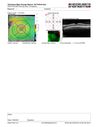
Neuroretinitis left eye - recovered with no therapy - possible B pertussisvu 369 fois57 year old female The patient has had blurred vision in the left eye and there is a haze across the top of the vision. This started about 2 weeks ago. The right eye is OK. She has sleep apnea and uses the machine. The left eye is becoming more and more painful for the last two weeks. The pain is like a stabbing in the eye that comes and goes. She also has some pain on eye movement. The patient has been going to Moffit for possible reconstructive surgery for the lymphedema.
PMHx – Sleep apnea, Breast Cancer, Adult lymphedema,
Meds: GabapentinÂ
VA OD: Dcc20/25-1
VA OS: Dcc20/100-2
AC quiet OU. 1 + NS cataract OU
IOP: TP: OD:15 OS:17
|
|

Branch Retinal Vein Occlusion with NVE and vitreous hemorrhage and non-perfusionvu 369 fois50 year old man The right eye was fine and then there was a sudden change in the vision. When he is outside looking at something bright there are thousands of dots with squiggly lines through them.Â
PMHx: Type II DM (1 year), HTN, AnemiaÂ
VA OD: sc20/40 PH20/32 NscJ7
VA OS: sc20/160 PH20/25 NscJ2
Myelinate RNFL in the left eye
|
|

Age-related macular degeneration - Geographic Atrophy - Intermittent CME left eye from CRVO, Anemia, Diabetesvu 369 fois79 year old female - 3 years post CRVO in the left eye (20/32 VA) with intermittent CME. Left eye is the better eye. Vision is stable since 2 years ago
PMHx: Type II DM x 20 years, Chronic Anemia, Hyperlipidemia
VA 20/200 OD, 20/50 OS (usually 20/40)
|
|
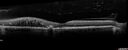
Nonproliferative Diabetic Retinopathy and Good Visionvu 369 fois59 year old man Vision is good. Just referred following annual eye examination
PMHx: Type II DM, HTN, Cholesterol,
MEDS: Lantus, Metformin, lisinopril, atorvastatin, gabapentin
VA 20/20 OU
2+ NS OU
Observation
|
|

Rapid macular atrophy with anti-VEGF therapy for myopic CNVMvu 368 fois64 year old female with 20/32 vision and distortion in the left and better eye. She was treated with monthly lucentis and her vision declined to 20/100. Medicine was stopped and she improved a little to 20/80.
|
|

Punctate Inner Choroidopathy and Choroidal Neovascular Membranevu 368 fois27 year old female with vision loss for 4 months. VA was 20/200. She had a CNVM which was extrafoveal with no subretinal fluid. Her vision improved over 6 months to 20/50 with Ranibizumab injections (she might have improved without treatment)
|
|

Choroidal Neovascular Membrane following Prior Central Serous Chorioretinopathy - OCT-Avu 368 fois67 year old female with CSR in the left eye first 20 years ago. She was managed with out therapy and has had a few episodes since. Now ther eis distortion in the left eye for a few days. She had a steroid shot in her shoulder 5 months ago.
VA 20/20 OD, 20/50 OS- ICG and OCT-A confirm CNVM in the left eye
|
|

Tilted Disc Syndromevu 368 fois56 year old female. Recent motor vehicle accident. Vision is 20/50 OD, 20/25 OS.
|
|

Proliferative Diabetic Retinopathy and vitreous hemorrhage left eyevu 368 fois44 year old man with type II diabetes for 14 years and no eye examinations in the last 5 years. VA 20/40 right eye and 20/60 left eye. Vision loss has been for about a week in the left eye.
|
|

Recurrent Toxoplasmosis Right eye then Left eye 8 years latervu 368 fois33 year old brazilian man. Initially presented with uveitis in the right eye in 2011. Then in the left eye in 2019. Since has had two more episodes.
|
|

Branch Retinal Vein Occlusion - Both Eyes - Edema OD and NVE OS - Severe Non-perfusion Both Eyesvu 368 fois83 year old man with vision loss for 3 months not sure which eye. VA 20/100 OD and 20/50 OS. Tried avastin once without much improvement in the right eye and then lost to follow-up. FA in the left eye shows leaking NVE
|
|
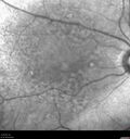
Intermediate Dry Age-Related Macular Degeneration - Soft Drusenvu 368 fois70-year-old Left eye worse than the right. Both eyes see distortion in the amsler grid for a few months
HTN, High Lipids, Osteoporosis
Meds: Lutein once daily, Calcium, Fish oil, Red Yeast Rice, CoQ10, Magnesium, MVI, Xanax
VA 20/16 OU
2+NS cataract
|
|

Dry AMD geographic atrophy and cystoid macular edemavu 368 fois79 year old vision is in for a checkup with no visual complaints in the left eye. This is her better eye. VA 20/200 OD, 20/50 OS. 3 years ago she had a CRVO in the left eye. She is also diabetic for 20 years, has carotid insufficiency and anemia. Left eye shows CME. This was not treated and the vision improved to 20/40 over the next year although mild edema persisted.
|
|

Central Retinal Artery Occlusion - Fluorescein Angiogram Video showing slow fillvu 368 fois63 year old man Sudden severe vision loss in the left eye for 3 days. Six months following macula off retinal detachment repair, Pneumatic then PPV, then PPV buckle. VA was 20/63 on 4/2021 (3 months post RD repair).
PMHx – HTN. Meds: HCT, Meloxicam
VA OD 20/25, OS HM
|
|
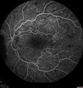
Pregnant - 25 weeks - new onset PDR and DME both eyes - Treated with PRP and ozurdexvu 368 fois29 year old female. Patient is 25 weeks pregnant and having flare up of retinopathy in both eyes. She states it is worse in the left. Patient has trouble with distant vision. At most recent visit, 1 years ago, VA was 20/20 OU. Two years ago anterior uveitis x 1 episode with normal labs (HLAB27, FTA, ACE, Lyme). 2017-2020 – Lucentis for DME left eye only.
PMHx: Type I DM since teenager, hyperlipidemia
BP was normal
|
|

vu 368 fois
|
|
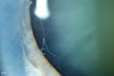
Pseudoexfoliation Material Peeling.vu 367 foisPatient comes in for regular eye exam. Last exam was 8-years ago. Complains of decrease in VA. VA was 20/30, right eye and 20/40, left eye. Slit lamp photography shows pseudoexfoliation peeling of the lens within 360-degrees. Pseudoexfoliation ring is present centrally. IOP was normal. Will revisit in 3-months for follow up.
|
|

Spontaneously Re-attached Asymptomatic Chronic Retinal Detachmentvu 367 fois47 year old female The vision has been gradually worsening for a few years. The two eyes have always been different. She has no knowledge of having had a retinal detachment - She now has mild CME.
VA OD: Dcc20/32 NscJ2
VA OS: Dcc20/20 NscJ1
|
|

Spontaneously Re-attached Asymptomatic Chronic Retinal Detachmentvu 367 fois47 year old female The vision has been gradually worsening for a few years. The two eyes have always been different. She has no knowledge of having had a retinal detachment - She now has mild CME.
VA OD: Dcc20/32 NscJ2
VA OS: Dcc20/20 NscJ1
|
|

Spontaneously Re-attached Asymptomatic Chronic Retinal Detachmentvu 367 fois47 year old female The vision has been gradually worsening for a few years. The two eyes have always been different. She has no knowledge of having had a retinal detachment - She now has mild CME.
VA OD: Dcc20/32 NscJ2
VA OS: Dcc20/20 NscJ1
|
|

Rapid macular atrophy with anti-VEGF therapy for myopic CNVMvu 367 fois64 year old female with 20/32 vision and distortion in the left and better eye. She was treated with monthly lucentis and her vision declined to 20/100. Medicine was stopped and she improved a little to 20/80.
|
|

Punctate Inner Choroidopathy and Choroidal Neovascular Membranevu 367 fois27 year old female with vision loss for 4 months. VA was 20/200. She had a CNVM which was extrafoveal with no subretinal fluid. Her vision improved over 6 months to 20/50 with Ranibizumab injections (she might have improved without treatment)
|
|

Macular Telangiectasi - Stage V - CNVM OUvu 367 fois60 year old female who has been treated with injections for 5 years starting in the left eye. She had no treatment 6 months prior to this visit and her vision has been getting worse. Her vision is 20/100 OD, 20/32 OS. Vision improved with intravitreal Avastin to 20/30 OU.
|
|

vu 367 fois
|
|

Retinitis Pigmentosa - X-linked Recessivevu 367 fois60 year old man The vision in the right eye is blurry recently. He has trouble with night vision for about 3 years. His brother has eye disease in Michigan.
VA OD: Dcc20/32-1 PHNI NccJ1-1
VA OS: Dcc20/20 NccJ1+-1
IOP: TP: OD:36 OS:18
He has pseudoexfoliative glaucoma in the right eye.
|
|

vu 367 fois
|
|
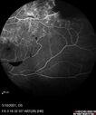
Branch Retinal Vein Occlusion with Neovascularization Elsewhere and Vitreous Hemorrhagevu 367 fois57 year old man New floaters in the left eye about a month ago. Now they are getting better. PMHx A-Fib on Pradaxa
VA 20/32 OD, 20/40 OS
1+ NS OU
BP 137/77, 200LBs, 5’8â€
PRP laser was done
|
|
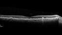
Intermediate Dry Age-Related Macular Degeneration - Soft Drusenvu 367 fois70-year-old Left eye worse than the right. Both eyes see distortion in the amsler grid for a few months
HTN, High Lipids, Osteoporosis
Meds: Lutein once daily, Calcium, Fish oil, Red Yeast Rice, CoQ10, Magnesium, MVI, Xanax
VA 20/16 OU
2+NS cataract
|
|

Amelanotic Choroidal Nevusvu 367 fois60 year old man. Nevus is stable over 6 years. Images are Photo, FAF, OCT, FA, ICG, and B-Scan
|
|

Amelanotic Choroidal Nevusvu 367 fois60 year old man. Nevus is stable over 6 years. Images are Photo, FAF, OCT, FA, ICG, and B-Scan
|
|

Normal Fluorescein Angiogram 01vu 367 fois
|
|
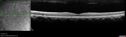
Ocular Lymphoma - Initially presented like auto-immune retinopathyvu 367 fois43 year old man: initial presentation - The vision in the left eye is blurry and opaque for about 2 weeks. In the central of the vision, when he covers the right eye, in the center of the left eye there is a film with a light in the middle. This has never happened before. He has not been sick for the last few months. A few weeks ago he saw waves on the floor when looking down but that has gone away. Since the vision loss started the vision has not changed much. The central vision is not getting better. Â
PMHx – Borderline HTN
VA OD: Dcc20/20
VA OS: Dcc20/40-1 IOP: TP: OD:15 OS:19
1+ NS
No uveitis
Labs normal. One positive antiretinal antibody - Arrestin
VA improved over 2 months to normal. Then the patient had 2 more episodes of self-limited vision loss (no treatment). Then 2 years later he presented with a diagnosis of CNS lymphoma and later developed subretinal deposits (last images)
|
|

Central Retinal Vein Occlusion with Macular Edema and Retinal Hemorrhages vu 366 foisFemale patient of 86-years of age presents a central retinal vein occlusion with macular edema in the right eye. Patient's VA was 20/70 in the right eye. Proceeded with laser treatment to the right eye.
|
|

vu 366 fois
|
|
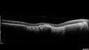
Macular Scars - Laser (1980s) Ocular Histoplasmosisvu 366 fois70 year old man who had laser at Wills, Wilmer and the Mayo clinic before better therapies were available for ocular histoplasmosis related CNVM
|
|

Angioid Streaks Scar and new CNVMvu 366 fois49 y/o WF with decreased vision x 1 month. Her OD is 20/400 and her OS is seeing 20/70. No cutaneous lesions or Ehlers-Danlos. The FP and FA show a predominant classic CNV (with fibrosis) OD and an Occult CNV OS with what appears to be a RAP lesion.
|
|

Acquired Toxoplasmosis Retinitis Para Foveal - Following Venison Consumptionvu 366 fois56 year old man who became very sick after eating venison at the end of December 2019. Presented 6 weeks later with a scotoma. Initial photos show presentation with 20/63 vision. Vision dropped to 5/200. All tests (including PCR of anterior chamber tap) were negative except serum toxoplasmosis IgG and IgM. Vision improved to 20/32 but the scotoma remained on oral trimetheprim/sulfa
|
|

Retinitis Pigmentosa - X-linked Recessivevu 366 fois60 year old man The vision in the right eye is blurry recently. He has trouble with night vision for about 3 years. His brother has eye disease in Michigan.
VA OD: Dcc20/32-1 PHNI NccJ1-1
VA OS: Dcc20/20 NccJ1+-1
IOP: TP: OD:36 OS:18
He has pseudoexfoliative glaucoma in the right eye.
|
|

Bilateral Acute Central Serous Chorioretinopathy - Evidence of old disease OSvu 366 fois64 year old man noticed in the right eye that is a black spot 4 days ago. Also vertical lines in his kitchen were a little wavy. He is not on any steroid medications. He has vertigo.
VA OD: Dcc20/25-2 NccJ1+-3
VA OS: Dcc20/32-1 PHNI NccJ5-1
IOP: TP: OD:12 OS:15
|
|

Bilateral Acute Central Serous Chorioretinopathy - Evidence of old disease OSvu 366 fois64 year old man noticed in the right eye that is a black spot 4 days ago. Also vertical lines in his kitchen were a little wavy. He is not on any steroid medications. He has vertigo.
VA OD: Dcc20/25-2 NccJ1+-3
VA OS: Dcc20/32-1 PHNI NccJ5-1
IOP: TP: OD:12 OS:15
|
|

Bilateral Acute Central Serous Chorioretinopathy - Evidence of old disease OSvu 366 fois64 year old man noticed in the right eye that is a black spot 4 days ago. Also vertical lines in his kitchen were a little wavy. He is not on any steroid medications. He has vertigo.
VA OD: Dcc20/25-2 NccJ1+-3
VA OS: Dcc20/32-1 PHNI NccJ5-1
IOP: TP: OD:12 OS:15
|
|
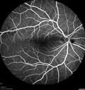
Elevated Episcleral Venous Pressure and Retinal Hemorrhages in both eyesvu 366 fois78 year old female with chronic red eye. The eyes are red all the time now. She is on Xeralto because of a history of blood clots in her lungs (2005 about). She has not had problems since. She has had floaters for many years. Ever since her eyes got funny she sees more floaters in both eyes. She had a brain scan over 6 months ago which was OK. She also had double vision when this first started which went away. She could not drive for a few months.
VA OD: Dcc20/32-1
VA OS: Dcc20/125
IOP: TP: OD:17 OS:16
Neuroimaging did not show a CC fistula
|
|
| 18117 photos sur 182 page(s) |
 |
|
|
|
|
137 | |
|
|
|
|


