|
|

Pigmented Choroidal Mass Right Eye - Melanoma vs. Nevus vs. Unilateral Melanocytic Proliferation - Initial Visit B Scan915 views71-year-old man is vision has been bothering him for about a month. He does not notice one eye particularly worse than the other. OD is 20/40, OS is 20/25 - Initial Visit
|
|

Retinal Tear with Laser915 views
|
|

Serous Retinal Detachment - Hypertensive Retinopathy - 165/120 - Fundus Autofluorescence (FAF)915 views48-year-old woman noticed when she was blinking there was a white flash in the left eye starting about a month ago. She sees it temporally. OD 20/16, OS 20/20. There is a serous retinal detachment superotemporal to the optic nerve falling short of the fovea. Her blood pressure 165/120.
|
|

Vitreomacular Traction - Macular Pucker - 20/50 Vision914 views75-year-old woman decreasing vision in the right eye a few months ago. She had her cataract removed and unfortunately her vision is still hazy in the right eye. Her left eye seems a little hazy from the cataract.
VISUAL ACUITY: OD 20/50, OS 20/20. IOP: OD 17, OS 18.
SLIT-LAMP EXAMINATION
|
|

Juxtafoveal Telangiectasis - MacTel - Group 2a - Stage IV - Pigment Plaque 89 Year Old Man - Nevus914 views89-year-old man has stage IV juxtafoveal retinal telangiectasis in both eyes with background diabetic retinopathy. His vision is stable since he was here last which is about 6 months ago. OD 20/50, OS 20/70.
|
|

Choroidal Melanoma914 viewsPost op Radioactive Implant with hard exudates surrounding the Melanoma with hemorrhage present.
|
|
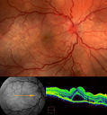
Vogt–Koyanagi–Harada Syndrome OD914 views43 yr old Italian Male with VKH in both eyes.
|
|

Diabetic Papillitis914 viewsPatient is about to undergo surgery for CNS Aneurysm. Patient woke up with little spots which never cleared up. Patients VA was 20/30-OD and 20/40-OS. Both eyes appeared to have disc edema with hemorrhages in the right eye. Ordered a CT of the brain to make sure the aneurysm didn't ruptured.
|
|

Ghost Cell Glaucoma914 viewsPost cataract surgery, patient presents with ghost cell in the anterior chamber after hemorrhage cleared. A small hyphema still is visible in the anterior chamber with a whitish ball appearance.
|
|

Unilateral Acute Idiopathic Maculopathy (UAIM)914 views1 week later eye looks better (after initial scan) 41 year old with serous retinal detachment OS and sudden vision loss. He had a viral prodrome. Coxscackie IgG was high but no acute infection was documented. Vision improved in about 2 months without therapy. - Right eye has spot nasal to the optic nerve
|
|

Ocular Trauma with Retinal Detachment913 viewsPatient suffered blunt force trauma to the eye resulting total retinal detachment. The retina is visible in the pupillary space.
|
|
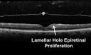
Lamellar Hole Epiretinal Proliferation - LHEP913 views
|
|

Papilledema - Spectral Domain OCT used for Diagnosis - 14 year old Child912 views14-year-old was with optic nerve swelling asymptomatic - picked up during routine eye exam. OD 20/20, OS 20/32
SD-OCT is used to differentiate optic disc edema from optic nerve head drusen (which are not yet calcified in children).
|
|

Glaucoma, Pseudoexfoliation Ring912 viewsSlit Lamp photography shows prominent pseudoexfoliation ring on the lens of the left eye. Patient will be followed up in 6-months...
|
|

Radioactive Plaque Placement for Choroidal Melanoma Transillumination of Globe to Localize Melanoma Arrow Points to Tumor Shadow911 views
|
|

Best Disease for 30 Years - 20/50 Vision both eyes - 61 Year Old Woman911 views61-year-old woman was diagnosed with Best’s disease about 30 years ago. She and her many family members were tested, both at the VA Hospital and then at USF. OD 20/50, OS 20/50
|
|

Gyrate Atrophy - 15 year old male - 20/25 vision both eyes - Positive Family History911 views15 year old male was diagnosed with “retinitis pigmentosa†at age seven. The patient reports that his night vision is poor. He reports he does not notice a visual field change. He reports that his maternal grandfather has lost vision and a cousin has lost vision as well. He reports he has an uncle with some visual changes.
The most significantly affected relative is a 26-year-old legally blind cousin.
20/25+ OD, 20/25 OS.
|
|

Acute Central Serous Retinopathy - CSR - Steroid Induced (probably) - Good Vision - Fundus HyperAutofluorescence Serous Retinal Detachment911 views42-year-old man was seen in the office on October 5, 2011. He had noticed starting in August after a course of antibiotic and steroids, that he developed new spots in his vision in the right eye. He may have had an episode like this sometime in the past. He did take steroids a few years ago and his vision did change at that time, but then returned.
VISUAL ACUITY: OD 20/32, OS 20/32. The OCT scan of the right eye confirms subretinal fluid.
|
|

Retinal Detachment Under Oil with Inferior Retinectomy910 views
|
|
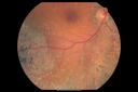
Psuedo-retinitis Pigmentosa - Bone Spicules One Eye - Probably Acute Zonal Occult Outer Retinopathy (AZOOR)910 views65-year-old woman has pseudoretinitis pigmentosa in the right eye only, most likely from acute zonal occult outer retinopathy. OD 20/25, OS 20/25
|
|

Retinal Arterial Macroaneurysm - Ruptured - Vitreous Hemorrhage (Fresh) 5/200 Vision910 views80-year-old woman has a retinal arterial macroaneurysm in the right eye I saw two years ago. She noticed the sudden onset of vision loss last night. OD is 5/200.
|
|

Idiopathic CNVM in 6 year old girl910 viewsCNVM in OD of a 6 yr old girl
|
|

Plaquenil (Hydroxycholorquine) Toxicity - Bull's Eye Maculopathy - 73 year old womand 25 Years therapy - 64 inches tall - 110 lbs909 views73-year-old woman has been on Plaquenil for twenty five years. She noticed loss of paracentral vision a few months ago. VISUAL ACUITY: OD 20/70, OS 20/30. Her color vision was 0 out 10 correct in both eyes and she also missed the controls.
|
|

Posterior Polar Annular Choroidal Dystrophy909 views68 year old woman with know macular irregularities for over 20 years. She has since been followed another 6 years and her vision remains 20/25.
|
|
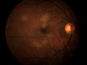
Acute Multifocal Placoid Pigment Epitheliopathy909 views
|
|

Polychromatic "Christmas Tree" Cataract909 viewsPatient comes in for annual eye exam. Eval on cataracts. VA is 20/20, right eye, 20/40, left eye. Slit Lamp exam shows polychromatic christmas tree cataract in the left eye.
|
|

Ghost Cell Glaucoma909 viewsPost cataract surgery, patient presents with ghost cell in the anterior chamber after hemorrhage cleared. A small hyphema still is visible in the anterior chamber with a whitish ball appearance.
|
|

Presumed Ocular Histoplasmosis with new CNVM in right and better eye - VA 20/80 OD; 20/800 OS909 viewsNew subretinal hemorrhage in better eye
|
|

Sclopteria Chorioretinitis - 16 Year Old Girl - Pellet Injury908 views
|
|

Retinoblastoma - Bilateral - Regressed - Chemotherapy, Thermal Therapy, Cryotherapy908 views10-year-old child was seen in the office on August 6, 2008. She was treated with a combination of chemotherapy at 10 months old as well as thermal therapy and cryotherapy for bilateral retinoblastoma tumors. She reportedly had nine tumors in the left eye and five tumors in the right eye
|
|

Mild Clinically Significant Diabetic Macular Edema908 views58-year-old woman diabetic for 20 years who has diabetic macular edema in both eyes. She has had waxing and waning edema in the past and I thought it might improve on its own but it did not. She notices her vision is still a little hazy.
VISUAL ACUITY: Vision OD is 20/30, OS is 20/25.
|
|

Acute Zonal Occult Outer Retinopathy907 views45-year-old man noticed a few years ago peripheral vision blurriness in the left eye and also some central vision loss. Previous to that, as far as he knows, the two eyes were okay.
VISUAL ACUITY: OD: 20/20; OS: 20/40.
|
|

Conjunctival Vascular Anomaly907 viewsPatient presents a vascular pattern of blood vessels @ 10 o'clock of the superior-temporal quadrant of the right eye. Patient has had flare ups with anterior scleritis in the past which is controlled by Methotrexate. Patient's VA is currently 20/40 with PH 20/30, right eye.
|
|
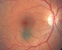
Choroidal Nevus in Macula 906 views
|
|

Asteroid Hyalosis After Vitrectomy (also reticular drusen)906 views
|
|

Punctate Inner Choroidopathy - PIC and Choroidal Neovascular Membrane - Right Eye - SD OCT906 views34-year-old woman with a change in vision in the right eye about eight days ago. Her vision was hazy and she saw waviness in the vision. She does have a history of aches and pains and she was diagnosed with Sjogren’s and 2 months ago she had some fevers and some sinus drainage and that went on for about two months. She is a moderate myope. OD 20/25, OS 20/20.
|
|

Acute Posterior Multifocal Placoid Pigment Epitheliopathy - APMPPE - Fluorescein Angiogram906 views 25-year-old man with one and half weeks ago he woke up with night chills. Also around that time his joints started hurting him. He also has headaches, although the headache at this point is not severe. His vision has been changing for about the last week and a half. He sees some spots in his vision and they are blurred spots. They are not in the central vision.
VISUAL ACUITY: OD 20/32, OS 20/32
|
|

CMV secondary to HIV906 views36 yr old Female Presents for reassessment of decreased vision after starting antiviral medication. OD Va @ onset was LP> OS Va stable 20/20.
|
|

Progressive Outer Retinal Necrosis 77 Year Old Woman with CLL (Acute Retinal Necrosis) (PORN - ARN)905 views77-year-old woman with CLL who had shingles on the left side of her face about 6 weeks ago then she developed a dendrite in the cornea which was treating about four weeks ago. She noticed severe vision loss in the left eye just a few days ago and you saw retinitis and she comes in because of that. Vision OD is 20/25, OS is hand motion
|
|

Pathology - Central Retinal Artery Occlusion - 6 weeks old - Inner Retinal Atrophy - Macular Pucker905 viewspatient died about 6 to 8 weeks after the artery occlusion, and he had no history of glaucoma. The inner retina atrophy, which looks very similar to that caused by COAG, is due to recent infarct.
|
|

Ruptured Retinal Arterial Macroaneurysm - Submacular Hemorrhage - Vision 20/400 never recovered - Hypertension 205/94904 views
|
|

Acute Annular Outer Retinopathy - VA loss 2 days Right Eye Only - 25 Year Old Woman- AZOOR - White Line Around Fresh Lesions904 viewsInitial visit (June 11, 2011): 2 days of vision loss right eye. Wedge suddenly of VA loss OD, too dardkto see through. No flashes or floaters. VA 20/20 OU.
|
|

telangiectasia904 views20/20 pt with blurry area in va
|
|

Myelinated Nerve Fiber Layer - Optic Nerve904 viewsRight eye subtle and inferior - left eye obvious
|
|

Macular Pucker Post-OP 20/30903 views
|
|
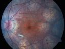
Gyrate Atrophy - 15 year old male - 20/25 vision both eyes - Positive Family History903 views15 year old male was diagnosed with “retinitis pigmentosa†at age seven. The patient reports that his night vision is poor. He reports he does not notice a visual field change. He reports that his maternal grandfather has lost vision and a cousin has lost vision as well. He reports he has an uncle with some visual changes.
The most significantly affected relative is a 26-year-old legally blind cousin.
20/25+ OD, 20/25 OS.
|
|

Fundus Flavimaculatus - Asymptomatic - Color Photo903 views45 year old man was told to come in because of ‘white spots on their retina’.
|
|

Sclopteria Chorioretinitis - 16 Year Old Girl - Pellet Injury902 views
|
|

Chorioretinal Scar with Radiating Chorioretinal Folds902 views79-year-old woman has wet age-related macular degeneration in both eyes. The left eye had intravitreal injections of Avastin starting in May of 2009. Her vision remains good in the left eye and poor in the right eye.
VISUAL ACUITY: OD 8/200, OS 20/60.
|
|

Congenital Achromatopsia 902 views26-year-old man was diagnosed with congenital achromatopsia by ERG as a child. He has family members who also have this disorder. OD 20/200, OS 20/200.
|
|

Drusen Maculopathy - Confluent Subfoveal Soft Hydrophobic Drusen - Early Fluorescein Angiogram Drusen do not yet Stain901 views73-year-old woman. She is diabetic for thirteen years. She is also on Coumadin for her heart disease.
VISUAL ACUITY: OD 20/25, OS 20/50.
|
|

Retinoma - Retinocytoma - B Scan Ultrasound901 viewsUltrasound shows shadowing from the calcifications in the tumor.
|
|

Retinal Tear with Vitreous Hemorrhage before and after Laser Photocoagulation901 views61-year-old man has noticed increased floaters in the right eye for about the last week. Vision OD is 20/160 There is 3+ vitreous hemorrhage with a retinal tear, about 2-disc diameters posterior to the equator at 10:00 o’clock over a retinal vessel.
|
|

ECTOPIC DISCIFORM SCAR/PERIPHERAL CHOROIDAL NEOVASCULAR MEMBRANE – EXUDATE –INFLAMMATION IN REACTION TO THE PERIPHERAL VASCULAR LESION 901 views53-year-old woman has noticed some blurred vision in the right eye for a few years. In retrospect, she thinks it is possible since she was a teenager the right eye was always the weaker eye and that the left eye has always been the better eye. VISUAL ACUITY: Vision OD is 20/400
|
|

Purtscher's Retinopathy901 views19 year old female after pregnancy
|
|

Macular Laser Scars901 views86 year old man who had Laser done from 2010 - 2018 for diabetic retinopathy. The right eye lost vision immediately after laser was done a few years ago. VA 1/200 OD; 20/25 OS
|
|

Central Serous Retinopathy - Acute900 views49-year-old man sees a spot in the central vision with a red halo for 2 weeks OD is 20/50, OS is 20/20
|
|

Toxoplasmosis - Inner Retinal - Recurrent - Treated with Bactrim DS 900 views43-year-old man has macular toxoplasmosis. He has responded nicely to Bactrim OS: 20/60
|
|
Acute Zonal Occult Outer Retinopathy900 views45-year-old man noticed a few years ago peripheral vision blurriness in the left eye and also some central vision loss. Previous to that, as far as he knows, the two eyes were okay.
VISUAL ACUITY: OD: 20/20; OS: 20/40.
|
|

Asteroid Hyalosis After Vitrectomy (also reticular drusen)900 views
|
|

Drusen Maculopathy - Confluent Subfoveal Soft Hydrophobic Drusen - AMD OU- OCT Line Scan Shows Thickening Under Retina with Retinal Thinning899 views73-year-old woman. She is diabetic for thirteen years. She is also on Coumadin for her heart disease.
VISUAL ACUITY: OD 20/25, OS 20/50.
|
|

Myopic Degeneration - Staphyloma - Atrophy899 views53-year-old man was a -15.00 myope. He is aphakic. His left eye also had a retinal detachment repair and unfortunately the left eye does not see well. He is on Timolol in both eyes for glaucoma. VISUAL ACUITY: OD 20/50, OS: Faint light perception.
|
|

Proliferative Diabetic Retinopathy and Mild Vitreous Hemorrhage899 views58-year-old woman has diabetic retinopathy in both eyes with neovascularization of the optic nerve, worse in the right eye than the left eye. Vision OD is 20/25, OS is 20/20
|
|

AION Color899 views
|
|
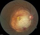
Choroidal Osteoma899 viewsYoung girl with vision loss from osteoma.
|
|

Foreign Body "Rust Ring" 899 viewsPatient complained of redness and tearing with white discharge. Patient was working with a grinding wheel on a vehicle. Slit lamp exam shows a rust ring barley off center. FBS was removed with Algiers Brush.
|
|

Outer Retinal Holes - Probably Solar Retinopathy (history is negative)899 views
|
|

Presumed Ocular Histoplasmosis with new CNVM in right and better eye - VA 20/80 OD; 20/800 OS899 viewsMacular scar 20 years old
|
|

Acute Zonal Occult Outer Retinopathy898 views45-year-old man noticed a few years ago peripheral vision blurriness in the left eye and also some central vision loss. Previous to that, as far as he knows, the two eyes were okay.
VISUAL ACUITY: OD: 20/20; OS: 20/40.
|
|

Branch Retinal Arterial Occlusion 20/20 Vision - Symptoms for 1 Day898 views
|
|

69 Year Old Woman Normal Fluorescein Angiogram898 views69-year-old woman OD 20/50 - OS 20/30 ATYPICAL PHOTOPSIAS
|
|

Best Disease for 30 Years - 20/50 Vision both eyes - 61 Year Old Woman898 views61-year-old woman was diagnosed with Best’s disease about 30 years ago. She and her many family members were tested, both at the VA Hospital and then at USF. OD 20/50, OS 20/50
|
|

Retinal Arterial Macroaneurysm - Ruptured and self Sealed - Vision Back to 20/80 after 6 Months898 views81-year-old woman vision improving 6 months following rupture of retinal arterial macroaneurysm. Vision OD is 20/80
|
|

Ghost Cell Glaucoma898 viewsPost cataract surgery, patient presents with ghost cell in the anterior chamber after hemorrhage cleared. A small hyphema still is visible in the anterior chamber with a whitish ball appearance.
|
|

Stellate Non-heredtiary Idiopathic Foveomacular Retinoschisis (SNIFR)898 views77 year old man who is healthy and 6'6" tall with 20/40 vision and no complaints. OCT shows diffuse retinoschisis.
|
|
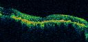
Bilateral Diffuse Uveal Melanocytic Proliferation - BDUMP - Paraneoplastic Syndrome897 views80-year-old man vision loss for one year. He died about one year after these photos from Metastatic Poorly Differentiated Large Cell Carcinoma of unknown primary. He was a smoker.
|
|

Juxtafoveal Telangiectasis - MacTel - Retinal Telangiectasis - Both Eyes - Crystals897 views61-year-old man has idiopathic juxtafoveal retinal telangiectasis type II in both eyes OD is 20/40, OS is 20/25
|
|

Shotgun wound to face with Perforating Pellet - Final Vision 20/400 897 views18-year-old had a gunshot wound to the left eye. He had surgery on December 31st (1 week post injury) and February 4th and his vision has remarkably done well.
VISUAL ACUITY: Vision OS is 3/200.
|
|

Three years after Injury - Optic Atrophy - Vision is 1/200 in each eye897 views
|
|
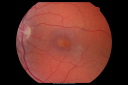
Congenital Achromatopsia 897 views26-year-old man was diagnosed with congenital achromatopsia by ERG as a child. He has family members who also have this disorder. OD 20/200, OS 20/200.
|
|

Morning Glory Syndrome, Optic Nerve897 viewsPatient comes in for cataract evaluation. VA 20/60, right eye and 20/150, left eye. Fundus photo shows "morning glory syndrome" of the optic nerve in the left eye.
|
|

Accordioning Crystalline Lens with loss of Posterior Capsule896 views71-year old male complains of blurred vision in the left eye. VA 20/40, right eye and 20/400, left eye without correction. Slit lamp exam shows Crystalline lens, both eyes. Right eye IOL is aligned and centered. Left eye shows an accordion of the crystalline lens. Retro Illumination shows the IOL bent inward in the left eye. There was a 6-diopter difference of astigmatism between the right and left eye. Patient will have surgery to correct the issue.
|
|

Macular possible Cone Dystrophy - Fundus Autofluorescence896 views
|
|

Melanosis Oculi and Choroidal Melanoma896 views88 year old female with 2.5 mm elevated choroidal melanoma and melanosis oculi. She also has geographic atrophy from AMD. The right eye is the better eye with 20/40 vision. After observation with growth, brachytherapy was done.
|
|

Eccentric Disciform Scar - Breakthrough Vitreous Hemorrhage - InferoTemporal Subretinal Fibrosis - Ectopic Choroidal Neovascular Membranes895 views88-year-old man has noticed for the last few months the vision in the right eye is foggy, like there is a film in the eye or a haze, His vision is 20/16 in each eye.
Right eye - inferotemporal periphery there is a hemorrhage at the equator at about 7:00 o’clock, which is about 4-disc diameters across and adjacent to that there is scarring.
Left eye - large disciform scar inferotemporally
|
|
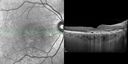
Serous Macular Detachment Both Eyes - Wet AMD vs. CSR - 62 Year Old Man - SD - OCT895 views
|
|

Retinal Arterial Macroaneurysm - Untreated Hypertension (250/135) - Serous Macular Detachment895 views82 year old asian woman. VA 20/80 Headaches and vision loss for 1 month. Negative medical history but has not been to the doctor for years. (no insurance)
|
|

Juxtapapillary CNVM and Serous Macular Detachment Wet AMD Rx Laser894 views85-year-old man OD is 20/25, OS is 20/100. IOP: OD 21, OS 22.
OS: Vertical C/D ratio is 0.1. There is a posterior vitreous separation. There is a hemorrhagic pigment epithelial detachment on the superior pole of the optic nerve, extending a disc-and-a-half diameter off the nerve, with adjacent exudates. There is a serous macular detachment involving the center of the fovea.
VA improved from 20/100 to 20/50 in four months from laser.
|
|

Bilateral Diffuse Uveal Melanocytic Proliferation - BDUMP - Paraneoplastic Syndrome894 views80-year-old man vision loss for one year. He died about one year after these photos from Metastatic Poorly Differentiated Large Cell Carcinoma of unknown primary. He was a smoker.
|
|

Ocular Histoplasmosis both Eyes - Laser Right Eye Only 894 views58-year-old woman who lost vision as a child in the left eye from ocular histoplasmosis. She used to play in her attic and she was told by her pulmonary doctor that is probably where she picked it up. She had laser in the right eye for a leaky lesion back in 1990. OD 20/25, OS 20/200.
|
|
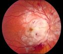
Purtscher's Retinopathy894 views19 year old female after pregnancy
|
|

Optic Nerve (Disc) Drusen - Spectral Domain Optical Coherence Tomography Line Scan894 views34 Year Old woman no headaches normal vision
|
|

NVD894 viewsDiabetic patient with NVD and NVE
|
|

Retinal Pigment Epithelial Dysgenesis893 views
|
|

69 Year Old Woman Normal OCT893 views69-year-old woman OD 20/50 - OS 20/30 ATYPICAL PHOTOPSIAS
|
|

Microaneurysms from Ocular Ischemic Syndrome (non-diabetic) Right Eye - Left eye CRAO 1 year ago893 views66-year-old woman lost vision in the left eye from a central retinal artery occlusion 1 year ago. Vision OD is fine. OD 20/25, OS 20/50
|
|

Horseshoe Retinal Tear with bridging blood vessel893 views
|
|

Peripheral Exudative Hemorrhagic Chorioretinopathy893 views70 year old man with recurrent peripheral choroidal hemorrhages - no treatment - b-scan shows hemorrhage shrinking over a month
|
|

Idiopathic Polypoidal Choroidal Vasculopathy - Fluorescein Angiogram893 views
|
|

Proliferative Diabetic Retinopathy - Moderate - mild NVD with NVE 893 viewsVenous beading, vascular loops and NVE are visible on photos
|
|
| 18117 files on 182 page(s) |
 |
|
|
|
|
14 | |
|
|
|
|


