|
|
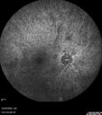
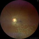
Ocular Ischemia from 100 percent right carotid occlusion303 views77 year old female the vision in the right eye got much worse. She has recently been getting more severe headaches. Her vision is now blank. She went to her old GP in Wisconsin who said it might be sinus pressure. Her chiropractor did an adjustment and the chiropractor checked the patient eyes and told her to go to the eye doctor. She has been feeling pressure around her eye since the air flight.
Specialty Meds (Initial): systane prn OU. Plavix, Combivent, ASA 325, Vitamin E, Zinc.
PSH – Carotid surgery in 2000 (20 years ago), Stent 11/2022, Mastectomy 2000.
VA OD: DccCF 3ft (eccentric) NccUnable
VA OS: Dcc20/25 NccJ1
IOP: TP: OD:18 OS:16
Psuedophakic OU
Right eye had rubeosis on the pupil margin and not in the angle
Further testing showed 100 percent occlusion of the right carotid artery. The left carotid artery was stented.
|
|

Syphilitic optic neuritis303 viewsTwo weeks ago she woke up with blurred vision in the left eye. She usually gets up at 5 AM. Then she noticed a gray spot in the left eye that did not go away. This is new for her. The gray spot is not getting any better or worse in the left eye. (4/4/23)
Systemic Meds: Lisinopril 40 mg. metformin hydrochloride 1000 MG. nifedipine 30mg
VA OD: sc20/80 PH20/50 NscJ10
VA OS: sc2'/200 Nsc20/400
IOP: TP: OD:15 OS:16
No AC or vitreous cells - later images was with second eye involved with uveitis. Patient received IV PCN and was lost to follow-up
|
|

Syphilitic optic neuritis303 viewsTwo weeks ago she woke up with blurred vision in the left eye. She usually gets up at 5 AM. Then she noticed a gray spot in the left eye that did not go away. This is new for her. The gray spot is not getting any better or worse in the left eye. (4/4/23)
Systemic Meds: Lisinopril 40 mg. metformin hydrochloride 1000 MG. nifedipine 30mg
VA OD: sc20/80 PH20/50 NscJ10
VA OS: sc2'/200 Nsc20/400
IOP: TP: OD:15 OS:16
No AC or vitreous cells - later images was with second eye involved with uveitis. Patient received IV PCN and was lost to follow-up
|
|
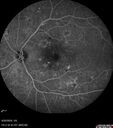
Severe NPDR303 views53 year old man with blurred vision (needs glasses).
Diabetic for 10 years with poor control
VA 20/32 OU
|
|

Cotton Wool Spots - Severe Anemia - Acute Lymphocytic Leukemia302 views23 year old just diagnosed with anemia and acute lymphocytic leukemia
: If he covers each eye neither eye is lacking. When he looks to the left he gets double vision when he looks to the right his vision is fine. This started about 2 weeks ago. Since it started it has been variable. He is on a bunch of medications for leukemia and he might be having high sugars. He noticed the vision change after his first blood transfusion in the hospital. He is currently very anemic.
VA OD: sc20/20 NscJ1+
VA OS: sc20/20 NscJ1+
|
|

Traction retinal detachment both eyes. Proliferative Diabetic Retinopathy302 views40 year old man came in with vision loss OD and rubeosis in both eyes. Was treated with Avastin monthly and laser OD then lost to follow-up for 6 months. Came back with table top traction in the left macula (second set of images). Vision had dropped OS from 20/20 to HM. He had PPV ERM peel OS and vision improved to 5/200.
OD stabilized to 20/20 with PRP laser. Now on 6 month checks.
|
|
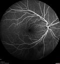
Subretinal Fluid - Inferior to the Nerve - Probable occult nerve pit302 views82 year old female. History of visual "discolorations" but cannot differentiate which eye. Denies loss in visual acuity, eye pain, or any systemic complaints. Referring doctor found fluid on the OCT scan
VA OD: Ncc20/25
VA OS: Ncc20/30
IOP: TP: OD:15 OS:13
|
|
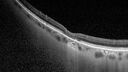
Torpedo Maculopathy - Small Lesion302 viewsAsymptomatic 77 year old female
|
|
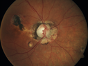
Macular Scars from Presumed Ocular Histoplasmosis Syndrome302 views85 year old man
He sometimes sees double vision. When he sees people walking they are sometimes double for about 6 months.
VA OD: sc20/25 PH20/20 NccJ3
VA OS: sc20/20-2 NccJ2
IOP: TP: OD:12 OS:14
|
|
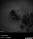
Macular Scars from Presumed Ocular Histoplasmosis Syndrome302 views85 year old man
He sometimes sees double vision. When he sees people walking they are sometimes double for about 6 months.
VA OD: sc20/25 PH20/20 NccJ3
VA OS: sc20/20-2 NccJ2
IOP: TP: OD:12 OS:14
|
|
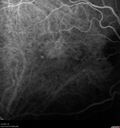
Asymptomatic exudative wet age-related macular degeneration302 views78 year old man with scar in the left eye being treated regularly and returns with new lesions in the right eye which is not symptomatic. Vision is 20/25 right eye and 20/125 left eye. The lesion was treated and responded to anti-VEGF therapy
|
|

Granulomatous Uveitis - Probable Sarcoid302 views28 year old white man with red-itch eyes and decreased vision for 3 months.
VA OD: 3/200 OS: 20/20
Right eye: granulomatous KP, trace cell, posterior synechiae and iris nodule,
Left eye: cicatricial upper lid changes, eye is quiet
|
|

Retinal detachment with choroidal effusions and RPE tear302 views83 year old man with chronic glaucoma presented with 10 days of vision loss in the left eye and a retinal detachment. He also has choroidal effusions and and RPE tear. He had a history of bladder cancer in remission. I did a biopsy of the choroidal lesions at the time of surgery which showed no cancer. Initial vision was HM and final vision was 20/80. The choroidal effusions may have occurred because of his age and prior history of glaucoma.
|
|

Retinal detachment with choroidal effusions and RPE tear302 views83 year old man with chronic glaucoma presented with 10 days of vision loss in the left eye and a retinal detachment. He also has choroidal effusions and and RPE tear. He had a history of bladder cancer in remission. I did a biopsy of the choroidal lesions at the time of surgery which showed no cancer. Initial vision was HM and final vision was 20/80. The choroidal effusions may have occurred because of his age and prior history of glaucoma.
|
|

Retinal detachment with choroidal effusions and RPE tear302 views83 year old man with chronic glaucoma presented with 10 days of vision loss in the left eye and a retinal detachment. He also has choroidal effusions and and RPE tear. He had a history of bladder cancer in remission. I did a biopsy of the choroidal lesions at the time of surgery which showed no cancer. Initial vision was HM and final vision was 20/80. The choroidal effusions may have occurred because of his age and prior history of glaucoma.
|
|

Severe Syphilitic Placoid Chorioretinitis302 views68 year old man 3 months ago the vision started fading in both eyes. Just sitting here he can see some light out of his right eye. The patient denies any recent illness or travel.
VA: OD: LP, OS: 4/200
IOP: 9, 13
Anterior segment: 1+ cell/ 2+ flare OU and 2+ NS and anterior vitreous cells
Labs Reactive RPR 1:256, FTA-ABS - reactive - Patient was admitted to hospital for IV PCN and recovered vision almost completely in the left eye and some in the right eye.
See line scans for recovery
|
|

Combined hamartoma of the retina and retinal pigment epithelium302 views8 year old boy failed vision screening at school. VA 20/20 OD, 20/40 OS
|
|

Punctate inner choroidopathy and CNVM302 views23 year old female noticed vision loss for 4 days.
VA 20/200 OD, 20/25 OS.
VA improved one month later to 20/40 with Avastin. She received a second injection and was lost to follow-up
|
|

Punctate inner choroidopathy and CNVM302 views23 year old female noticed vision loss for 4 days.
VA 20/200 OD, 20/25 OS.
VA improved one month later to 20/40 with Avastin. She received a second injection and was lost to follow-up
|
|
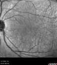
Acute Syphilitic Posterior Placoid Chorioretinitis302 views40 year old man About 10 days ago the patient woke up with a dark circle in the middle of the left eye that blocks his vision. He thought it might be from fatigue. The month of March was very stressful. After a few days the spot in the left eye got darker and he could not drive at night. During the last week while not going to work and sleeping more his vision is better but not normal. He has never had vision loss in the past. Â
PMHx: Benign, Meds: Multivitamin
VA OD: sc20/16 NscJ1+VA OS: sc20/32+2 NscJ4
IOP: TP: OD:12 OS:12
|
|

Stage V (neovascular) Macular Telangiectasia - Fluorescein302 views67 year old female with vision in only the left eye. She has mild CNVM which is treated with Avastin and does well over several years.
|
|

Central retinal artery occlusion and paracentral acute middle maculopathy and emboli302 views86 year old female with sudden vision loss a week ago and imaging shows PAMM lesions and emboli. Her vision is 20/200 in the left eye
|
|

Central retinal artery occlusion and paracentral acute middle maculopathy and emboli302 views86 year old female with sudden vision loss a week ago and imaging shows PAMM lesions and emboli. Her vision is 20/200 in the left eye
|
|

Central retinal artery occlusion and paracentral acute middle maculopathy and emboli302 views86 year old female with sudden vision loss a week ago and imaging shows PAMM lesions and emboli. Her vision is 20/200 in the left eye
|
|

Retinal Arterial Macroaneursym302 views69 year old female with vision loss for a few months in the right eye. Images show a macroaneurysm in the right eye. The lesion had bled and FA showed no leakage. No treatment was done. VA improved from 20/40 - 20/32
|
|
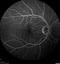
Branch Retinal Vein Occlusion - Foveal Ischemia Neovascularization Non perfusion302 views38 year old patient with a BRVO who had PRP laser for vitreous hemorrhage. Vision remained 20/160 from foveal ischemia
|
|

BRVO with late onset venous macroaneurysm302 viewsThe vision improved on presentation from 20/160 - 20/32 with Avastin, Then a few years later, he developed a venous Macroaneurysm which was lasered . Avastin shots were then stopped.
|
|

Macular ischemia left eye in type I diabetic (enlarged foveal avascular zone)302 views29 year old female with type I diabetes since age 5. Left eye has enlarged FAZ with vision of 20/20 OD and 20/60 OS. Both eyes have CSME. The left eye did have focal laser
|
|

central retinal vein occlusion - a lot of hemorrhage - on coumadin302 views84 year old man with severe vision loss left eye for 2 weeks. VA 5/200 OS - Patient on Warfarin for valve replacement.
|
|

301 views
|
|

Cotton Wool Spots - Severe Anemia - Acute Lymphocytic Leukemia301 views23 year old just diagnosed with anemia and acute lymphocytic leukemia
: If he covers each eye neither eye is lacking. When he looks to the left he gets double vision when he looks to the right his vision is fine. This started about 2 weeks ago. Since it started it has been variable. He is on a bunch of medications for leukemia and he might be having high sugars. He noticed the vision change after his first blood transfusion in the hospital. He is currently very anemic.
VA OD: sc20/20 NscJ1+
VA OS: sc20/20 NscJ1+
|
|

Traction retinal detachment both eyes. Proliferative Diabetic Retinopathy301 views40 year old man came in with vision loss OD and rubeosis in both eyes. Was treated with Avastin monthly and laser OD then lost to follow-up for 6 months. Came back with table top traction in the left macula (second set of images). Vision had dropped OS from 20/20 to HM. He had PPV ERM peel OS and vision improved to 5/200.
OD stabilized to 20/20 with PRP laser. Now on 6 month checks.
|
|
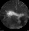
Traction retinal detachment both eyes. Proliferative Diabetic Retinopathy301 views40 year old man came in with vision loss OD and rubeosis in both eyes. Was treated with Avastin monthly and laser OD then lost to follow-up for 6 months. Came back with table top traction in the left macula (second set of images). Vision had dropped OS from 20/20 to HM. He had PPV ERM peel OS and vision improved to 5/200.
OD stabilized to 20/20 with PRP laser. Now on 6 month checks.
|
|

Subretinal Fluid - Inferior to the Nerve - Probable occult nerve pit301 views82 year old female. History of visual "discolorations" but cannot differentiate which eye. Denies loss in visual acuity, eye pain, or any systemic complaints. Referring doctor found fluid on the OCT scan
VA OD: Ncc20/25
VA OS: Ncc20/30
IOP: TP: OD:15 OS:13
|
|
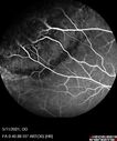
Retinal Angioma301 views53 year old female with no-symptoms and no significant medical history (or family history)
|
|
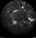
Severe Proliferative Diabetic Retinopathy and Good Vision301 views49 year old man with vision loss in the right eye for 2 weeks. VA 20/40 OD, 20/63 OS. DM x 15 years and HTN.
|
|
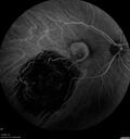
Choroidal Neovascular Membrane (CNVM) secondary to laser scar from TTT for choroidal melanoma 20 years earlier301 views71-year old female - Vision loss in the right eye for 1 week. She had transpupillary thermotherapy 20 years ago in the right eye for choroidal melanoma.
VA 4/200 OD, 20/20 OS
|
|

Alcohol Amblyopia - Toxic Optic Neuropathy301 views59 year old man HPI: CC: Blurred Vision OU. Duration of Problem: about 7 months. Location: central vision. Context Onset/Aggravation: Started after using Accutane for 1 month. Associated Symptoms: no flashes or floaters.
HX TECH: EA
VA OD: sc20/100 PH20/80+1 Nsc20/400 PAM20/50+2
VA OS: sc20/100 PH20/80-2 NscJ16 PAM20/50+1
IOP 24 OD, 26 OS
B-12 and Folate were normal - Presumed ETOH amblyopia - improved to 20/63,20/80 after one year of B-12 supplements
IOP: TP: OD:24 OS:26 Time:02:08 PM
|
|

Macular Scars from Presumed Ocular Histoplasmosis Syndrome301 views85 year old man
He sometimes sees double vision. When he sees people walking they are sometimes double for about 6 months.
VA OD: sc20/25 PH20/20 NccJ3
VA OS: sc20/20-2 NccJ2
IOP: TP: OD:12 OS:14
|
|

Bilateral Diffuse Melanocytic Proliferation (BDUMP)301 views67 year old man
The patient started noticing dimming of the vision in November of 2020. He had rapidly worsening cataracts. He had the cataract surgery done in both eyes in April of 2021. The vision did not improve with the cataract surgery. On June 1 of 2021 he had immunotherapy for his renal cell carcinoma and then developed severe vision loss. The patient noticed that one of the commercials for nivolumab (checkpoint inhibitor targeting programmed death receptor-1) said it may cause vision loss. He recalled that he started the nivolumab in October of 2020 and had vision loss starting then.  A retinal evaluation showed subretinal fluid and vision loss.  The peripheral vision and central vision were pretty good. The vision did seem to get better off the nivolumab. Then when the patient returned to Florida in September and now the vision is getting worse. The patient did not have any treatments in either eye. The patient is now off the oncology treatment. He was then started on a TKI cabometex for the cancer which was dosed in mid June through mid July just for a month. It was stopped because of a skin rash and reactions. Â
VA OD: Dcc20/200-2 PHNI Ncc20/800
VA OS: Dcc20/63 PHNI NccJ7 He has been off the checkpoint inhibitor for a year.
|
|
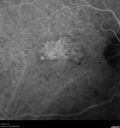
Asymptomatic exudative wet age-related macular degeneration301 views78 year old man with scar in the left eye being treated regularly and returns with new lesions in the right eye which is not symptomatic. Vision is 20/25 right eye and 20/125 left eye. The lesion was treated and responded to anti-VEGF therapy
|
|
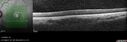
Granulomatous Uveitis - Probable Sarcoid301 views28 year old white man with red-itch eyes and decreased vision for 3 months.
VA OD: 3/200 OS: 20/20
Right eye: granulomatous KP, trace cell, posterior synechiae and iris nodule,
Left eye: cicatricial upper lid changes, eye is quiet
|
|
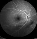
Retinal detachment with choroidal effusions and RPE tear301 views83 year old man with chronic glaucoma presented with 10 days of vision loss in the left eye and a retinal detachment. He also has choroidal effusions and and RPE tear. He had a history of bladder cancer in remission. I did a biopsy of the choroidal lesions at the time of surgery which showed no cancer. Initial vision was HM and final vision was 20/80. The choroidal effusions may have occurred because of his age and prior history of glaucoma.
|
|

Choroidal Effusions following IOL exchange301 views67 year old man 10 years ago had retinal detachment with buckle, vitrectomy, laser, gas. Vision was 20/25 post-op. 2 months ago had sudden vision loss in the right eye from a dislocated PCIOL (in the bag). 3 weeks ago had a vitrectomy, remove IOL and scleral suture fixation of Akreos AO60 PCIOL with gortex sutures. IOP was 12 on post-op day 1 with a vitreous hemorrhage. Post-op day 7, vitreous hemorrhage cleared.
PMhx: HTN, High Cholesterol.
Med: Amlodipine, ASA, Atorvastatin, Metoprolol
Vision 20/200,
|
|

Severe Syphilitic Placoid Chorioretinitis301 views68 year old man 3 months ago the vision started fading in both eyes. Just sitting here he can see some light out of his right eye. The patient denies any recent illness or travel.
VA: OD: LP, OS: 4/200
IOP: 9, 13
Anterior segment: 1+ cell/ 2+ flare OU and 2+ NS and anterior vitreous cells
Labs Reactive RPR 1:256, FTA-ABS - reactive - Patient was admitted to hospital for IV PCN and recovered vision almost completely in the left eye and some in the right eye.
See line scans for recovery
|
|
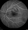
Central Serous Retinopathy - Female on steroids301 views35 year old female Noticed blurred vision when waking up in the morning. Also, vision blacks out for a few seconds when she gets up quickly. About 3 months ago she had a history of intermittent vomiting.
VA 20/20 OD, 20/25 OS
MRI normal, MRA showed lateral sinus thrombosis. Lupus anticoagulant was positive
|
|
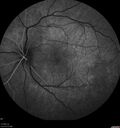
Acute Syphilitic Posterior Placoid Chorioretinitis301 views40 year old man About 10 days ago the patient woke up with a dark circle in the middle of the left eye that blocks his vision. He thought it might be from fatigue. The month of March was very stressful. After a few days the spot in the left eye got darker and he could not drive at night. During the last week while not going to work and sleeping more his vision is better but not normal. He has never had vision loss in the past. Â
PMHx: Benign, Meds: Multivitamin
VA OD: sc20/16 NscJ1+VA OS: sc20/32+2 NscJ4
IOP: TP: OD:12 OS:12
|
|

Stage V (neovascular) Macular Telangiectasia - Fluorescein301 views67 year old female with vision in only the left eye. She has mild CNVM which is treated with Avastin and does well over several years.
|
|

Stage V (neovascular) Macular Telangiectasia - Fluorescein301 views67 year old female with vision in only the left eye. She has mild CNVM which is treated with Avastin and does well over several years.
|
|

Choroidal Hemorrhage following intraocular lens exchange surgery301 views68 year old man Patient is 1 week post IOL exchange with sutured Akreos PCIOL with PST Kenalog. IOP was 12 on POD 1 and 21 today (POD 7). Preop the IOP was 17.
He has a history of retinal detachment repair 12 years ago with PPV, Buckle, Laser and Gas.
VA OD: 20/200, OS: 20/50. IOP 21, 19
|
|

Retinal Arterial Macroaneursym301 views69 year old female with vision loss for a few months in the right eye. Images show a macroaneurysm in the right eye. The lesion had bled and FA showed no leakage. No treatment was done. VA improved from 20/40 - 20/32
|
|

Syphilitic optic neuritis301 viewsTwo weeks ago she woke up with blurred vision in the left eye. She usually gets up at 5 AM. Then she noticed a gray spot in the left eye that did not go away. This is new for her. The gray spot is not getting any better or worse in the left eye. (4/4/23)
Systemic Meds: Lisinopril 40 mg. metformin hydrochloride 1000 MG. nifedipine 30mg
VA OD: sc20/80 PH20/50 NscJ10
VA OS: sc2'/200 Nsc20/400
IOP: TP: OD:15 OS:16
No AC or vitreous cells - later images was with second eye involved with uveitis. Patient received IV PCN and was lost to follow-up
|
|

central retinal vein occlusion - a lot of hemorrhage - on coumadin301 views84 year old man with severe vision loss left eye for 2 weeks. VA 5/200 OS - Patient on Warfarin for valve replacement.
|
|

Advanced age-related macular degeneration301 views87 year old female. Severe macular bleed 15 years ago OD. Now with disciform scar. Left eye has progressing geographic atrophy.
VA OD: HM, OS: 5/200. IOP normal OU
|
|
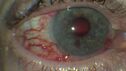
Elevated Episcleral Venous Pressure and Retinal Hemorrhages in both eyes300 views78 year old female with chronic red eye. The eyes are red all the time now. She is on Xeralto because of a history of blood clots in her lungs (2005 about). She has not had problems since. She has had floaters for many years. Ever since her eyes got funny she sees more floaters in both eyes. She had a brain scan over 6 months ago which was OK. She also had double vision when this first started which went away. She could not drive for a few months.
VA OD: Dcc20/32-1
VA OS: Dcc20/125
IOP: TP: OD:17 OS:16
Neuroimaging did not show a CC fistula
|
|
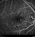
Wet age-related macular degeneration - Classic Juxtafoveal Choroidal Neovascular Membrane300 views80 year old female with mild vision loss. 20/40 OD, 20/25 OS. Avastin controlled disease. VA improved to 20/25 after 2 injections. At 1 year she is getting every 2 months treatment
|
|

Retinal Angioma300 views53 year old female with no-symptoms and no significant medical history (or family history)
|
|

Bilateral Choroidal Tumors - Metastatic Breast Cancer300 views51 year old female. First 3 sets of images show lesions growing despite systemic chemotherapy. She eventually had external beam radiation x 15 sessions and the tumors shrunk in the last two sets of images
|
|

Neuroretinitis left eye - recovered with no therapy - possible B pertussis300 views57 year old female The patient has had blurred vision in the left eye and there is a haze across the top of the vision. This started about 2 weeks ago. The right eye is OK. She has sleep apnea and uses the machine. The left eye is becoming more and more painful for the last two weeks. The pain is like a stabbing in the eye that comes and goes. She also has some pain on eye movement. The patient has been going to Moffit for possible reconstructive surgery for the lymphedema.
PMHx – Sleep apnea, Breast Cancer, Adult lymphedema,
Meds: GabapentinÂ
VA OD: Dcc20/25-1
VA OS: Dcc20/100-2
AC quiet OU. 1 + NS cataract OU
IOP: TP: OD:15 OS:17
|
|

Non-exudative wet AMD - OCT angiogram300 views85 year old female with minimal visual complaints.
VA OD: sc20/80+1 PHNI NccJ3
VA OS: sc20/40-2 PH20/32+1 NccJ3
IOP: TP: OD:14 OS:16
|
|

Retinal Arterial Macroaneurysm treated with Laser300 views89 year old female with peripapillary CNVM treated with Lucentis. Vision was 20/25 OS 3 weeks ago. She noticed vision loss OS. Vision had dropped to 20/60. She had a fresh MA and was treated with laser.
|
|

Granulomatous Uveitis - Probable Sarcoid300 views28 year old white man with red-itch eyes and decreased vision for 3 months.
VA OD: 3/200 OS: 20/20
Right eye: granulomatous KP, trace cell, posterior synechiae and iris nodule,
Left eye: cicatricial upper lid changes, eye is quiet
|
|

Severe Syphilitic Placoid Chorioretinitis300 views68 year old man 3 months ago the vision started fading in both eyes. Just sitting here he can see some light out of his right eye. The patient denies any recent illness or travel.
VA: OD: LP, OS: 4/200
IOP: 9, 13
Anterior segment: 1+ cell/ 2+ flare OU and 2+ NS and anterior vitreous cells
Labs Reactive RPR 1:256, FTA-ABS - reactive - Patient was admitted to hospital for IV PCN and recovered vision almost completely in the left eye and some in the right eye.
See line scans for recovery
|
|

Severe Syphilitic Placoid Chorioretinitis300 views68 year old man 3 months ago the vision started fading in both eyes. Just sitting here he can see some light out of his right eye. The patient denies any recent illness or travel.
VA: OD: LP, OS: 4/200
IOP: 9, 13
Anterior segment: 1+ cell/ 2+ flare OU and 2+ NS and anterior vitreous cells
Labs Reactive RPR 1:256, FTA-ABS - reactive - Patient was admitted to hospital for IV PCN and recovered vision almost completely in the left eye and some in the right eye.
See line scans for recovery
|
|

Combined hamartoma of the retina and retinal pigment epithelium300 views8 year old boy failed vision screening at school. VA 20/20 OD, 20/40 OS
|
|

Acute Syphilitic Posterior Placoid Chorioretinitis300 views40 year old man About 10 days ago the patient woke up with a dark circle in the middle of the left eye that blocks his vision. He thought it might be from fatigue. The month of March was very stressful. After a few days the spot in the left eye got darker and he could not drive at night. During the last week while not going to work and sleeping more his vision is better but not normal. He has never had vision loss in the past. Â
PMHx: Benign, Meds: Multivitamin
VA OD: sc20/16 NscJ1+VA OS: sc20/32+2 NscJ4
IOP: TP: OD:12 OS:12
|
|

Severe diabetic tractional retinal detachment one year following diagnosis of type II Diabetes Mellitus300 views44 year old man The vision in the left eye started to decline about 4 days ago. Prior to that the left eye was the better eye. He was diagnosed with diabetes a year ago.Â
VA OD: Dcc20/50-1 OS: Dcc20/125-2, TP: OD:12 OS:13
Following two surgeries in each eye, silicone oil remaining forever, monthly anti-VEGF for severe DME - BCVA OU 20/200 at 3 years
|
|

Branch Retinal Vein Occlusion and worsened cystoid macular edema following cataract surgery300 views51 year old female The patient noticed gradual vision loss in both eyes starting possibly after her last cataract surgery in May of 2022 (3 months ago). She had ECCE 3/2022 OD and 5/2022 OS. She does not have diabetes. She had glaucoma shunts placed about 2017. She has been on latanoprost and timolol. She is now getting ready to start Combigan and Bromfenac. She is also staring xidra for dry eye. She stopped the latanorpost on 8/15/22. She has had macular edema before for which she had intraocular injections which worked. She has had no injections lately. When she was getting treated for macular edema in the past she had an IOP of 60.
Medical Hx: Hypertension, Systemic. Glaucoma. Arthritis. Vertigo. Stroke.
Systemic Meds: Trimfya. NAPROXEN. LISINOPRIL.
VA OD: sc20/63-1
VA OS: sc20/50
IOP: TP: OD:12 OS:12
|
|

Choroidal Hemorrhage following intraocular lens exchange surgery300 views68 year old man Patient is 1 week post IOL exchange with sutured Akreos PCIOL with PST Kenalog. IOP was 12 on POD 1 and 21 today (POD 7). Preop the IOP was 17.
He has a history of retinal detachment repair 12 years ago with PPV, Buckle, Laser and Gas.
VA OD: 20/200, OS: 20/50. IOP 21, 19
|
|

Birdshot chorioretinitis with macular edema - positive HLAA29300 views67 year old female with 'vitreous hemorrhage' in the right eye for a year. Extensive uveitis work up done but now with HLA A29. HLA A29 was positive. She has not responded to kenalog injections (posterior) and will be started on systemic therapy soon
|
|

Central retinal artery occlusion and paracentral acute middle maculopathy and emboli300 views86 year old female with sudden vision loss a week ago and imaging shows PAMM lesions and emboli. Her vision is 20/200 in the left eye
|
|

Pigment epithelial detachment with cholesterol crystals300 viewsOnion sign
|
|

Pigment epithelial detachment with cholesterol crystals300 viewsOnion sign
|
|
Branch Retinal Vein Occlusion - Foveal Ischemia Neovascularization Non perfusion300 views38 year old patient with a BRVO who had PRP laser for vitreous hemorrhage. Vision remained 20/160 from foveal ischemia
|
|

Macular pucker in the left eye300 viewsVision was 20/125 pre-op and 20/25 post-op.
|
|

BRVO with late onset venous macroaneurysm300 viewsThe vision improved on presentation from 20/160 - 20/32 with Avastin, Then a few years later, he developed a venous Macroaneurysm which was lasered . Avastin shots were then stopped.
|
|

Twig (Macular) Branch Retinal Vein Occlusion with no symptoms300 views73 year old female with 20/25 vision and no symptoms
|
|

Pseudophakic cystoid macular edema (CME)300 views66 year old female 4 months following cataract surgery. Never had very good vision after surgery. On PF QID for one month. VA 20/50 in both eyes.
|
|

CNV outcomes299 viewsFibrotic scar in choroidal neovascularization after anti-VEGF treatment
|
|
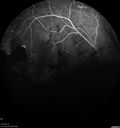
PDR and Vitreous hemorrhage - Second FA is following PRP laser299 views42 year old man (date 2018): Prior to 2 months ago the vision in the right eye was good. This started with a glob in the vision and then the vision got better. Then the string came back down and is getting bigger and bigger. The last time he had bleeding in the left eye he had surgery in 2009. The left eye is now doing OK other than there is a cataract. The right eye had some laser treatment done in 2009. The vitrectomy and laser were done at William Beaumont.
VA OD: sc3'/200 NscUnable
VA OS: sc20/40 NscJ2-1
|
|
PDR and Vitreous hemorrhage - Second FA is following PRP laser299 views42 year old man (date 2018): Prior to 2 months ago the vision in the right eye was good. This started with a glob in the vision and then the vision got better. Then the string came back down and is getting bigger and bigger. The last time he had bleeding in the left eye he had surgery in 2009. The left eye is now doing OK other than there is a cataract. The right eye had some laser treatment done in 2009. The vitrectomy and laser were done at William Beaumont.
VA OD: sc3'/200 NscUnable
VA OS: sc20/40 NscJ2-1
|
|

Proliferative Diabetic Retinopathy with Panretinal Photocoagulation299 views57 year old female Type 1 DM with prior laser about 10 years ago in Chicago.
HTN, Thyroid cancer and removal, Cardiovascular disease
Meds: ASA 325 mg /day, etc
Recent new floaters in the right eye:
VA 20/50 OD, 20/40 OS
|
|

Sectoral Retinitis Pigmentosa - Asymptomatic299 views56-year-old female - The patient was in for an annual checkup because of her diabetes. No visual complaints (no night vision trouble)
PMHx DM sine 2015, Asthma, Thyroid Disease, HTN
MEDS: Metformin, Levothyroxine, Lisinopril,, Inhaler
ROS Negative
VA OD: sc20/32-1 VA OS: sc20/32-1
IOP: TP: OD:18 OS:17
293 Invitae genetic panel - negative
|
|
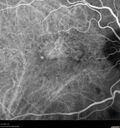
Asymptomatic exudative wet age-related macular degeneration299 views78 year old man with scar in the left eye being treated regularly and returns with new lesions in the right eye which is not symptomatic. Vision is 20/25 right eye and 20/125 left eye. The lesion was treated and responded to anti-VEGF therapy
|
|
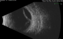
Retinal detachment with choroidal effusions and RPE tear299 views83 year old man with chronic glaucoma presented with 10 days of vision loss in the left eye and a retinal detachment. He also has choroidal effusions and and RPE tear. He had a history of bladder cancer in remission. I did a biopsy of the choroidal lesions at the time of surgery which showed no cancer. Initial vision was HM and final vision was 20/80. The choroidal effusions may have occurred because of his age and prior history of glaucoma.
|
|

299 views
|
|
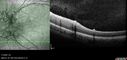
Combined hamartoma of the retina and retinal pigment epithelium299 views8 year old boy failed vision screening at school. VA 20/20 OD, 20/40 OS
|
|
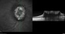
Papilledema Lateral Sinus Thrombosis299 views35 year old female Noticed blurred vision when waking up in the morning. Also, vision blacks out for a few seconds when she gets up quickly. About 3 months ago she had a history of intermittent vomiting.
VA 20/20 OD, 20/25 OS
MRI normal, MRA showed lateral sinus thrombosis. Lupus anticoagulant was positive
|
|
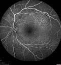
Acute Syphilitic Posterior Placoid Chorioretinitis299 views40 year old man About 10 days ago the patient woke up with a dark circle in the middle of the left eye that blocks his vision. He thought it might be from fatigue. The month of March was very stressful. After a few days the spot in the left eye got darker and he could not drive at night. During the last week while not going to work and sleeping more his vision is better but not normal. He has never had vision loss in the past. Â
PMHx: Benign, Meds: Multivitamin
VA OD: sc20/16 NscJ1+VA OS: sc20/32+2 NscJ4
IOP: TP: OD:12 OS:12
|
|

Asymptomatic progressing retinal detachment 27 year old man299 viewsThis detachment was followed for one month, progressed, and was repaired with a scleral buckle.
|
|

Branch Retinal Vein Occlusion and worsened cystoid macular edema following cataract surgery299 views51 year old female The patient noticed gradual vision loss in both eyes starting possibly after her last cataract surgery in May of 2022 (3 months ago). She had ECCE 3/2022 OD and 5/2022 OS. She does not have diabetes. She had glaucoma shunts placed about 2017. She has been on latanoprost and timolol. She is now getting ready to start Combigan and Bromfenac. She is also staring xidra for dry eye. She stopped the latanorpost on 8/15/22. She has had macular edema before for which she had intraocular injections which worked. She has had no injections lately. When she was getting treated for macular edema in the past she had an IOP of 60.
Medical Hx: Hypertension, Systemic. Glaucoma. Arthritis. Vertigo. Stroke.
Systemic Meds: Trimfya. NAPROXEN. LISINOPRIL.
VA OD: sc20/63-1
VA OS: sc20/50
IOP: TP: OD:12 OS:12
|
|

Toxoplasmosis right eye with diffuse retinal vascular whitening PCR confirmed299 views66 year old man The patient was doing fine until about 2-3 months ago. He woke up with shadows and spots in the right eye. Every blue moon he gets some pain in the right eye. He has been getting treated with eye drops in the right eye. Since this happened the vision is mostly the same. Sometimes he sees better than other times. He has not been treated with oral medications. Â
Medical history: none – Medications: none, ROS negative
VA OD: sc20/63-1 OS: sc20/20
IOP: TP: OD:15 OS:20Â
Negative labs: ACE, Lysozyme, RPR, HLA B27, Quant TB Gold, ESR (slight elevation of CRP)
PCR anterior chamber: negative herpes virus (HSV, VZV, CMV)
Positive for toxoplasmosis
|
|

Toxoplasmosis right eye with diffuse retinal vascular whitening PCR confirmed299 views66 year old man The patient was doing fine until about 2-3 months ago. He woke up with shadows and spots in the right eye. Every blue moon he gets some pain in the right eye. He has been getting treated with eye drops in the right eye. Since this happened the vision is mostly the same. Sometimes he sees better than other times. He has not been treated with oral medications. Â
Medical history: none – Medications: none, ROS negative
VA OD: sc20/63-1 OS: sc20/20
IOP: TP: OD:15 OS:20Â
Negative labs: ACE, Lysozyme, RPR, HLA B27, Quant TB Gold, ESR (slight elevation of CRP)
PCR anterior chamber: negative herpes virus (HSV, VZV, CMV)
Positive for toxoplasmosis
|
|

Choroidal Hemangioma Requiring PDT repeatedly299 views64 year old man. Diagnosed with hemangioma in 2015. PDT in 2016. Images are from 2020 when fluid started to reaccumulate. Needed PDT repeated two more times.
PDT treatments: [1] 2/19/2015 -Â Pavan (IOPÂ problem after PDT)Â AND [2] 11/2/21 PDT (7 mm temporal to fovea) - SMCÂ AND [3] 7/22/22 PDT 7.5 mm Temporal to the fovea
|
|
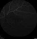
Branch Retinal Vein Occlusion - Foveal Ischemia Neovascularization Non perfusion299 views38 year old patient with a BRVO who had PRP laser for vitreous hemorrhage. Vision remained 20/160 from foveal ischemia
|
|

central retinal vein occlusion - a lot of hemorrhage - on coumadin299 views84 year old man with severe vision loss left eye for 2 weeks. VA 5/200 OS - Patient on Warfarin for valve replacement.
|
|
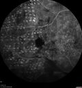
PDR and Vitreous hemorrhage - Second FA is following PRP laser298 views42 year old man (date 2018): Prior to 2 months ago the vision in the right eye was good. This started with a glob in the vision and then the vision got better. Then the string came back down and is getting bigger and bigger. The last time he had bleeding in the left eye he had surgery in 2009. The left eye is now doing OK other than there is a cataract. The right eye had some laser treatment done in 2009. The vitrectomy and laser were done at William Beaumont.
VA OD: sc3'/200 NscUnable
VA OS: sc20/40 NscJ2-1
|
|
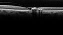
Macular Telangiectasis (MacTel) Both Eye - Stage IV - Pigment Spots298 views63 year old female with Type II DM x 4 years. Vision is 20/40 both eyes.
|
|

Retinal Angioma298 views53 year old female with no-symptoms and no significant medical history (or family history)
|
|
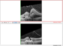
Choroidal Neovascular Membrane (CNVM) secondary to laser scar from TTT for choroidal melanoma 20 years earlier298 views71-year old female - Vision loss in the right eye for 1 week. She had transpupillary thermotherapy 20 years ago in the right eye for choroidal melanoma.
VA 4/200 OD, 20/20 OS
|
|
| 18014 files on 181 page(s) |
 |
|
|
|
|
149 | |
|
|
|
|


