|
|
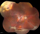
Rhegmatogenous Retinal Detachmentvu 831 foisPatient comes in for second opinion on RD, right eye. VA is NLP in the right eye. Fundus photography shows inferior retinal detachment with holes and subretinal fibrosis. No further surgery is suggested at this time.
|
|
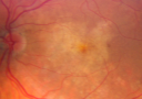
Pattern Dystrophy - Congenital Deafness - Diabetes - Developmental Delay - Fundus Photo Left Eye - Yellow Submacular Depositsvu 830 fois45-year-old woman congenitally deaf. She has diabetes for the last two years. She also has developed mental delay and memory problems. OD 20/200, OS 20/50. She has sluggish pupillary responses in both eyes.
|
|

Ruptured Retinal Arterial Macroaneurysm - Submacular Hemorrhage - Vision 20/400 never recovered - Hypertension 205/94vu 830 fois
|
|

Metastasis Choroidal Melanoma vu 830 fois71-year old female who has undergone chemotherapy treatment for breast cancer. Choroidal Melanoma was from metastasis from her breast cancer. VA is 20/20 as the tumor is in the superior aspect above the optic nerve.
|
|

Classic Subfoveal Choroidal Neovascular Membrane - 20/400 Vision - 6 - 12 months old lesionvu 829 fois78-year-old woman six months or a year ago decreasing vision in the right eye. OD 20/400. Fluorescein angiography shows a classic subfoveal choroidal neovascular membrane in the right eye 3 disc-diameters across, which has brisk leakage. The left eye shows staining drusen.
|
|

Toxocariasisvu 829 fois15-year-old one year ago had pink eye then her vision has been abnormal. She recently went for a drivers’ test and failed, OD: 20/80; OS: 20/20.
|
|
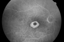
Plaquenil Toxicity - Bulls Eye Maculopathyvu 829 fois70-year-old woman with systemic Lupus erythematosus and clotting problems. She was on the Plaquenil for about eight years and then off the Plaquenil for the last eight years because she developed macular toxicity. Although her vision was hazy, it was stable. Recent deceased vision left eye: OD 20/60, OS 20/100. IOP: OD 18, OS 19.
|
|

Atypical Birdshot Chorioretinitis and Retinal Arterial Macroaneurysm Right Eye - HLA-B29 Positivevu 829 fois55-year-old woman has multifocal choroiditis in both eyes because she was HLA A-29 positive. She has presumptive birdshot chorioretinopathy. She has not noticed any vision changes. OD 20/40, OS 20/20.
|
|

Sturge-Weber Encephalotigeminal Angiomatosis - Facial Hemangioma and Asymptomatic Ipsilateral Diffuse Choroidal Hemangiomavu 829 fois61-year-old man with Sturge-Weber syndrome with a hemangioma on the left side of his face.
VISUAL ACUITY: Vision OD is 20/50, PH 20/30; OS 20/80, PH 20/30. IOP: OD 16, OS 19.
|
|

Retinal Arterial Macroaneurysm - Normal Vision (2 Years before rupture)vu 829 fois78-year-old woman has a retinal arterial macroaneurysm in the right eye. VA 20/25 (pre-rupture photos)
|
|

Uveitis and Retinal Whitening - 04-27-12 - Prior to PPV OD - Possible Primary Ocular Lymphoma vu 829 fois
|
|

Bilateral Retinal Arteriol Occlusions - Possible Susac Syndrome vu 828 fois80-year-old woman one month ago had vision loss and a vascular occlusion in the right eye. Vision loss occurred in the left eye about 9 years ago with cotton wool spots. Patients has a 30 year history of tinnitis.
|
|
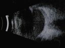
Nanopthalmous (Microphthalmous) Chorioretinal Folds and Choroidal Thickening Both Eyes B-Scan Ultrasoundvu 828 fois
|
|

Acute Annular Outer Retinopathy for 3 Months - 25 YO Female OCT Movievu 828 fois25-year-old female who, about three months ago, put on her sunglasses and noticed a smudge in the vision and at that point she noticed the right eye was hazy. She had a cold a week or two before the vision loss, but she didn’t have any fever. She has no focal neurological problems in her past history. VA 20/20 OU
|
|

Peripheral Retinal Degenerationvu 828 foisPatient comes in with double vision. VA was 20/20 in both eyes. Fundus exam shows Retinal Degenerative changes in both eyes. Offer to correct double vision with temporary Fresnel prism
|
|
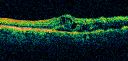
branch retinal vein occlusion - oct line scanvu 827 fois70 year old woman with 20/80 vision from a branch retinal vein occlusion and macular edema. She has had laser treatment and intravitreal steroids. Her vision with further laser improved to 20/50.
|
|
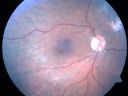
Chronic Retinal Detachment and Macular hole Post-Opvu 827 fois51-year-old woman about 3 or 4 months ago she noticed some flashing lights. Her vision became hazy and dark, especially when she was covering her left eye. OD 2/200, OS 20/20. Post op vision 20/200.
|
|

Classic Subfoveal Choroidal Neovascular Membrane - 20/400 Vision - 6 - 12 months old lesionvu 827 fois78-year-old woman six months or a year ago decreasing vision in the right eye. OD 20/400. Fluorescein angiography shows a classic subfoveal choroidal neovascular membrane in the right eye 3 disc-diameters across, which has brisk leakage. The left eye shows staining drusen.
|
|

Fungal Endophthalmitisvu 827 fois65-year-old woman had chronic endophthalmitis in the left eye 1 day post-op vitrectomy.
|
|

Retinal Torsion after Macula Off Retinal Detachment Repair with Inferior PVRvu 827 fois37 year old with torsion of the retina after macula off retinal detachment repair with inferior PVR and membranes dragging the fovea inferiorly.
|
|

Papilledema post-spinal tap va 20/25vu 827 fois24-year-old woman was seen in the office on 2/23/2010. She has had noticed positional blurring of her vision when she bends over since around November. When she bends over and stands up straight, she notices her vision is blurred. Vision OD is 20/100, PH 20/40; OS is 20/80, PH 20/30. IOP: 19 OU.
vision was 20/25 after tap
|
|

Hemicentral Retinal Vein Occlusion Vision 20/60 vu 827 fois84-year-old man has a hemicentral retinal vein occlusion in the right eye. His vision has declined since then.
VISUAL ACUITY: Vision OD is 20/60.
|
|

Acute Posterior Multifocal Placoid Pigment Epitheliopathy - APMPPE - Indocyanine Green Angiogramvu 827 fois 25-year-old man with one and half weeks ago he woke up with night chills. Also around that time his joints started hurting him. He also has headaches, although the headache at this point is not severe. His vision has been changing for about the last week and a half. He sees some spots in his vision and they are blurred spots. They are not in the central vision.
VISUAL ACUITY: OD 20/32, OS 20/32
|
|

Primary Ocular Lymphoma - White Mass Left Eye - 112712 vu 827 fois
|
|

Uveitis and Cystoid Macular Edema - Probably Sarcoidosis - Posterior Synechia and Koeppe Nodulevu 827 foisVA 20/63 OD 20/50 OS - 3 months vision loss - second episode by history of uveitis
|
|

PVR_preopvu 827 foisScott Pautler, MD
|
|

Proliferative Sickle Retinopathy - Hemoglobin SCvu 826 fois53-year-old woman with a new spider-like floater in the right eye for about six days. She sees one big floater and another little one. She does have hemoglobin SC hemoglobinopathy. VISUAL ACUITY: Vision OD is 20/20; OS is 20/50, PH is 20/20.
|
|

Anomalous Branch Retinal Artery vu 826 fois59-year-old man OD 20/25, OS 20/25. TORTUOUS RETINAL ARTERY – LEF EYE
(POSSIBLE GROUP 2 WYBURN-MASON SYNDROME
POSSIBLE ACEPHALIC MIGRAINES )
|
|

Congenital Toxoplasmosis with non-macular Scars Both Eyes - Recent Reactivation (recurrence) Right Eyevu 826 fois34-year-old man has congenital toxoplasmosis with four week history of foggy vision and new floaters in the right eye. OD is 20/40, OS 20/30
|
|

Idiopathic Cystoid Macular Edema - CME - Responded to Subtenons Kenalog - Color Photovu 825 fois49-year-old man had a flu two weeks ago the right eye has been throbbing and his vision in the right eye has been foggy. OD 20/80, OS 20/40. IOP: 14 OU.
|
|

Occult Maculopathy - Thin Fovea on OCT and Normal Color VA, Photos, FA VA 20/80 OUvu 825 fois45-year-old man Normal FA
|
|

Best Vettiliform Dystrophy with CNVM-2vu 825 fois
|
|
vu 825 fois
|
|

Gyrate Atrophy - 15 year old male - 20/25 vision both eyes - Positive Family Historyvu 824 fois15 year old male was diagnosed with “retinitis pigmentosa†at age seven. The patient reports that his night vision is poor. He reports he does not notice a visual field change. He reports that his maternal grandfather has lost vision and a cousin has lost vision as well. He reports he has an uncle with some visual changes.
The most significantly affected relative is a 26-year-old legally blind cousin.
20/25+ OD, 20/25 OS.
|
|

Dehydration Injury to Retina after Macula-On Retinal Detachment Repair - Also Subretinal Perfluoronvu 824 fois18 month ago he had a scleral buckle, vitrectomy, laser and gas for macula on retinal detachment. Since he notices blind spots in the center of the left eye/ He also notices a dense, inferior visual field defect. OD 20/20, OS 20/50
|
|

Acute Zonal Occult Outer Retinopathyvu 823 fois45-year-old man noticed a few years ago peripheral vision blurriness in the left eye and also some central vision loss. Previous to that, as far as he knows, the two eyes were okay.
VISUAL ACUITY: OD: 20/20; OS: 20/40.
|
|

Hypertensive Retinopathy with Choroidal Ischemia Right Eye and Vision 8/200 - Blood Pressure 240/120 mmHg IgA Nephropathyvu 823 fois39-year-old man two days ago he noticed poor vision in his right eye OD: 8/200; OS: 20/20. BP is 240/120
|
|
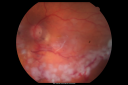
Retina Attached Under Oil after oil exchange, perfluoron, laservu 823 fois
|
|

Superior Retinal Detachment - Bullous - Macula-offvu 823 fois70-year-old man noticed a shadow creeping over the vision in the left eye and he no longer sees well out of that eye. He had cataract surgery two years ago and a YAG capsulotomy six months ago.
VISUAL ACUITY: OD 20/20, OS 1/200
|
|

Hyperpigmentation of the Retinal Pigment Epithelium Right Eye (Treated wet AMD OS)vu 823 fois85-year-old man has wet age-related macular degeneration in the left eye treated for one year most recently one year ago. OD is 20/30, OS is 20/60
|
|

Acute Central Serous Retinopathy - CSR - Steroid Induced (probably) - Good Vision - 6 months later - vision Better - FAF shows small abnormalityvu 823 fois42-year-old man has central serous retinopathy in the right eye with serous retinal detachment. 6 month follow-up Vision is better.
VISUAL ACUITY: OD 20/20.
|
|

Optic Nerve Hypoplasia - 43 Year Old Man - declining visionvu 823 fois43-year-old man was diagnosed with optic nerve hypoplasia at age fifteen at the National Institute of Health. His vision seems to be declining lately.
Vision OD is 20/80, OS is 20/120
|
|

Pseudoexfoliation Material Peeling.vu 823 foisPatient comes in for regular eye exam. Last exam was 8-years ago. Complains of decrease in VA. VA was 20/30, right eye and 20/40, left eye. Slit lamp photography shows pseudoexfoliation peeling of the lens within 360-degrees. Pseudoexfoliation ring is present centrally. IOP was normal. Will revisit in 3-months for follow up.
|
|

Sarcoid Multifocal Choroiditis - Posterior Uveitisvu 823 fois66 year old man with biopsy proven (by dentist) sarcoidosis. Problems with recurrent vitreous cells responsive to PST kenalog injections. VA 20/32 OU
|
|
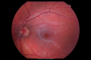
Multiple Evanescent White Dot Syndrome - Atypical Yellow Foveal Spot Right Eyevu 822 fois12-year-old had decreased vision in the left eye from reported amblyopia since around the year 2000. His right eye was doing fine until two days ago, when he had sudden severe vision loss. He sees a blurred spot in the middle of the vision in the right eye wherever he looks.
OD 20/60 with eccentric movement, OS 20/200. Pinhole 20/70. IOP: OD 14, OS 10.
|
|

Partial PVD - Posterior Hyaloid Gap Over Center of Maculavu 822 foisThis image illustraites how sometimes, when the posterior vitreous separates from the retina, a small area of vitreous is left behind. One theory holds that macular puckers can form from that left behind vitreous
|
|

Vision Loss, Blood, and Cartwheel CNVM OD with no fluid on OCT Scanvu 822 fois86-year-old man has had changing vision. He says his vision has been gradually declining since I saw him in June. You saw a hemorrhage in the eye and suggested he comes in here for an evaluation. He is on Combigan and Travatan in the right eye for his glaucoma.
VISUAL ACUITY: Vision OD is 6/200 OS is 8/200 The macula in the right eye is wet even though interestingly the OCT does not show subretinal fluid.
|
|

Proliferative Diabetic Retinopathy - Fresh and Old Scatter (Panretinal) Photocoagulation (Laser) - Neovascularization is regressingvu 822 fois1 month after initial laser
|
|

DRIL - Disorganization Retinal Inner Layersvu 822 foisFollowing treatment for BRVO vision stayed at 3/200 despite resolution of edema
|
|

Subfoveal CNVM wet AMD late fa full imagevu 821 foisShe has noticed decreased vision in the left eye. It is hard to say for how long, maybe a few months. She had her last eye exam about a year ago. She thoughts perhaps it was cataracts, but you saw a problem with the macula and suggested she come here for evaluation.
VISUAL ACUITY: Vision OD is 20/25, OS is 2/200
|
|

Idiopathic Cystoid Macular Edema - CME - Responded to Subtenons Kenalog - Fellow (uninvolved) Eye Nearly Normal Dark Center FAvu 821 fois49-year-old man had a flu two weeks ago the right eye has been throbbing and his vision in the right eye has been foggy. OD 20/80, OS 20/40. IOP: 14 OU.
|
|

Proliferative Diabetic Retinopathy both eyes Type I Diabetic for 25 yearsvu 821 fois47-year-old woman with diabetes for 25 years and decreased vision for 3 months. OD 20/40, OS 20/40
|
|

Central Retinal Artery Occlusion less than 24 hours - 69 year old man VA light perceptionvu 821 fois
|
|

Optic Nerve Pit Retinal Detachment - Macular Detachment - 8 Year Old - 5/200 Vision OSvu 821 fois8-year-old child on two school exams that his vision was poor in the left eye and his last normal exam was in preschool.
VISUAL ACUITY: OD 20/30, OS 5/200. OS: Vertical C/D ratio is 0.2. There is a whitening on the temporal edge of the optic nerve consistent with an optic disc pit and there is a serous macular detachment.
|
|

Hyperpigmentation of the Retinal Pigment Epithelium Right Eye (Treated wet AMD OS)vu 821 fois85-year-old man has wet age-related macular degeneration in the left eye treated for one year most recently one year ago. OD is 20/30, OS is 20/60
|
|

Glaucomatous optic nerve with severe cuppingvu 821 fois
|
|
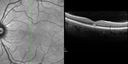
Acute Macular Neuroretinopathy - (AMN) - Spot in Vision OS - 40 Year old Femalevu 821 foisSD OCT shows disruption of the outer retina nasal to the fovea
|
|

Subfoveal CNVM wet AMD fellow eyevu 820 foisShe has noticed decreased vision in the left eye. It is hard to say for how long, maybe a few months. She had her last eye exam about a year ago. She thoughts perhaps it was cataracts, but you saw a problem with the macula and suggested she come here for evaluation.
VISUAL ACUITY: Vision OD is 20/25, OS is 2/200
|
|

Basal Laminar (cuticular) Drusen both eyes 60 Year Old Womanvu 820 fois60-year-old woman with minimal vision loss. Vision OD is 20/30 OS 20/40
|
|
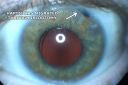
Chronic Pseudophakic Cystoid Macular Edema - Haptic Migrated into Anterior Chamber Through Peripheral Iridotomyvu 820 fois57-year-old man with congenital nystagmus. He had cataract surgery done 30 years ago and has decreased vision left eye for 6 months. OD is 20/50, OS is 20/100
|
|

Proliferative Diabetic Retinopathy both Eyes Neovascularization of the Discvu 819 fois63-year-old woman has been diabetic for twenty one years who sees new floaters in the right eye. OD 20/40, OS 20/40.
|
|

Closed Funnel Retinal Detachment Under Silicone Oil - NLP Visionvu 819 fois15-year-old slowly developing cataract in the left eye over about the last three years. His cataract was already dense when it was removed August 3rd of 2009. The preoperative ultrasound suggested the retina might be abnormal and then once the retina was detached, it was clear that he had a retinal detachment. Because of the amount of scarring the retina was impossible to unfold and it was reattached in a closed funnel formation.
VISUAL ACUITY: OD 20/30, OS: NLP.
|
|

Stargardt's Disease - Macular Dystrophy - 69 Year Old African American Male - 20/200 both eyesvu 819 fois69-year-old man has had vision loss in both eyes starting 20 years ago. He was diagnosed at one point with Stargardt’s macular dystrophy. When I saw him, he had retinal atrophy in each eye.
Vision is 20/200 in each eye
|
|

Central Serous Retinopathy Acute vu 819 fois42-year-old man was seen in the office on October 5, 2011. He had noticed starting in August after a course of antibiotic and steroids, that he developed new spots in his vision in the right eye. He may have had an episode like this sometime in the past. He did take steroids a few years ago and his vision did change at that time, but then returned.
VISUAL ACUITY: OD 20/32, OS 20/32
|
|

Confluent Soft Drusen with Subetinal Fluid - Right Eyevu 819 foisAsymptomatic 85 year old man. OCT shows pocket of subfoveal fluid in the center of the macula. This could be mechanical from the drusen preventing the retina from settling down. Testing did not reveal a CNVM and subsequent OCT 6 weeks later showed no change in structure.
|
|

Chronic Central Serous Retinopathy treated with Photodynamic Laser and Visudynevu 818 fois48-year-old man has had waxing and waning vision in the left eye for the last year from central serous retinopathy. OD is 20/16, OS is 20/16 - 2 months post PDT therapy with Visudyne.
|
|

Central Retinal Artery Occlusion with Cilioretinal Sparing - Acute - FA Very Slow Filling CRAvu 818 fois 77-year-old man was doing fine until midnight of last night when he noticed sudden severe vision loss in the left eye. VISUAL ACUITY: OD 20/20, OS 2/200.
|
|
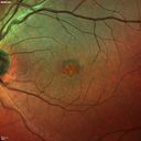
Best Disease - Heidelberg False Colorvu 818 fois
|
|

Retinal Detachmentvu 817 fois
|
|

Serpiginouse Choroiditis (Chorioretinitis) - Acute Right Eye - Old Left Eye VA 20/25 OD , 20/50 OSvu 817 fois63-year-old woman has serpiginous choroiditis (date - March 2011). The right eye has not been previously involved, and then she noticed new onset floaters in the right eye for the last two weeks. Her vision in the right eye is hazy because of that.
VISUAL ACUITY: Vision OD is 20/25, OS is 20/50
|
|

Shotgun Injury 2.5 Years later - 10/200vu 817 foison 4/5/2011. This pleasant 19-year-old had a gunshot injury to the face in December of 2008. He has had several surgeries and his vision is remarkably good. He is aphakic. He is not wearing his contact much but he notices no change. If anything, his vision is a little better.
VISUAL ACUITY: Vision OS is 10/200.
|
|

Progressive Outer Retinal Necrosis 77 Year Old Woman with CLL (Acute Retinal Necrosis) (PORN - ARN)vu 817 fois77-year-old woman with CLL who had shingles on the left side of her face about 6 weeks ago then she developed a dendrite in the cornea which was treating about four weeks ago. She noticed severe vision loss in the left eye just a few days ago and you saw retinitis and she comes in because of that. Vision OD is 20/25, OS is hand motion
|
|

DUSN - Diffuse Unilateral Subacute Neuroretinitis - Nematodevu 817 fois
|
|
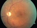
Proliferative Diabetic Retinopathy and Mild Vitreous Hemorrhagevu 817 fois58-year-old woman has diabetic retinopathy in both eyes with neovascularization of the optic nerve, worse in the right eye than the left eye. Vision OD is 20/25, OS is 20/20
|
|

FA: CRAOvu 817 foisCRAO with cilio artery perfusion
|
|

Equatorial Drusenvu 817 fois61 year old female with normal visoin
|
|

Retinal Torsion after Macula Off Retinal Detachment Repair with Inferior PVRvu 816 fois37 year old with torsion of the retina after macula off retinal detachment repair with inferior PVR and membranes dragging the fovea inferiorly.
|
|

Acute Central Serous Chorioretinopathy with Symptoms in Hazy Vision Both Eyesvu 816 fois48-year-old man with hazy vision in the right eye for about three days. He has a little more of a headache than usual. He is under a fair amount of stress. He does not take any steroids that he knows of. He does use a nasal spray, which he is not sure if that is a steroid spray or not. He is going to call us with that medicine. I checked his blood pressure today is was 115/95.
VISUAL ACUITY: OD 20/15, OS 20/25
|
|
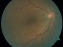
ECTOPIC DISCIFORM SCAR/PERIPHERAL CHOROIDAL NEOVASCULAR MEMBRANE – EXUDATE –INFLAMMATION IN REACTION TO THE PERIPHERAL VASCULAR LESION vu 816 fois53-year-old woman has noticed some blurred vision in the right eye for a few years. In retrospect, she thinks it is possible since she was a teenager the right eye was always the weaker eye and that the left eye has always been the better eye. VISUAL ACUITY: Vision OD is 20/400
|
|

C3F8 Gas Bubble for retinal tear/ Pneumatic Retinopexyvu 816 fois
|
|

Blood Strand through Anterior Chambervu 816 fois
|
|
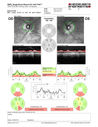
Normal Tension Glaucoma - Nerve Fiber Layer Scan is difficult to read because of artifact from peripapillary atrophyvu 816 fois90 Year old man: IOP 9 OD, 11 OS
|
|

Malignant Hypertension - Cotton Wool Spots - Elschnig Spots - Optic Nerve Edema vu 816 foisMacular Scan Shows Cotton Wool Spots and Nerve Swelling (on Left)
|
|

Embolic CRAO colorvu 816 fois
|
|

Melanoma - Large Ciliary Body A scan shows medium to low internal reflectivityvu 815 fois
|
|
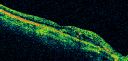
Atypical Birdshot Chorioretinitis and Retinal Arterial Macroaneurysm Right Eye - HLA-B29 Positivevu 815 fois55-year-old woman has multifocal choroiditis in both eyes because she was HLA A-29 positive. She has presumptive birdshot chorioretinopathy. She has not noticed any vision changes. OD 20/40, OS 20/20.
|
|

Hypertensive Retinopathy with Choroidal Ischemia Right Eye and Vision 8/200 - Blood Pressure 240/120 mmHg IgA Nephropathyvu 815 fois39-year-old man two days ago he noticed poor vision in his right eye OD: 8/200; OS: 20/20. BP is 240/120
|
|

Macular Pucker - Diabetic - Severe non-foveal Edema OSvu 815 fois
|
|

Acute Posterior Multifocal Placoid Pigment Epitheliopathy - APMPPE - Fluorescein Angiogramvu 815 fois 25-year-old man with one and half weeks ago he woke up with night chills. Also around that time his joints started hurting him. He also has headaches, although the headache at this point is not severe. His vision has been changing for about the last week and a half. He sees some spots in his vision and they are blurred spots. They are not in the central vision.
VISUAL ACUITY: OD 20/32, OS 20/32
|
|

Old Branch Retinal Vein Occlusion- Severe CME - Pucker - Diabetic Fundus Photovu 814 fois61-year old man has diabetic retinopathy in the right eye. He also had a macular pucker and macular edema. I did a vitrectomy on September 5th. His vision was initially improving. His vision in the right eye seems to be a little more foggy.
VISUAL ACUITY: OD: 20/200
|
|

Gyrate Atrophy - 15 year old male - 20/25 vision both eyes - Positive Family Historyvu 814 fois15 year old male was diagnosed with “retinitis pigmentosa†at age seven. The patient reports that his night vision is poor. He reports he does not notice a visual field change. He reports that his maternal grandfather has lost vision and a cousin has lost vision as well. He reports he has an uncle with some visual changes.
The most significantly affected relative is a 26-year-old legally blind cousin.
20/25+ OD, 20/25 OS.
|
|

Macular Hole - Asymptomatic - 20/400 Visionvu 814 fois75-year-old woman in a recent exam it was noticed her vision in the left eye had substantially declined. She is not clear as to when that exactly happened. She had not noticed a vision change in the left eye. VISUAL ACUITY: OD 20/30, OS 20/400.
|
|

Macular Hole OCT - 1 year post Surgery - Closed Hole - Vision 20/30 (Pre-op 20/100)vu 814 fois62-year-old woman 1 year post surgery with 20/30 vision.
|
|

Bilateral Retinal Arteriol Occlusions - Possible Susac Syndrome vu 813 fois80-year-old woman one month ago had vision loss and a vascular occlusion in the right eye. Vision loss occurred in the left eye about 9 years ago with cotton wool spots. Patients has a 30 year history of tinnitis.
|
|

Retinal Angiomatous Proliferation - Type III choroidal neovascular membranevu 813 fois83 year old female with wet AMD - 20/160 VA- Vision improved to 20/40 with 3 injections of anti-VEGF drugs. OCT shows break in RPE and FA and ICG show PED with hot spot on ICG
|
|

Subfoveal CNVM wet AMD early fa full imagevu 812 foisShe has noticed decreased vision in the left eye. It is hard to say for how long, maybe a few months. She had her last eye exam about a year ago. She thoughts perhaps it was cataracts, but you saw a problem with the macula and suggested she come here for evaluation.
VISUAL ACUITY: Vision OD is 20/25, OS is 2/200
|
|

69 Year Old Woman Normal FAvu 812 fois69-year-old woman OD 20/50 - OS 20/30 ATYPICAL PHOTOPSIAS
|
|

Shotgun Injury 2.5 Years later - 10/200vu 812 foison 4/5/2011. This pleasant 19-year-old had a gunshot injury to the face in December of 2008. He has had several surgeries and his vision is remarkably good. He is aphakic. He is not wearing his contact much but he notices no change. If anything, his vision is a little better.
VISUAL ACUITY: Vision OS is 10/200.
|
|

Optic Atrophy Left Eye and Benign Pituitary Tumor - VA 20/20 Right Eye 20/160 Left Eyevu 812 fois58-year-old man has had gradually declining vision in the left eye over about the last 18 months. Vision OD is 20/20, OS is 20/160
OCT SCAN: The OCT scan of the nerve fiber layer in the right eye shows a thickness of 88 microns, the left eye 59 microns.
MRI did show a suprasellar mass which is affecting the optic chiasm and is responsible for the vision loss. The tumor was resected and was a benign pituitary tumor.
|
|
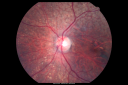
Asteroid Hyalosis After Vitrectomy (also reticular drusen)vu 812 fois
|
|
| 17965 photos sur 180 page(s) |
 |
|
|
|
|
17 | |
|
|
|
|


