|
|
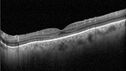
Mild Cone Dystrophy - CRX Mutation85 x angesehen65 year old female with minimal symptoms and a mild cone dystrophy.
|
|
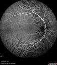
Acute Retinal Pigment Epitheliitis (Krills disease)85 x angesehen15 year old with sudden central vision loss in one eye. The vision loss persisted though for at least 6 months (which may be a laser injury)
|
|
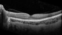
Chronic macula-splitting demarcated retinal detachment85 x angesehen35 year old female with 20/25 vision and noticing something wrong with the superior vision in the right eye for a long time.
|
|
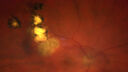
Recurrent toxoplasmosis with elevated IOP84 x angesehen48 year old man with 4 episodes over 40 years of vision loss from toxoplasmosis. His IOP was 44 on presentation.
|
|
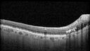
Peripapillary CNVM No Treatment84 x angesehen85 year old with peripapillary CNVM watched for many years.
|
|
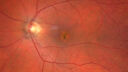
Pattern Dystrophy with Collapsed Vitelliform84 x angesehen74 year old female with vision loss in eye with collapsed vitelliform lesion.
|
|
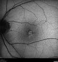
Pattern Dystrophy with Collapsed Vitelliform84 x angesehen74 year old female with vision loss in eye with collapsed vitelliform lesion.
|
|
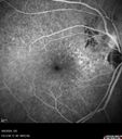
Peripapillary CNVM No Treatment83 x angesehen85 year old with peripapillary CNVM watched for many years.
|
|
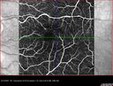
MacTel with foveal atrophy in both eyes83 x angesehen62 year old female with 20/50 OD, 20/40 OS.
|
|
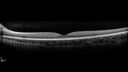
Optic Disc Drusen in a child (non Calcified)83 x angesehenFluorescein angiography shows no leakage
|
|
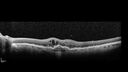
Conversion to Wet AMD after Izervay83 x angesehen83 year old man whose left eye had minimal GA and converted from dry to wet AMD after a single Izervay treatment
|
|
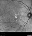
Conversion to Wet AMD after Izervay82 x angesehen83 year old man whose left eye had minimal GA and converted from dry to wet AMD after a single Izervay treatment
|
|
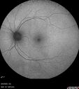
Cotton Wool Spots following cardio-pulmonary bypass82 x angesehenThe patient had emergency thoracotomy about 3 weeks ago he had an aortic dissection with a root aneurysm. It had dissected down to the iliacs. The patient had a thoracotomy and aortic valve replacement and large aortic graft.
|
|
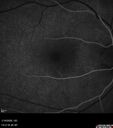
Acute Retinal Pigment Epitheliitis (Krills disease)82 x angesehen15 year old with sudden central vision loss in one eye. The vision loss persisted though for at least 6 months (which may be a laser injury)
|
|
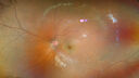
Choroidal hemangioma 10 year old black male81 x angesehen10 year old black child The patient has been wearing glasses which for 3 years and the left eye has had a changing prescription.
3/13/23: OD: +1.00; OS: +5.50
7/18/24: OD: +1.00; OS: +8.00 -1.25 x 015
2/3/25: (Cycloplegic): OD: +2.00; OS: 11.50+1.50 x 120
She had an MRI scan was not diagnostic.
VA OD:20/16
OS: 20/30-2
B-scan 5.1 mm H x 12.3 mm A x 15.0 mm T highly reflective mass centered just temporal to the macula involving the entire macula).
Seen at Wills and observation was decided because of no macular fluid
|
|
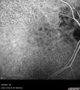
Peripapillary CNVM No Treatment81 x angesehen85 year old with peripapillary CNVM watched for many years.
|
|
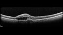
Conversion to Wet AMD after Izervay81 x angesehen83 year old man whose left eye had minimal GA and converted from dry to wet AMD after a single Izervay treatment
|
|
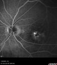
Conversion to Wet AMD after Izervay81 x angesehen83 year old man whose left eye had minimal GA and converted from dry to wet AMD after a single Izervay treatment
|
|
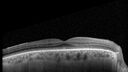
Mild Cone Dystrophy - CRX Mutation81 x angesehen65 year old female with minimal symptoms and a mild cone dystrophy.
|
|
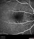
Acute Retinal Pigment Epitheliitis (Krills disease)81 x angesehen15 year old with sudden central vision loss in one eye. The vision loss persisted though for at least 6 months (which may be a laser injury)
|
|
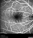
Acute Retinal Pigment Epitheliitis (Krills disease)81 x angesehen15 year old with sudden central vision loss in one eye. The vision loss persisted though for at least 6 months (which may be a laser injury)
|
|
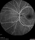
Cotton Wool Spots following cardio-pulmonary bypass80 x angesehenThe patient had emergency thoracotomy about 3 weeks ago he had an aortic dissection with a root aneurysm. It had dissected down to the iliacs. The patient had a thoracotomy and aortic valve replacement and large aortic graft.
|
|
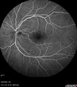
Cotton Wool Spots following cardio-pulmonary bypass80 x angesehenThe patient had emergency thoracotomy about 3 weeks ago he had an aortic dissection with a root aneurysm. It had dissected down to the iliacs. The patient had a thoracotomy and aortic valve replacement and large aortic graft.
|
|
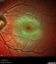
Acute Retinal Pigment Epitheliitis (Krills disease)80 x angesehen15 year old with sudden central vision loss in one eye. The vision loss persisted though for at least 6 months (which may be a laser injury)
|
|

Chronic macula-splitting demarcated retinal detachment80 x angesehen35 year old female with 20/25 vision and noticing something wrong with the superior vision in the right eye for a long time.
|
|
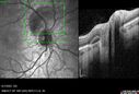
Traumatic Peripapillary Subretinal Hemorrhage80 x angesehen16 year old hit in eye with finger during basketball. Vision is 20/25 and there is a peripapillary hemorrhage.
|
|
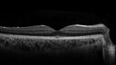
Acute Retinal Pigment Epitheliitis (Krills disease)79 x angesehen15 year old with sudden central vision loss in one eye. The vision loss persisted though for at least 6 months (which may be a laser injury)
|
|
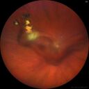
Recurrent toxoplasmosis with elevated IOP78 x angesehen48 year old man with 4 episodes over 40 years of vision loss from toxoplasmosis. His IOP was 44 on presentation.
|
|
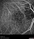
Peripapillary CNVM No Treatment78 x angesehen85 year old with peripapillary CNVM watched for many years.
|
|
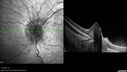
Optic Disc Drusen in a child (non Calcified)78 x angesehenFluorescein angiography shows no leakage
|
|
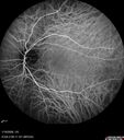
Acute Retinal Pigment Epitheliitis (Krills disease)78 x angesehen15 year old with sudden central vision loss in one eye. The vision loss persisted though for at least 6 months (which may be a laser injury)
|
|
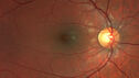
Acute Retinal Pigment Epitheliitis (Krills disease)78 x angesehen15 year old with sudden central vision loss in one eye. The vision loss persisted though for at least 6 months (which may be a laser injury)
|
|
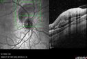
Traumatic Peripapillary Subretinal Hemorrhage78 x angesehen16 year old hit in eye with finger during basketball. Vision is 20/25 and there is a peripapillary hemorrhage.
|
|
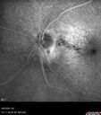
Peripapillary CNVM No Treatment77 x angesehen85 year old with peripapillary CNVM watched for many years.
|
|
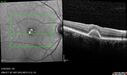
Pattern Dystrophy with Collapsed Vitelliform77 x angesehen74 year old female with vision loss in eye with collapsed vitelliform lesion.
|
|
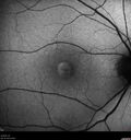
Pattern Dystrophy with Collapsed Vitelliform76 x angesehen74 year old female with vision loss in eye with collapsed vitelliform lesion.
|
|
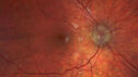
Optic Disc Drusen in a child (non Calcified)76 x angesehenFluorescein angiography shows no leakage
|
|
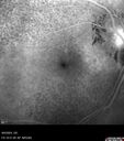
Peripapillary CNVM No Treatment75 x angesehen85 year old with peripapillary CNVM watched for many years.
|
|
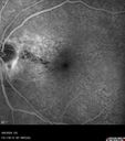
Peripapillary CNVM No Treatment75 x angesehen85 year old with peripapillary CNVM watched for many years.
|
|
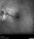
Peripapillary CNVM No Treatment75 x angesehen85 year old with peripapillary CNVM watched for many years.
|
|
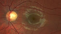
Acute Retinal Pigment Epitheliitis (Krills disease)75 x angesehen15 year old with sudden central vision loss in one eye. The vision loss persisted though for at least 6 months (which may be a laser injury)
|
|
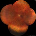
Chronic macula-splitting demarcated retinal detachment75 x angesehen35 year old female with 20/25 vision and noticing something wrong with the superior vision in the right eye for a long time.
|
|
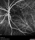
Peripapillary CNVM No Treatment74 x angesehen85 year old with peripapillary CNVM watched for many years.
|
|
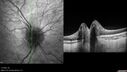
Optic Disc Drusen in a child (non Calcified)74 x angesehenFluorescein angiography shows no leakage
|
|
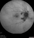
Cotton Wool Spots following cardio-pulmonary bypass74 x angesehenThe patient had emergency thoracotomy about 3 weeks ago he had an aortic dissection with a root aneurysm. It had dissected down to the iliacs. The patient had a thoracotomy and aortic valve replacement and large aortic graft.
|
|
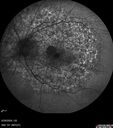
Elmiron (pentasosan polysulfate) Macular Toxicity74 x angesehen77 year old female who had been on Elmiron for 20 years and stopped it 2 years ago
|
|
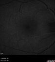
Acute Retinal Pigment Epitheliitis (Krills disease)74 x angesehen15 year old with sudden central vision loss in one eye. The vision loss persisted though for at least 6 months (which may be a laser injury)
|
|
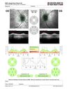
Optic Disc Drusen in a child (non Calcified)73 x angesehenFluorescein angiography shows no leakage
|
|

Optic Disc Drusen in a child (non Calcified)73 x angesehenFluorescein angiography shows no leakage
|
|
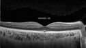
Acute Retinal Pigment Epitheliitis (Krills disease)73 x angesehen15 year old with sudden central vision loss in one eye. The vision loss persisted though for at least 6 months (which may be a laser injury)
|
|
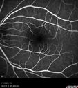
Acute Retinal Pigment Epitheliitis (Krills disease)73 x angesehen15 year old with sudden central vision loss in one eye. The vision loss persisted though for at least 6 months (which may be a laser injury)
|
|
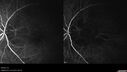
Peripapillary CNVM No Treatment72 x angesehen85 year old with peripapillary CNVM watched for many years.
|
|
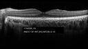
Acute Retinal Pigment Epitheliitis (Krills disease)72 x angesehen15 year old with sudden central vision loss in one eye. The vision loss persisted though for at least 6 months (which may be a laser injury)
|
|
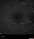
Acute Retinal Pigment Epitheliitis (Krills disease)72 x angesehen15 year old with sudden central vision loss in one eye. The vision loss persisted though for at least 6 months (which may be a laser injury)
|
|
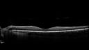
Traumatic Peripapillary Subretinal Hemorrhage72 x angesehen16 year old hit in eye with finger during basketball. Vision is 20/25 and there is a peripapillary hemorrhage.
|
|
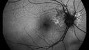
Peripapillary CNVM No Treatment71 x angesehen85 year old with peripapillary CNVM watched for many years.
|
|
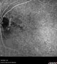
Peripapillary CNVM No Treatment71 x angesehen85 year old with peripapillary CNVM watched for many years.
|
|
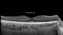
Acute Retinal Pigment Epitheliitis (Krills disease)71 x angesehen15 year old with sudden central vision loss in one eye. The vision loss persisted though for at least 6 months (which may be a laser injury)
|
|
Conversion to Wet AMD after Izervay70 x angesehen83 year old man whose left eye had minimal GA and converted from dry to wet AMD after a single Izervay treatment
|
|
Cotton Wool Spots following cardio-pulmonary bypass69 x angesehenThe patient had emergency thoracotomy about 3 weeks ago he had an aortic dissection with a root aneurysm. It had dissected down to the iliacs. The patient had a thoracotomy and aortic valve replacement and large aortic graft.
|
|
Acute Retinal Pigment Epitheliitis (Krills disease)69 x angesehen15 year old with sudden central vision loss in one eye. The vision loss persisted though for at least 6 months (which may be a laser injury)
|
|
Acute Retinal Pigment Epitheliitis (Krills disease)68 x angesehen15 year old with sudden central vision loss in one eye. The vision loss persisted though for at least 6 months (which may be a laser injury)
|
|

AZOOR68 x angesehen
|
|
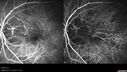
Peripapillary CNVM No Treatment67 x angesehen85 year old with peripapillary CNVM watched for many years.
|
|
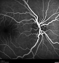
Optic Disc Drusen in a child (non Calcified)67 x angesehenFluorescein angiography shows no leakage
|
|
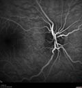
Optic Disc Drusen in a child (non Calcified)66 x angesehenFluorescein angiography shows no leakage
|
|

Auto-immune retinopathy54 x angesehen73 year old man with mild vision loss and history of melanoma now with CLL
|
|

Neovacular Glaucoma from Proliferative Diabetic Retinopathy53 x angesehenSevere non-perfusion and NVG from PDR
|
|

Neovacular Glaucoma from Proliferative Diabetic Retinopathy53 x angesehenSevere non-perfusion and NVG from PDR
|
|

Retinal detachment with dialysis post-trauma51 x angesehen10 year old female who at age 10 was hit in the right eye with a football and had retinal whitening and hemorrhage. Now with asymptomatic dialysis RD
|
|

PDR and Vitreous Hemorrhage with inadequate PRP (nonperfusion)51 x angesehen36 year old with recent vision loss. Diabetes since age 8. Had light PRP done previously. VA 20/25, 20/80
|
|

AZOOR49 x angesehen
|
|

Auto-immune retinopathy48 x angesehen73 year old man with mild vision loss and history of melanoma now with CLL
|
|

Neovacular Glaucoma from Proliferative Diabetic Retinopathy46 x angesehenSevere non-perfusion and NVG from PDR
|
|

Neovacular Glaucoma from Proliferative Diabetic Retinopathy46 x angesehenSevere non-perfusion and NVG from PDR
|
|

AZOOR46 x angesehen
|
|

AZOOR45 x angesehen
|
|

CRAO and old BRAO same eye - interesting images45 x angesehen66 year old man with vision loss for one day in his better eye. Images show CRAO with cilioretinal artery sparing and absence of retinal whitening superotemporally from prior BRAO
|
|

Retinal detachment with dialysis post-trauma44 x angesehen10 year old female who at age 10 was hit in the right eye with a football and had retinal whitening and hemorrhage. Now with asymptomatic dialysis RD
|
|

Neovacular Glaucoma from Proliferative Diabetic Retinopathy43 x angesehenSevere non-perfusion and NVG from PDR
|
|

PDR and Vitreous Hemorrhage with inadequate PRP (nonperfusion)42 x angesehen36 year old with recent vision loss. Diabetes since age 8. Had light PRP done previously. VA 20/25, 20/80
|
|

PDR and Vitreous Hemorrhage with inadequate PRP (nonperfusion)42 x angesehen36 year old with recent vision loss. Diabetes since age 8. Had light PRP done previously. VA 20/25, 20/80
|
|

Auto-immune retinopathy41 x angesehen73 year old man with mild vision loss and history of melanoma now with CLL
|
|

AZOOR41 x angesehen
|
|

AZOOR41 x angesehen
|
|

PDR and Vitreous Hemorrhage with inadequate PRP (nonperfusion)41 x angesehen36 year old with recent vision loss. Diabetes since age 8. Had light PRP done previously. VA 20/25, 20/80
|
|

Pachychoroid Pigment Epitheliopathy40 x angesehen34 year old man with normal vision was told he might have pattern dystrophy or early onset AMD. VA 20/20 OU. Findings - very thick choroid and small PEDs
|
|

Pachychoroid Pigment Epitheliopathy40 x angesehen34 year old man with normal vision was told he might have pattern dystrophy or early onset AMD. VA 20/20 OU. Findings - very thick choroid and small PEDs
|
|

Neovacular Glaucoma from Proliferative Diabetic Retinopathy39 x angesehenSevere non-perfusion and NVG from PDR
|
|

Stickles-12yearold-Post-Laser (1)39 x angesehen
|
|

Neovacular Glaucoma from Proliferative Diabetic Retinopathy38 x angesehenSevere non-perfusion and NVG from PDR
|
|

AZOOR38 x angesehen
|
|

Neovacular Glaucoma from Proliferative Diabetic Retinopathy37 x angesehenSevere non-perfusion and NVG from PDR
|
|

Neovacular Glaucoma from Proliferative Diabetic Retinopathy37 x angesehenSevere non-perfusion and NVG from PDR
|
|

Pachychoroid Pigment Epitheliopathy37 x angesehen34 year old man with normal vision was told he might have pattern dystrophy or early onset AMD. VA 20/20 OU. Findings - very thick choroid and small PEDs
|
|

Pachychoroid Pigment Epitheliopathy37 x angesehen34 year old man with normal vision was told he might have pattern dystrophy or early onset AMD. VA 20/20 OU. Findings - very thick choroid and small PEDs
|
|

PDR and Vitreous Hemorrhage with inadequate PRP (nonperfusion)37 x angesehen36 year old with recent vision loss. Diabetes since age 8. Had light PRP done previously. VA 20/25, 20/80
|
|

PDR and Vitreous Hemorrhage with inadequate PRP (nonperfusion)37 x angesehen36 year old with recent vision loss. Diabetes since age 8. Had light PRP done previously. VA 20/25, 20/80
|
|

Auto-immune retinopathy36 x angesehen73 year old man with mild vision loss and history of melanoma now with CLL
|
|

Neovacular Glaucoma from Proliferative Diabetic Retinopathy36 x angesehenSevere non-perfusion and NVG from PDR
|
|
| 18117 Dateien auf 182 Seite(n) |
 |
|
|
|
|
|
|
|
180 | |
|


