|
|

Stargardt's Juvenille Macular Dystrophy - Fundus Flavimaculatus - Famlial1962 views55-year-old woman was seen in the office on 11/18/08. She has a half sister with Stargardt’s disease and seven other siblings who are fine. 20/120 OD, 20/160 OS.
|
|

Myelinated Nerve Fiber Layer Right Eye (white area)1940 views62-year-old man myelinated nerve fiber layer in the right eye. OD 20/25, OS 20/30.
|
|
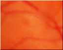
Acute Posterior Vitreous Detachment - Weiss Ring - Vogt Ring1939 views
|
|
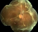
TRD w/ fibrovascular proliferation and sub retinal PVR OD1932 views41 year old african american female with tractional retinal detachment, fibrovascular proliferation, old vitreous hemorrhage, disc neovascularization, and proliferative vitreoretinopathy. Pt has a vision of 20/200
|
|

80 year old woman with Reticular Drusen (pseudodrusen) 20/30 Vision1924 views80 year old woman with asteroid hyalosis and macular degeneration.
OPHTHALMIC EXAMINATION: Visual acuity with correction 20/30-3 OD and 20/30-2
|
|

Retinal Tear After Laser1919 views65-year-old woman new onset floaters for 1 week. She initially saw flashing lights and then the flashing lights subsided and then she had the floaters VISUAL ACUITY: OD 20/25, OS 20/50
|
|

Reticular Macular Disease (Pseudo-drusen) Both Eyes - Wet AMD OS - Dry AMD OD SD-OCT1919 views84-year-old woman has wet age-related macular degeneration in the left eye and dry macular degeneration in the right eye. She takes the eye vitamins and her vision is stable since she was treated three months ago with Avastin. OD 20/50, OS 20/32
|
|

Papilledema - Spectral Domain OCT used for Diagnosis - 14 year old Child - Line Scan over Nerve Shows Retinal Edema1897 views14-year-old was with optic nerve swelling asymptomatic - picked up during routine eye exam. OD 20/20, OS 20/32
SD-OCT is used to differentiate optic disc edema from optic nerve head drusen (which are not yet calcified in children).
|
|
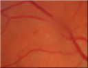
Acute Posterior Vitreous Detachment - Weiss Ring - Vogt Ring1895 views
|
|
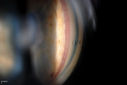
Gonioscopy, Neovascularization in the Angle1893 viewsNeovascularization in the right eye, temporally. VA is 20/40, right eye with IOP at 25. Patient will proceed with Avastin injection to the right eye.
|
|

Anterior Ischemic Optic Neuropathy - Non-Arteritic - AION1871 views72-year-old woman sudden vision loss in the right eye about four days ago. She also has noticed that things are dim out of that eye. She has no headaches. She does feel a little tired. She has no pain in her jaw when she chews and she has no fevers.
OD is 20/80, PH 20/25; OS is 20/25
|
|

Macular Scar1869 viewsFundus photography with Auto Fluorescent shows macular scar centrally, right eye. VA is 20/200 in the right eye.
|
|

Cilio-Retinal Artery Occlusion with "Box Carring" and Cherry Red Spot1861 viewsCilioretinal Artery Occlusion with Cherry Red Spot
|
|

Best Disease - Left Eye Treated Previously, Elsewhere, with PDT and Avastin1854 views11-year-old has Best’s disease that runs in his family on his mothers side. At another retina specialists office, he had photodynamic laser in April of 2009 and Avastin subsequently in June of 2009. Unfortunately with that his vision is substantially declining. OD 20/30, OS 20/100
|
|

OU Stereo Pair Optic Disc Drusen1854 viewsText book example of OU optic discs with drusen in stereo.
|
|

Plaquenil Toxicity - Bulls Eye Maculopathy1853 views70-year-old woman with systemic Lupus erythematosus and clotting problems. She was on the Plaquenil for about eight years and then off the Plaquenil for the last eight years because she developed macular toxicity. Although her vision was hazy, it was stable. Recent deceased vision left eye: OD 20/60, OS 20/100. IOP: OD 18, OS 19.
|
|

Coats' Disease in 14 Year Old Boy Treated with Cryotherapy and Avastin1847 views14 Year Old Boy with Coats Disease
|
|

Background Diabetic Retinopathy1842 viewsPatient with diabetes for over 11-years comes in with blurred vision. Blood sugar control is very poor. VA 20/50- right eye, 20/25-left eye. Fundus exam shows hard exudates with Circinate Rings and edema. Micro-aneurysms and retinal hemorrhage are present in both eyes. Patient will come back for laser treatment.
|
|
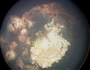
Retinoblastoma1839 views
|
|

Retinal Arterial Macroaneurysm - Recurrent Hemorrhage 6 months post laser1834 views6 months post laser: Her vision had improved, but then three weeks ago it worsened again. She is not on any blood thinners. She wasn’t doing any heavy lifting or straining.
VISUAL ACUITY: OD 3/200
|
|
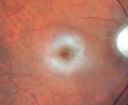
Central Retinal Artery Occlusion - 2 months old by history1832 views77-year-old man was doing fine. His left eye was a little worse than the right eye, and then in the 2 months ago he had sudden severe vision loss in the right eye. Vision OD is 3/200; OS is 20/60. IOP: OD 30, OS 15.
OD: Vertical C/D ratio is 0.4. The macula is white from ischemia. The arteries are narrow.
Photos confirm clinical findings.
OCT SCAN: OCT scan of the right eye shows retinal thickening, consistent with macular edema associated with a central retinal artery occlusion. The left eye has severe inferior retinal thinning, consistent with an old hemicentral retinal artery occlusion in the left eye. The fovea in the left eye is a little bit thin but not too bad.
IMPRESSION: 1. CENTRAL RETINAL ARTERY OCCLUSION RIGHT EYE
2 OLD HEMICENTRAL RETINAL ARTERY OCCLUSION LEFT EYE
3 DIABETES WITHOUT RETINOPATHY
4 ELEVATED INTRAOCULAR PRESSURE RIGHT EYE STATUS POST INJECTION RIGHT EYE A WEEK AGO
DISCUSSION: I explained to the patient that the right eye unfortunately has had a central retinal artery and unfortunately we do not have anything to treat that. His vision will probably improve some over time. He needs to be watched though. It is possible he is developing rubeotic glaucoma, although it is probable the intraocular pressure is high from the injection last week. I asked him to restart the Alphagan, to stop the other two drops, and to return here in three weeks to make sure his pressure is more acceptable.
At this point, having lost almost all the retinal circulation in the right eye and half of the retinal circulation in the left eye, he only has half a retina in the left eye he is working off of. I told him it is very important he keep seeing you and anything that can be done to keep his blood pressure low and his cholesterol reasonable would be helpful to what remains of his retinal circulation.
|
|

Papilledema - Spectral Domain OCT used for Diagnosis - 14 year old Child - Nerve Fiber Layer Scan shows swelling1821 views14-year-old was with optic nerve swelling asymptomatic - picked up during routine eye exam. OD 20/20, OS 20/32
SD-OCT is used to differentiate optic disc edema from optic nerve head drusen (which are not yet calcified in children).
|
|

Retinitis Pigmentosa1809 viewsPatient comes in with cloudy vision. VA is 20/200 in both eyes. Patient has had retinal pigmentosa since 1986. Patient to consider cataract surgery to help her visual symptoms...
|
|

Proliferative Diabetic Retinopathy - Preretinal Hemorrhage1805 viewsPreretinal Hemorrhage secondary to Proliferative Diabetic Retinopathy (PDR)
James L. Perron, C.R.A.
|
|

Fundus Flavimaculatus - Stargardt Disease - 20/50 OD 20/200 OS 61 Year old Color Photo1799 views61-year-old decreasing vision for about the last five years. OD 20/50, OS 20/200.
Pisciform Lesions and Macular Atrophy
|
|

Papilledema - Spectral Domain OCT used for Diagnosis - 14 year old Child - Line Scan over Nerve Shows Retinal Edema1796 views14-year-old was with optic nerve swelling asymptomatic - picked up during routine eye exam. OD 20/20, OS 20/32
SD-OCT is used to differentiate optic disc edema from optic nerve head drusen (which are not yet calcified in children).
|
|
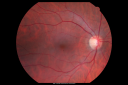
1787 views
|
|

Normal Spectral Domain Optical Coherence Tomography (SD-OCT)1783 views48-year-old woman has had a problem with light and dark adaptations. She notices when she sees a point source of light like a cell phone in a dimly lit room and then she turns the cell phone off, that subsequently her vision is very dark until she goes back into a well lighted area. OD 20/20, OS 20/25.
|
|

Gonioscopy; Scattered Peripheral Anterior Synechiae1783 viewsPatient comes in for evaluation for glaucoma. Patient also has a history of Uveitis. Last flare up was back in 1990. Patient's VA was 20/30, Right eye and 20/40-1, Left eye. Slit Lamp Gonioscopy reveals iris bow with scattered PAS around the angles of the anterior chamber. You can also see pigmentation in the trabecular meshwork. Patient will follow up in 3-months.
|
|

360-Degree Scleral Buckle1780 viewsPatient comes in with double vision from recent retinal detachment surgery. Optos shows 360 degree scleral buckle with retina attached. No holes or breaks visible. Patient had fresnel prism added on to lens to correct the double vision.
|
|

Central Retinal Artery Occlusion1772 views60 year old male with LP vision due to extensive blood flow loss.
|
|

Acute Cytomegalovirus Retinitis1768 views45 y/o male presents with newly diagnosed AIDS and retinal infiltrates.
|
|

Aplastic anemia1765 views29 y.o.m. with aplastic anemia OD VA 20/25.
|
|
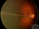
1764 viewsPHPV - right eye - optic nerve
|
|
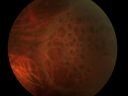
Retinoschisis with Multiple Outer Layer Holes1759 viewsRetinoschisis with Multiple Outer Layer Holes
|
|
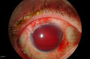
Hyphema1758 viewsHyphema
|
|

Berlin's Edema - Post Bungee Cord Injury Right Eye1757 views20/50 vision - 2 days post-injury
|
|

Anterior Ischemic Optic Neuropathy - Non-Arteritic - AION1756 views72-year-old woman sudden vision loss in the right eye about four days ago. She also has noticed that things are dim out of that eye. She has no headaches. She does feel a little tired. She has no pain in her jaw when she chews and she has no fevers.
OD is 20/80, PH 20/25; OS is 20/25
|
|

Plaquenil Toxicity both Eyes - Partial Bull's Eye - Discontinued 6 Years ago SD-OCT (Spectral domain optical coherence tomography)1754 views82-year-old woman was on Plaquenil from 1976 from 2005, 200 mg a day. It was discontinued because of abnormal visual fields 6 years ago. OD 20/32, OS 20/40
|
|
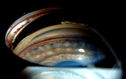
Gonioscopy- Pigmentary Glaucoma Suspect1747 viewsPatient with no family history of glaucoma, comes in with elevated IOP. During Gonioscopy exam. brown pigment overlying the trabecular meshwork. Also, trans-illumination defects on the iris.
|
|

Normal Fundus Autofluorescence1746 views48-year-old woman has had a problem with light and dark adaptations. She notices when she sees a point source of light like a cell phone in a dimly lit room and then she turns the cell phone off, that subsequently her vision is very dark until she goes back into a well lighted area. OD 20/20, OS 20/25.
|
|
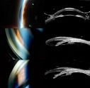
Angle Closure/ UBM & Gonioscopy1745 viewsPatient comes in a day after dilation with severe headache and nausea. Patient's IOP was 64 in the right eye. Patient ended up having angle closure attack. Promoted severe nausea and eye pain. Peripheral Iridotomy was performed and IOP dropped slowly. Patient was followed up the next morning and will be seen 2-days after that.
|
|

Giant Retinal Tear1742 views
|
|

Toxoplasmosis Macular Scar (acquired not congenital) 31 Year Old Man- Recent Vision Change - Creeping Scar1740 views31-year-old man had an episode of toxoplasmosis in the left eye, where unfortunately it adversely affected his central vision that was back in 2004 (7 years ago)
He did notice recently a change in his visual function. He plays softball, he was getting hit by the ball and having trouble seeing and he said that is how he noticed the toxoplasmosis when it first occurred and he was concerned it might have recurred.
OD 20/16, OS 20/80.
|
|

Amelanotic Choroidal Nevus 1736 views45-year-old man OD 20/20, OS 20/20. 5x5 mm flat, yellow choroidal lesion just inferonasal to the optic nerve, two disc-diameters from the nerve with scalloped edges. It looks like a confluent drusen, but it is not elevated.
|
|

1736 viewsAdult male with trauma to the right eye and orbital floor fracture. Hemorrhage with Choroidal Rupture.
|
|
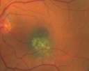
Macular Choroidal Nevus1734 views78-year-old woman has choroidal nevus in the macula of the left eye. You asked her to come in here for an evaluation and nevus was noted in the past about 7 years ago. She has not noticed any change in vision in that eye.
VISUAL ACUITY: OU 20/30.
|
|

Psuedo-retinitis Pigmentosa - Bone Spicules One Eye - Probably Acute Zonal Occult Outer Retinopathy (AZOOR)1734 views65-year-old woman has pseudoretinitis pigmentosa in the right eye only, most likely from acute zonal occult outer retinopathy. OD 20/25, OS 20/25
|
|

CHRPE - Gardner Syndrome - FAMILIAL COLORECTAL POLYPOSIS - 11 year old female1732 views11-year-old child with CHRPE OS has six undescended adult teeth and also she has had a cyst removed from her arm pit and she has three more in her arm pit and one on her face and she is seeing a dermatologist for that in the near future. The mother reports no health problems, however, the patient’s grandmother does have a history of intestinal polyps, as does the patient’s grandmother’s brothers son.
|
|

Normal Spectral Domain Optical Coherence Tomography (SD-OCT)1732 views48-year-old woman has had a problem with light and dark adaptations. She notices when she sees a point source of light like a cell phone in a dimly lit room and then she turns the cell phone off, that subsequently her vision is very dark until she goes back into a well lighted area. OD 20/20, OS 20/25.
|
|

Ozurdex Implant in AC1728 views79 yr old male presents with Blurry vison with slight discomfort x 1 in OD. Pt is S/P Ozurdex Implant x 2 mos which has now surfaced in the Anterior Chamber.
|
|

Diabetic Macular Edema and Hypertensive Retinopathy - Circinate Exudate (Ring Exudate) 1726 views55-year-old woman diabetic for fifteen years and high blood pressure, especially over the last few months. She has had problems with nosebleeds, headaches, and there has been some difficulty bringing her blood pressure down. She said now the blood pressure is under control but it was running 220/120 mmHg for some time. Her vision in the right eye has been poor for four weeks with a spot in the central vision and both eyes have been blurred.
VISUAL ACUITY: Vision OD is 20/160, OS is 20/60
|
|

Optic Nerve Drusen - - Fundus Autofluorescence - FAF1726 views72-year-old woman has had optic nerve drusen for sometime and has visual field abnormalities. OD 20/32, OS 20/32.
|
|

Chorioretinal Scar1720 views21 year old male with a chorioretinal scar OD
|
|

Normal Spectral Domain Optical Coherence Tomography (SD-OCT) - Prefoveal Vitreous Liquifaction1716 views48-year-old woman has had a problem with light and dark adaptations. She notices when she sees a point source of light like a cell phone in a dimly lit room and then she turns the cell phone off, that subsequently her vision is very dark until she goes back into a well lighted area. OD 20/20, OS 20/25.
|
|

Fundus Flavimaculatus - Stargardt Disease - 20/50 OD 20/200 OS 61 Year old Fundus Autofluorescence os1715 views61-year-old decreasing vision for about the last five years. OD 20/50, OS 20/200.
Pisciform Lesions and Macular Atrophy
|
|

Optic Nerve Head Drusen Right Eye1711 views57-year-old woman has optic nerve drusen in both eyes.
VISUAL ACUITY: OU 20/20.
|
|
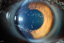
"Jelly Bumps" Soft Contact Lens1705 viewsYoung female patient comes in wearing overnight contact lenses. Complains of irritation in both eyes. Denies sleeping in SCL. Slit lamp exam shows little jelly bumps underneath the contact lens in both eyes. Was issued a new pair and lectured on handling them.
|
|
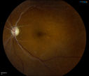
Retinal Artery Occlusion1704 viewsPatient comes in for eval on artery occlusions in both eyes. VA is 20/400, right eye and NLP, left eye. Fundus photos show paniretinal scars in the right eye with arterial narrowing and the left eye has arterial narrowing as well.
|
|

Malignant Hypertension - Cotton Wool Spots - Elschnig Spots - Optic Nerve Edema 1703 views
|
|

Choroidal Nevus with Overlying Drusen1702 views48-year-old woman Vision OD is 20/16, OS is 20/20.
|
|

Reticular peripheral pigment degeneration1701 views
|
|

Asteroid Hyalosis - Benign Calcified Deposits in the Vitreous1699 views75 year old woman with 20/30 vision and dense asteroid haylosis
|
|
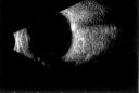
Optic Nerve Head Drusen B-Scan 1699 views57-year-old woman has optic nerve drusen in both eyes.
VISUAL ACUITY: OU 20/20.
|
|

Acute Posterior Multifocal Placoid Pigment Epitheliopathy - APMPPE - Fundus Photo - Color1699 views 25-year-old man with one and half weeks ago he woke up with night chills. Also around that time his joints started hurting him. He also has headaches, although the headache at this point is not severe. His vision has been changing for about the last week and a half. He sees some spots in his vision and they are blurred spots. They are not in the central vision.
VISUAL ACUITY: OD 20/32, OS 20/32
|
|

Clinically Significant Diabetic Macular Edema both Eyes with Exudates1697 views56-year-old woman with diabetes for twelve years. She has noticed blurring of the vision in the right eye for about six months. OD is 20/70, OS is 20/25.
|
|

Diabetic Macular Edema and Hypertensive Retinopathy - Circinate Exudate (Ring Exudate) 1697 views55-year-old woman diabetic for fifteen years and high blood pressure, especially over the last few months. She has had problems with nosebleeds, headaches, and there has been some difficulty bringing her blood pressure down. She said now the blood pressure is under control but it was running 220/120 mmHg for some time. Her vision in the right eye has been poor for four weeks with a spot in the central vision and both eyes have been blurred.
VISUAL ACUITY: Vision OD is 20/160, OS is 20/60
|
|

Punctate Inner Choroidopathy - PIC and Choroidal Neovascular Membrane - Right Eye - Color Fundus Photograph1692 views34-year-old woman with a change in vision in the right eye about eight days ago. Her vision was hazy and she saw waviness in the vision. She does have a history of aches and pains and she was diagnosed with Sjogren’s and 2 months ago she had some fevers and some sinus drainage and that went on for about two months. She is a moderate myope. OD 20/25, OS 20/20.
|
|

Pigmented Peripheral Retinal Degeneration1691 views42-year old male comes in for routine eye exam and to follow up on peripheral retinal degeneration in both eyes. VA is 20/20, right eye and 20/25, left eye. Patient is asymptomatic with no visual complaints.
|
|

Weiss Ring -Vogt Ring - Posterior Vitreous Detachment1684 viewsChronic Floater - 10 years
|
|

Notching of Optic Nerve Rim Inferiorly1683 viewsProgression of glaucoma in the left eye. Past 3-years, developed a notch inferior to the optic disc in the left eye.
|
|

Ectopia Lentis - Marfan's Syndrome - Intermittent Pupillary Block Glaucoma Right Eye1681 views41-year-old woman decreased vision right eye and intermittent pupillary block glaucoma for 6 months from a dislocated natural lens in the eye, probably associated with Marfan syndrome.Vision OD is 20/160, PH 20/40; OS is 20/30, PH 20/25
|
|

Acute Central Serous Retinopathy - Classic Smokestack on Fluorescein Angiogram 1680 views34-year-old man noticed about five days ago decreased vision in the right eye. He was having difficulty playing video games and seeing with that eye. He thought maybe his refractive error was shifting.
VISUAL ACUITY: Vision OD is 20/40, OS is 20/16
|
|

Gonioscopy; Scattered Peripheral Anterior Synechiae1680 viewsPatient comes in for evaluation for glaucoma. Patient also has a history of Uveitis. Last flare up was back in 1990. Patient's VA was 20/30, Right eye and 20/40-1, Left eye. Slit Lamp Gonioscopy reveals iris bow with scattered PAS around the angles of the anterior chamber. You can also see pigmentation in the trabecular meshwork. Patient will follow up in 3-months.
|
|

Psuedo-retinitis Pigmentosa - Bone Spicules One Eye - Probably Acute Zonal Occult Outer Retinopathy (AZOOR)1678 views65-year-old woman has pseudoretinitis pigmentosa in the right eye only, most likely from acute zonal occult outer retinopathy. OD 20/25, OS 20/25
|
|

CHRPE - Gardner Syndrome - FAMILIAL COLORECTAL POLYPOSIS - 11 year old female1677 views11-year-old child with CHRPE OS has six undescended adult teeth and also she has had a cyst removed from her arm pit and she has three more in her arm pit and one on her face and she is seeing a dermatologist for that in the near future. The mother reports no health problems, however, the patient’s grandmother does have a history of intestinal polyps, as does the patient’s grandmother’s brothers son.
|
|

Melanoma1677 viewsYoung male with large Melanoma
|
|
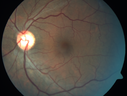
Normal Fellow Eye (Berlin's Edema)1674 views
|
|

Basal Laminar Drusen - Cuticular Drusen - Equatorial Drusen - 70 Year Old Woman - Macular Photo Right Eye1672 views70 year old woman has had macular drusen since her thirties and she said her sister has a similar problem. Recently the vision in the left eye has substantially declined. OD 20/16, OS 10/200.
|
|

Retinal Tear Before Laser1671 views65-year-old woman new onset floaters for 1 week. She initially saw flashing lights and then the flashing lights subsided and then she had the floaters VISUAL ACUITY: OD 20/25, OS 20/50
|
|
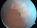
Commotio Retinae - One Day following Basketball to Eye1666 views43-year-old man was playing basketball last night. He was hit straight in the left eye with the ball. Initially he had darkness in his inferior vision and now he has darkness inferonasally in the left eye.
Vision OD is 20/16, vision OS is 20/32
|
|

Fuchs Heterochromic Iridocyclitis - Left eye has inflammation1662 viewsThe right eye is darker than the left eye which has chronic iritis
|
|

Malignant Hypertension - Cotton Wool Spots - Elschnig Spots - Optic Nerve Edema 1661 viewsElschnig Spots are pigment spots in periphery
|
|

Reticular Macular Disease - Reticular Pseudodrusen and Soft Macular Drusen - 57 Year Old Woman1658 views57-year-old woman was seen in the office on 2/22/2011. She has macular drusen in both eyes. I saw her back in 2006. Since then she does notice slight increased distortion. OD is 20/25, OS is 20/25
|
|

Inferotemporal and Inferonasal BRAO1648 viewsBranch Retinal Artery Occlusions - Multiple
|
|

Peripheral Bullous Retinoschisis Fundus Photo (superotemporal)1644 views64-year-old man with peripheral retinal problem noted during comprehensive eye examination. OD 20/32, OS 20/25
|
|

Melanoma1638 views40 Year old male
|
|

Malignant Hypertension - Cotton Wool Spots - Elschnig Spots - Optic Nerve Edema 1636 views
|
|

Floating Vitreous Cyst1633 viewsFloating Vitreous Cyst
|
|

Subhyloid Hem with PDR1633 views
|
|

Choroidal Hemorrhage1630 viewsHemorrhagic Choroidal Effusion
|
|

360-Degree's Pseudoexfoliation1628 viewsFollow-up on glaucoma. Patient has sever pseudoexfoliation in the right eye. VA is 20/25 with IOP at 27. Patient will return in 6-months for re-evaluation.
|
|
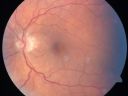
Anterior Ischemic Optic Neuropathy - Non-Arteritic - AION1627 views72-year-old woman sudden vision loss in the right eye about four days ago. She also has noticed that things are dim out of that eye. She has no headaches. She does feel a little tired. She has no pain in her jaw when she chews and she has no fevers.
OD is 20/80, PH 20/25; OS is 20/25
|
|

Proliferative Diabetic Retinopathy both Eyes Neovascularization of the Disc1623 views63-year-old woman has been diabetic for twenty one years who sees new floaters in the right eye. OD 20/40, OS 20/40.
|
|

Central Serous Retinopathy Acute - Indocyanine Green Angiogram - Leakage Choriocapillaris 1621 views42-year-old man was seen in the office on October 5, 2011. He had noticed starting in August after a course of antibiotic and steroids, that he developed new spots in his vision in the right eye. He may have had an episode like this sometime in the past. He did take steroids a few years ago and his vision did change at that time, but then returned.
VISUAL ACUITY: OD 20/32, OS 20/32
|
|

Swollen Nerve with Hemorraghe1619 views
|
|

late phase mushroom cloud, acute CSR1617 viewsFocus is on the peri-foveal vessels illustrates the extremely bullous RD from this mushroom cloud of leakage. The RD was substantially improved after 2 weeks of oral rifampin. Visual acuity improved from 20/200 to 20/70 OD.
|
|

Drusen Maculopathy - Confluent Subfoveal Soft Hydrophobic Drusen - Yellow Drusen in Center of the Macula1614 views
|
|

Acute Central Serous Retinopathy - Classic Smokestack on Fluorescein Angiogram - Antimated .GIF1611 views34-year-old man noticed about five days ago decreased vision in the right eye. He was having difficulty playing video games and seeing with that eye. He thought maybe his refractive error was shifting.
VISUAL ACUITY: Vision OD is 20/40, OS is 20/16
|
|

Hemi-retinal Vein Occlusion1607 views81 year old female seen in 2009 for a Hemi-retinal vein Occlusion OS with vision of 20/25.
|
|
| 18117 files on 182 page(s) |
2 |  |
|
|
|
|
|
|
|
|
|


