|
|

69 Year Old Woman Normal Fluorescein Angiogram766 views69-year-old woman OD 20/50 - OS 20/30 ATYPICAL PHOTOPSIAS
|
|

Twig Branch Retinal Vein Occlusion - Recurrent Macular Edema with extended interval Lucentis Therapy after first 6 monthly Injections766 views39-year-old man has a macular branch retinal vein occlusion with macular edema. He had an excellent response to Lucentis for his first 6 treatments. He was last here 6 weeks ago and his vision seems OK to him. OD 20/40.
|
|
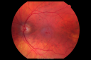
Juxtafoveal Telangiectasis - Stage 5 Left Eye - New Blood and Neovascularization MacTel 766 views60-year-old man has juxtafoveal retinal telangiectasis recent vision loss OD 20/50, OS 20/70 : Hemorrhage OS and new SRNVM
|
|

Acute Macular Neuroretinopathy - (AMN) - Spot in Vision OS - 40 Year old Female766 viewsICG angiogram is normal
|
|

Coloboma - optic nerve, choroid, and retina - left eye766 views80 year old man with glaucoma in the right eye. The left eye has a coloboma of the optic nerve, choroid and retina.
VA 20/25 OD, 20/32 OS
|
|

Bilateral Retinal Arteriol Occlusions - Possible Susac Syndrome 765 views80-year-old woman one month ago had vision loss and a vascular occlusion in the right eye. Vision loss occurred in the left eye about 9 years ago with cotton wool spots. Patients has a 30 year history of tinnitis.
|
|

Pathology - Central Retinal Artery Occlusion - 6 weeks old - Inner Retinal Atrophy765 viewspatient died about 6 to 8 weeks after the artery occlusion, and he had no history of glaucoma. The inner retina atrophy, which looks very similar to that caused by COAG, is due to recent infarct.
|
|
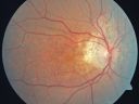
Angioid Streaks - Pseudoxanthoma Elasticum - Supressed CNVM765 views54-year-old woman had angioid streaks and macular degeneration in both eyes. She has had intravitreal Avastin in the right eye about every three months and her vision in that eye is good.
VISUAL ACUITY: Vision OD is 20/30, OS is 2/200
|
|

Eccentric Disciform Scar - Breakthrough Vitreous Hemorrhage - InferoTemporal Subretinal Fibrosis - Ectopic Choroidal Neovascular Membranes765 views88-year-old man has noticed for the last few months the vision in the right eye is foggy, like there is a film in the eye or a haze, His vision is 20/16 in each eye.
Right eye - inferotemporal periphery there is a hemorrhage at the equator at about 7:00 o’clock, which is about 4-disc diameters across and adjacent to that there is scarring.
Left eye - large disciform scar inferotemporally
|
|

765 views
|
|

Punctate Inner Choroidopathy - PIC and Choroidal Neovascular Membrane - Right Eye - InfraRed Photo765 views34-year-old woman with a change in vision in the right eye about eight days ago. Her vision was hazy and she saw waviness in the vision. She does have a history of aches and pains and she was diagnosed with Sjogren’s and 2 months ago she had some fevers and some sinus drainage and that went on for about two months. She is a moderate myope. OD 20/25, OS 20/20.
|
|
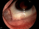
Displaced Scleral Buckle765 viewsDisplaced Scleral Buckle in OD of a 75 yr old male
|
|

Central Areolar Choroidal Dystrophy765 viewsPatient wanted second opinion for atrophic macular degeneration. VA is 20/400, right eye and 20/100. left eye. Patient has very poor vision and also hearing impaired. Fundiscopic exam reveals very atrophy in the macula. FAF shows a central hole to the choroid with no neovascularization present.
|
|

Cartwheel CNVM - Classic - wet AMD - evidence of possible old CSR765 views75 y.o Decreased Vision right eye for one year. VA OD: sc20/125+2 OS 20/25. Autofluorescence and SD OCT show evidence of old serous retinal detachment superior to the fovea suggesting a history of CSR. The macular lesion on FA shows a classic cartwheel appearance
|
|

Proliferative Diabetic Retinopathy both Eyes Neovascularization of the Disc764 views63-year-old woman has been diabetic for twenty one years who sees new floaters in the right eye. OD 20/40, OS 20/40.
|
|

Retinal Arterial Macroaneurysm - Ruptured and self-sealed after hemorrhage764 views90-year-old woman has vision loss in the right eye for 6 weeks. OD 8/200, OS 20/50. Initial Visit
|
|

Non-Foveal Geographic Atrophy Both Eyes764 views81-year-old man has age-related macular degeneration in both eyes. Vision OD is 20/40, OS is 20/80.
|
|

Streptococcus mitis Endophthalmitis following Intravitreal Avastin for wet AMD764 views88 Year Old Man treated with Avastin 02/28/13. Next day developed pain and severe vision loss. Treated with Intravitreal Vancomycin 1mg/0.1 cc; Ceftazidime 2mg/0.1cc; Decadron 0.4mg/0.1cc. Heavy growth Strep 2 days later from culture. Reinjected with Vancomycin 2 days after first Vancomyin. Now seeing Light Perception.
|
|

Posterior Uveal Effusion764 views
|
|

Choroidal Folds Hyperope (+5.00)764 views
|
|

Subretinal Hemorrhage - Commotio Retinae - Macula - hit with baseball764 views15 year old with VA 20/60 3 days after being hit with baseball in the right eye
|
|

Cat Scratch - Neuroretinitis OD AND retinitis OS - Bartonella henselae positive764 views27 year old female with exposure to cats and prior fever. Reports with recent vision loss right eye. She was treated with doxycycline and within 2 weeks the right eye looks better. Then she skipped all follow-up visits. The right eye presented 20/200 and the left 20/16
|
|

Diabetic Macular Edema and Hypertensive Retinopathy - Circinate Exudate (Ring Exudate) 763 views55-year-old woman diabetic for fifteen years and high blood pressure, especially over the last few months. She has had problems with nosebleeds, headaches, and there has been some difficulty bringing her blood pressure down. She said now the blood pressure is under control but it was running 220/120 mmHg for some time. Her vision in the right eye has been poor for four weeks with a spot in the central vision and both eyes have been blurred.
VISUAL ACUITY: Vision OD is 20/160, OS is 20/60
|
|

Diabetic Macular Edema and Hypertensive Retinopathy - Circinate Exudate (Ring Exudate) 763 views55-year-old woman diabetic for fifteen years and high blood pressure, especially over the last few months. She has had problems with nosebleeds, headaches, and there has been some difficulty bringing her blood pressure down. She said now the blood pressure is under control but it was running 220/120 mmHg for some time. Her vision in the right eye has been poor for four weeks with a spot in the central vision and both eyes have been blurred.
VISUAL ACUITY: Vision OD is 20/160, OS is 20/60
|
|

Acute Central Serous Retinopathy (Chorioretinopathy) Right Eye763 views45-year-old man decreased vision in the right eye starting about two days ago. VISUAL ACUITY: Vision OD is 20/60, OS is 20/20.
|
|

Diabetic Papillitis763 viewsPatient is about to undergo surgery for CNS Aneurysm. Patient woke up with little spots which never cleared up. Patients VA was 20/30-OD and 20/40-OS. Both eyes appeared to have disc edema with hemorrhages in the right eye. Ordered a CT of the brain to make sure the aneurysm didn't ruptured.
|
|

Asymptomatic Juxtapapillary Choroidal Neovascular Membrane - Reticular Macular Disease (Wet AMD) - Heidelberg Color763 views
|
|
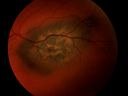
Melanoma763 views
|
|

Anemic Retinopathy763 viewsMyelodysplasia - HgB 8 and very low platelet count
|
|

Diabetic Retinopathy - Non-Perfusion - Possible Central Retinal Vein Occlusion762 views68-year-old woman OD 20/70, OS 20/200 CYSTOID MACULAR EDEMA, POSSIBLE EITHER OCULAR ISCHEMIC SYNDROME IN THE RIGHT EYE OR OCCULT CENTRAL RETINAL VEIN OCCLUSION IN THE RIGHT EYE,S EVERE NON-PerfUSION WITH DIABETIC RETINOPATHY IN THE LEFT EYE.
|
|
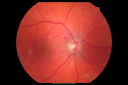
Hypertensive Retinopathy with Choroidal Ischemia Right Eye and Vision 8/200 - Blood Pressure 240/120 mmHg IgA Nephropathy762 views39-year-old man two days ago he noticed poor vision in his right eye OD: 8/200; OS: 20/20. BP is 240/120
|
|

Progressive Outer Retinal Necrosis 77 Year Old Woman with CLL (Acute Retinal Necrosis) (PORN - ARN)762 views77-year-old woman with CLL who had shingles on the left side of her face about 6 weeks ago then she developed a dendrite in the cornea which was treating about four weeks ago. She noticed severe vision loss in the left eye just a few days ago and you saw retinitis and she comes in because of that. Vision OD is 20/25, OS is hand motion
|
|

Anomalous Branch Retinal Artery 762 views59-year-old man OD 20/25, OS 20/25. TORTUOUS RETINAL ARTERY – LEF EYE
(POSSIBLE GROUP 2 WYBURN-MASON SYNDROME
POSSIBLE ACEPHALIC MIGRAINES )
|
|

Cilioretinal artery occlusion and Ophthalmic Artery Occlusion with Relative Sparing of the Central Retinal Artery762 views80-year-old woman who says she was looking in the mirror, covered her right eye and noticed she could not see well out of the left eye. That just happened yesterday. Her last eye check was a few years ago. She also had a carotid endarterectomy on the right side in 2006 shortly after coronary bypass surgery. OD is 20/40, OS is 20/200.
|
|

Choroidal Melanoma (Medium) Exudative Retinal Detachment 762 views87-year-old man noticed a new onset floater a few months ago. OD is 20/40, OS is 20/30
|
|

Equatorial Drusen762 views76-year-old man with equatorial drusen
VISUAL ACUITY: Vision OD is 20/40, OS is 20/25.
|
|
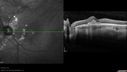
Punctate Inner Choroidopathy - Recurrent CNVM 4 years after initial treatment Left Eye - SD OCT762 viewsJune 7, 2012. This pleasant 35-year-old woman noticed decreasing vision in the left eye recently. She returns now noticing the vision in the left eye is declining. OD 20/50, OS 20/80. OS: Vertical C/D ratio is 0.3. There is a scar just inferonasal to the fovea, which has grown some towards the fovea with a little blood just under the fovea.
|
|

Uveitis and Cystoid Macular Edema - Probably Sarcoidosis762 viewsVA 20/63 OD 20/50 OS - 3 months vision loss - second episode by history of uveitis
|
|
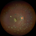
Multifocal Choroiditis and Subretinal Fibrosis with new Subretinal Neovascular Membrane762 views26 year old African American Girl Saw me at age 10 (16 years ago) with acute multifocal choroiditis and panuveitis which evolved into subretinal fibrosis. She was initially treated with 3 years of methotrexate. Vision ended up 20/20 in both eyes. Her eyes have been inactive since and she was lost to follow-up.
A week ago she noticed sudden decreased vision in the right eye. Within the past few weeks there are more floaters in the right eye.
VA OD: sc6’/200 PHNI
VA OS: sc20/16
|
|

Multiple Evanescent White Dot Syndrome - Acute Idiopathic Blind Spot Enlargement761 views33-year-old woman one week ago she awoke and saw a red spots flashing out of her right eye. She notices them to the right side, which is the temporal visual field out of the right eye, then they changed to bursts of white lights without colors and then she notices straight ahead a couple spots. She has headaches in the back of her head. OD 20/20, OS 20/20
|
|

Best Disease for 30 Years - 20/50 Vision both eyes - 61 Year Old Woman761 views61-year-old woman was diagnosed with Best’s disease about 30 years ago. She and her many family members were tested, both at the VA Hospital and then at USF. OD 20/50, OS 20/50
|
|

Gyrate Atrophy - 15 year old male - 20/25 vision both eyes - Positive Family History761 views15 year old male was diagnosed with “retinitis pigmentosa†at age seven. The patient reports that his night vision is poor. He reports he does not notice a visual field change. He reports that his maternal grandfather has lost vision and a cousin has lost vision as well. He reports he has an uncle with some visual changes.
The most significantly affected relative is a 26-year-old legally blind cousin.
20/25+ OD, 20/25 OS.
|
|

Macular Scar with Retinal Pigment Epithelial Hyperplasia - Hyperpigmentation - Wet Macular Degeneration761 viewsMacular Scar in age related macular degeneration with pathology showing retinal pigment epithelial hyperplasia at the edge of the scar. Histopathogy, microscopic slide, pathology.
|
|

fellow eye with subclinical areas of leakage761 views
|
|

Punctate Inner Choroidopathy - PIC and Choroidal Neovascular Membrane - Right Eye - Color Fundus Photograph761 views34-year-old woman with a change in vision in the right eye about eight days ago. Her vision was hazy and she saw waviness in the vision. She does have a history of aches and pains and she was diagnosed with Sjogren’s and 2 months ago she had some fevers and some sinus drainage and that went on for about two months. She is a moderate myope. OD 20/25, OS 20/20.
|
|

Peripheral Exudative Hemorrhagic Chorioretinopathy761 views70 year old man with recurrent peripheral choroidal hemorrhages - no treatment - b-scan shows hemorrhage shrinking over a month
|
|
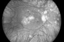
Multifocal Choroiditis and Subretinal Fibrosis - 32 yo Female Old Scar OD760 views32-year-old woman vision loss in the right eye associated with macular scarring and multifocal choroiditis in 1999 with new vision loss in left eye: OD 20/400, OS 20/50.
|
|

Central Serous Retinopathy - Acute760 views49-year-old man sees a spot in the central vision with a red halo for 2 weeks OD is 20/50, OS is 20/20
|
|

Acute Central Serous Chorioretinopathy with Symptoms in Hazy Vision Both Eyes760 views48-year-old man with hazy vision in the right eye for about three days. He has a little more of a headache than usual. He is under a fair amount of stress. He does not take any steroids that he knows of. He does use a nasal spray, which he is not sure if that is a steroid spray or not. He is going to call us with that medicine. I checked his blood pressure today is was 115/95.
VISUAL ACUITY: OD 20/15, OS 20/25
|
|

Retinal Pigment Epithelial Dysgenesis760 views
|
|

Progressive Outer Retinal Necrosis 77 Year Old Woman with CLL (Acute Retinal Necrosis) (PORN - ARN)760 views77-year-old woman with CLL who had shingles on the left side of her face about 6 weeks ago then she developed a dendrite in the cornea which was treating about four weeks ago. She noticed severe vision loss in the left eye just a few days ago and you saw retinitis and she comes in because of that. Vision OD is 20/25, OS is hand motion
|
|

Juxtafoveal Telangiectasis - After Hemorrhage Absorbed Right Eye with Avastin Injections760 views73-year-old woman has juxtafoveal retinal telangiectasis in both eyes. The right eye has stage 5 disease. Her last Avastin treatment in the right eye was in 6 months ago. The right eye has been the worst of her two eyes for years.
VISUAL ACUITY: Vision OD is 20/200, OS is 20/50.
|
|

Basal Laminar Drusen - Cuticular Drusen - 19 Year Old with Lupus Nephritis760 views19-year-old. She has a history of developing severe systemic lupus erythematosus in 2001. She was in renal failure and had biopsy proven Class 2 lupus nephritis. She is currently on Plaquenil 400 mg for six years, as well as Prednisolone orally, every other day. OD 20/20, OS 20/25.
|
|

Angioid Streaks - Peau D'Orange - Pseudoxanthoma Elasticum - Asymptomatic Extrafoveal Choroidal Neovascular Membrane760 views60-year-old woman has pseudoxanthoma elasticum since 2008. Her sister has the same problem and lost vision in both eyes. She had a problem with blood vessel growth in the left eye for which she had injections of Avastin in August, September, and October of 2011 (5 months ago). Subsequent to that, the macula has been dry and you have just been watching her carefully. She is in Florida for a short time but will ultimately be spending nine months out of the year here. Both of her eyes have good vision at
|
|
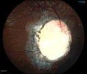
760 viewsYoung 19-year old female with Charge Syndrome comes in for eye exam. VA is 20/400, right eye, 20/30, left eye. Fundus photography shows coloboma covering the macula and pushing the optic nerve in the right eye.
|
|

Retinoblastoma - Bilateral - Regressed - Chemotherapy, Thermal Therapy, Cryotherapy759 views10-year-old child was seen in the office on August 6, 2008. She was treated with a combination of chemotherapy at 10 months old as well as thermal therapy and cryotherapy for bilateral retinoblastoma tumors. She reportedly had nine tumors in the left eye and five tumors in the right eye
|
|

Clinically Significant Diabetic Macular Edema both Eyes with Exudates759 views56-year-old woman with diabetes for twelve years. She has noticed blurring of the vision in the right eye for about six months. OD is 20/70, OS is 20/25.
|
|

Serous Macular Detachment Left Eye - Possible Hypertensive Choroidopathy v. Central Serous Retinopathy759 views59-year-old woman decreasing vision in the left eye for about two days. She sees a spot in front of her vision. It is a yellow spot which is just straight ahead and moves with her eye. She does have high blood pressure. She checks her blood pressure at home, and even at home her blood pressure runs very high, with the systolic running around 200 and the diastolic around 100, although occasionally it does return to the normal range.
VISUAL ACUITY: Vision OD is 20/60, OS is 20/30
|
|

Macular Scars - Laser (1980s) Ocular Histoplasmosis759 views70 year old man who had laser at Wills, Wilmer and the Mayo clinic before better therapies were available for ocular histoplasmosis related CNVM
|
|
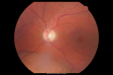
Choroidal Melanoma - Exudative Retinal Detachment 82 Year Old Man 759 views82-year-old man who had 3 falls 2 weeks ago. After the falls he started checking his vision and noticed there was a veil over the left eye, which he had seen for about two weeks in the superior visual field. OD 20/25, OS 20/40
(patient was medically never well enough for brachytherapy and died 6 months later from heart disease)
|
|

Geographic Atrophy 20/160 Vision759 views84-year-old woman has age-related macular degeneration in both eyes with foveal atrophy. She also has a central retinal vein occlusion
in the left eye for which she had intravitreal Kenalog and finally chorioretinal anastomosis laser a year
ago. VISUAL ACUITY: Vision OD is 20/160, OS is 7/200.
|
|

New Subfoveal Classic CNVM OS - Wet AMD - Macular Scar Congenital Toxoplasmosis Right Eye 759 views77-year-old woman as had poor vision in the right eye since birth from congenital toxoplasmosis of her macula in the right eye. Two to three weeks ago when she noticed vision loss in the left eye. VISUAL ACUITY: OD 2/200, OS 20/200.
|
|

Dehydration Injury to Retina after Macula-On Retinal Detachment Repair - Also Subretinal Perfluoron759 views18 month ago he had a scleral buckle, vitrectomy, laser and gas for macula on retinal detachment. Since he notices blind spots in the center of the left eye/ He also notices a dense, inferior visual field defect. OD 20/20, OS 20/50
|
|
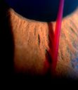
Blood strand into the anterior chamber759 views
|
|

Probable Measles Retinopathy759 views52-year old female wanted second opinion on retinal eval. Patient had PK and PDT in the left eye. VA was 20/30, right eye was count fingers. Pinhole was NI, both eyes. Patient's mother had measles. Notice pigment changes in both the color and FAF photo.
|
|

Angioid Streaks and Macular Degeneration - CNVM OS - Band Keratopathy 759 views79-year-old man has wet age-related macular degeneration in the left eye, for which he is having Lucentis treatment. OD 20/40, Pinhole 20/32. OS 20/160, Pinhole 20/50. He has band keratopathy sparing the visual axis in both eyes. ICG shows angioid streaks both eye.
|
|

FA showing NVE, NVD, non- perfusion left eye inferonasally with neovascularization at the margin of the non-perfusion759 views
|
|

Fundus Flavimaculatus - Asymptomatic - Fundus Autofluorescence759 views45 year old man was told to come in because of ‘white spots on their retina’. - FAF image shows that spots are strongly autofluorescent
|
|

Microaneursyms and Macular Edema Not Diabetic759 views85 year old man has been tested about 2-3 times and is always negative for diabetes
|
|

Retinal Vasculopathy associated with Facioscapulohumeral Muscular Dystrophy OCT right eye line scan758 views59-year-old woman had a blood pressure spike to 170/90 and was on medications for that. She does have facioscapulohumeral muscular dystrophy, which runs in her family. She has a cousin with the same disorder.
VISUAL ACUITY: Her vision is 20/25 OU. IOP: OD 21, OS 18.
There is no posterior vitreous separation, telangiectatic vessels near the macula. There are patchy retinal hemorrhages. There is no edema or proliferation.
|
|

Optic Nerve Pit Retinal Detachment - Macular Detachment - 8 Year Old - 5/200 Vision OS758 views8-year-old child on two school exams that his vision was poor in the left eye and his last normal exam was in preschool.
VISUAL ACUITY: OD 20/30, OS 5/200. OS: Vertical C/D ratio is 0.2. There is a whitening on the temporal edge of the optic nerve consistent with an optic disc pit and there is a serous macular detachment.
|
|

Hyperpigmentation of Retinal Pigment Epithlium in Dry AMD (hyperplasia)758 views87-year-old woman has wet age-related macular degeneration in the left eye. She had treatment up north but there has been a gap in therapy and the vision in the left eye is poor. Vision OD is 20/25, OS is 20/400
|
|

Diabetic Retinopathy - Foveal Laser Scar Right Eye758 views74-year-old woman has diabetic retinopathy in both eyes. She has had focal laser in her right eye done elsewhere in the mid 90’s and unfortunately the laser was fairly close to the center and as time progressed she developed foveal atrophy around the scars. She has noticed her vision declining since she was here last a year ago. She does take Travatan in both eyes at night for glaucoma.
VISUAL ACUITY: OD 20/200, OS 20/40.
|
|

Juvenile Xanthogranuloma758 viewsPresented with hyphaema after minor trauma
|
|

PXE with Angioid Streaks (4-up for comparison)758 viewsPXE with angioid streaks OU, 4-up for comparison between 2010 and 2011 - angioid streaks are stable.
|
|

Macular possible Cone Dystrophy758 views
|
|

Vitreous Traction over retinal vessel758 views79 year old man. No visual complaints.
|
|

Diabetic and Pseudophakic Cystoid Macular Edema Laser Photo - Focal Laser757 views56-year-old woman has diabetic retinopathy in both eyes and macular edema. She is recently pseudophakic. OD 20/80, OS 20/200.
|
|

Diabetic Retinopathy - Non-Perfusion - Possible Central Retinal Vein Occlusion757 views68-year-old woman OD 20/70, OS 20/200 CYSTOID MACULAR EDEMA, POSSIBLE EITHER OCULAR ISCHEMIC SYNDROME IN THE RIGHT EYE OR OCCULT CENTRAL RETINAL VEIN OCCLUSION IN THE RIGHT EYE,S EVERE NON-PerfUSION WITH DIABETIC RETINOPATHY IN THE LEFT EYE.
|
|
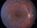
X-Linked Juvenille Retinoschisis 10 Year Old Child treated with Trusopt757 views10-year-old child has x-linked juvenile retinoschisis VA 20/60, OS is 20/100. Macula and Vision improved with Trusopt drops.
|
|

Macular Fold - 40 years post retinal detachment repair - Glaucoma 757 views79-year-old woman has had a long-standing macular fold in the right eye associated with retinal detachment repair in 1970. (40 years ago) She has a macular scar in that eye. She has optic atrophy in both eyes from glaucoma. Vision OD is hand motion, OS is 20/50
|
|

Central Retinal Vein Occlusion and Laser Induced Chorioretinal Anastomosis757 views84-year-old woman has age-related macular degeneration in both eyes with foveal atrophy. She also has a central retinal vein occlusion
in the left eye for which she had intravitreal Kenalog and finally chorioretinal anastomosis laser a year
ago. VISUAL ACUITY: Vision OD is 20/160, OS is 7/200
|
|
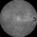
InfraRed Image - Enhanced S Cone Syndrome - Goldmann Favre - 757 views55-year-old woman while in college her vision was poor even with glasses and she sought evaluation for that. She was told after she had an electroretinogram at USF 15 years ago, that she had something with her blue cones. She does have poor night vision, but her reading vision is pretty good.
VISUAL ACUITY: OD 20/40, OS 20/40
|
|

Reticular Macular Disease (Pseudo-drusen) Both Eyes - Wet AMD OS - Dry AMD OD 757 views84-year-old woman has wet age-related macular degeneration in the left eye and dry macular degeneration in the right eye. She takes the eye vitamins and her vision is stable since she was treated three months ago with Avastin. OD 20/50, OS 20/32
|
|
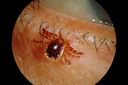
Tick on Lower Lid757 viewsPt. came in for cataract surgery and a tick was found on the lower lid. Pt. had no idea the tick was attached and did not know how long it had been there. Tick was photographed and then removed by physician.
|
|

PXE with Angioid Streaks757 viewsMontage photography OS of a patient that has PXE with angioid streaks.
|
|

Corneal Abrasion with Foreign Body Present 757 viewsER patient comes in with corneal abrasion in the left eye which was getting worst. VA 20/25. Slit lamp exam showed corneal abrasion superiorly at 2-o'clock. Flipped eyelid and foreign body appeared on the upper tarsal plate. Foreign body was removed.
|
|
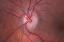
Acute non arteritic anterior ischemic optic neuropathy left eye - Color Photo757 views66 y.o. woman spot in central vision in left eye started about 1 week ago since then it is not changing. VA OD: sc20/25 OS: sc20/200
|
|
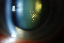
Polychromatic "Christmas Tree" Cataract757 viewsPatient comes in for annual eye exam. Eval on cataracts. VA is 20/20, right eye, 20/40, left eye. Slit Lamp exam shows polychromatic christmas tree cataract in the left eye.
|
|

Autosomal Dominant Optic Atrophy - Father757 views52 year old man Vision has not been correctable for many years.
EXTENDED HPI: The patient has been seen for a while and his vision is not normal. Â
VA OD: sc20/100 PH20/50-1 Nsc20/400
VA OS: sc20/63 PH20/50-2 NscJ16
IOP: TP: OD:10 OS:9 Time:02:29 PM
Two siblings and his son have the same problem
|
|

Classic Subfoveal Choroidal Neovascular Membrane - 20/400 Vision - 6 - 12 months old lesion756 views78-year-old woman six months or a year ago decreasing vision in the right eye. OD 20/400. Fluorescein angiography shows a classic subfoveal choroidal neovascular membrane in the right eye 3 disc-diameters across, which has brisk leakage. The left eye shows staining drusen.
|
|
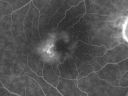
Group 2a Juxtafoveal Telangiectasis - MacTel - Idiopathic Juxtafoveal Macular Telangiectasia - Macular Hole756 views80-year-old man has juxtafoveal retinal telangiectasis in both eyes. He also has a true full-thickness macular hole in the left eye. The vision OD is 20/40, OS is 20/160
Group 2a Juxtafoveal Retinal Telangiectasis (telangiectasia), MacTel
|
|

Calcified Drusen - Dry Age-related Macular Degeneration - Geographic Atrophy756 views87 Year old woman with dry age-related macular degeneration in both eyes.With both eyes open she is fine, but when she closes the left eye, things are wavier.
VISUAL ACUITY: Vision OD is 20/100, OS is 20/40.
|
|

Indocyanine Green Angiogram - Enhanced S Cone Syndrome - Goldmann Favre - 756 views55-year-old woman while in college her vision was poor even with glasses and she sought evaluation for that. She was told after she had an electroretinogram at USF 15 years ago, that she had something with her blue cones. She does have poor night vision, but her reading vision is pretty good.
VISUAL ACUITY: OD 20/40, OS 20/40
|
|

Twig Branch Retinal Vein Occlusion - Recurrent Macular Edema with extended interval Lucentis Therapy after first 6 monthly Injections756 views39-year-old man has a macular branch retinal vein occlusion with macular edema. He had an excellent response to Lucentis for his first 6 treatments. He was last here 6 weeks ago and his vision seems OK to him. OD 20/40.
|
|
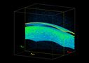
3-D HD OCT Image of Soft Contact Lens over Cornea756 views
|
|

Acute non arteritic anterior ischemic optic neuropathy left eye - SD Optical Coherence Tomography Line Scan756 views66 y.o. woman spot in central vision in left eye started about 1 week ago since then it is not changing. VA OD: sc20/25 OS: sc20/200
|
|

Outer Retinal Holes - Probably Solar Retinopathy (history is negative)756 views
|
|

Serpiginouse Choroiditis (Chorioretinitis) - Acute Right Eye - Old Left Eye VA 20/25 OD , 20/50 OS755 views63-year-old woman has serpiginous choroiditis (date - March 2011). The right eye has not been previously involved, and then she noticed new onset floaters in the right eye for the last two weeks. Her vision in the right eye is hazy because of that.
VISUAL ACUITY: Vision OD is 20/25, OS is 20/50
|
|

Eccentric Disciform Scar - Breakthrough Vitreous Hemorrhage - InferoTemporal Subretinal Fibrosis - Ectopic Choroidal Neovascular Membranes755 views88-year-old man has noticed for the last few months the vision in the right eye is foggy, like there is a film in the eye or a haze, His vision is 20/16 in each eye.
Right eye - inferotemporal periphery there is a hemorrhage at the equator at about 7:00 o’clock, which is about 4-disc diameters across and adjacent to that there is scarring.
Left eye - large disciform scar inferotemporally
|
|
| 18005 files on 181 page(s) |
 |
|
|
|
|
22 | |
|
|
|
|


