|
|

Asteroid Hyalosis - Asymptomatic755 views80-year-old woman her vision is not bothering her, you noticed a problem with the retina. Her left eye tests worse than the right eye today, but she says that it has been like that for a long time and she is not bothered by any floaters or change in central vision.
VISUAL ACUITY: OD is 20/25, OS 20/40
|
|

Choroidal Melanoma Medium755 views33-year-old man with a history of decreased in the right eye. He had blurring in the eye about a year ago. He reports that it improved spontaneously until it was “nearly resolvedâ€. He said he had some persistent distortion, but that over the past couple of weeks he has noticed this dark spot recurring in the right eye, the left eye is unaffected. He does not have any flashes, floaters or change in his peripheral visual acuity. 20/80 OD, 20/16 OS. No improvement with pinhole. IOP: 12 OD, 11 OS.
POSTERIOR SEGMENT EXAMINATION: Shows a clear view with no PVD in either eye. C/D ratio is 0.2 OU. Large retinal vessels are unremarkable. The macula of the right eye has a brown choroidal lesion with pigment overlying the surface and orange spots along its lateral surface. The left macula is normal.
B-SCAN: B-scan ecography of the ultrasound of the right eye shows a mass about 5 mm high with a moderate to low internal reflectivity on A-scan echogram. There is serous fluid inferiorly on the echogram
|
|

Toxoplasmosis - Inner Retinal - Recurrent - Treated with Bactrim DS 755 views43-year-old man has macular toxoplasmosis. He has responded nicely to Bactrim OS: 20/60
|
|

Bilateral Diffuse Uveal Melanocytic Proliferation - BDUMP - Paraneoplastic Syndrome755 views80-year-old man vision loss for one year. He died about one year after these photos from Metastatic Poorly Differentiated Large Cell Carcinoma of unknown primary. He was a smoker.
|
|

Best Disease for 30 Years - 20/50 Vision both eyes - 61 Year Old Woman755 views61-year-old woman was diagnosed with Best’s disease about 30 years ago. She and her many family members were tested, both at the VA Hospital and then at USF. OD 20/50, OS 20/50
|
|

Non-Arteritic Anterior Ischemic Optic Neuropathy - Peripapillary Choroidal Ischemia755 views68-year-old woman noticed central vision loss in the right eye about a week ago. It is getting a little bit better. OD 20/160, OS 20/25.
|
|

Non-arteritic Anterior Ischemic Optic Neuropathy following Intravitreal Injection 755 views81-year-old woman has had injections in both eyes for wet age-related macular degeneration. 2 weeks ago the left eye was treated. Less than a week ago she noticed a change in vision in the left eye. She had a dull left frontal headache and noticed her vision was hazy as if there was a veil over the vision. OD is 20/25, OS is 20/25. IOP: OD 18, OS 26. The left eye has a 3+ APD
|
|

Acute Posterior Multifocal Placoid Pigment Epitheliopathy - APMPPE - Fundus Photo - Color755 views 25-year-old man with one and half weeks ago he woke up with night chills. Also around that time his joints started hurting him. He also has headaches, although the headache at this point is not severe. His vision has been changing for about the last week and a half. He sees some spots in his vision and they are blurred spots. They are not in the central vision.
VISUAL ACUITY: OD 20/32, OS 20/32
|
|

Peripheral Exudative Hemorrhagic Chorioretinopathy755 views70 year old man with recurrent peripheral choroidal hemorrhages - no treatment - b-scan shows hemorrhage shrinking over a month
|
|

Primary Ocular Lymphoma - White Mass Left Eye - 112712 755 views
|
|

Fundus Flavimaculatus - Asymptomatic - Multi-Color (false color) Photo755 views45 year old man was told to come in because of ‘white spots on their retina’.
|
|

Polypoidal Choroidal Vasculopathy with Pulsating ICG polyp left eye755 views
|
|

Unilateral Acute Idiopathic Maculopathy (UAIM)755 viewsVision has improved back to 20/20 without any therapy. 1 month following vision loss
|
|

Multifocal Choroiditis and Subretinal Fibrosis - 32 yo Female FA New Lesion OS754 views32-year-old woman vision loss in the right eye associated with macular scarring and multifocal choroiditis in 1999 with new vision loss in left eye: OD 20/400, OS 20/50.
|
|
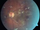
Diabetic Patient with Macular Edema and Blood Pressure 200/95754 views49-year-old decreasing vision over the last year. OD is 20/80, OS 20/80. blood pressure which was 200/95.
|
|

Disciform Macular Scar Both Eyes from Wet Macular Degeneration754 views86-year-old woman has disciform scars OD: 20/400; OS: 20/100
|
|
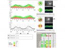
Optic Atrophy Left Eye and Benign Pituitary Tumor - VA 20/20 Right Eye 20/160 Left Eye754 views58-year-old man has had gradually declining vision in the left eye over about the last 18 months. Vision OD is 20/20, OS is 20/160
OCT SCAN: The OCT scan of the nerve fiber layer in the right eye shows a thickness of 88 microns, the left eye 59 microns.
MRI did show a suprasellar mass which is affecting the optic chiasm and is responsible for the vision loss. The tumor was resected and was a benign pituitary tumor.
|
|

Macular Hole - Asymptomatic - 20/400 Vision754 views75-year-old woman in a recent exam it was noticed her vision in the left eye had substantially declined. She is not clear as to when that exactly happened. She had not noticed a vision change in the left eye. VISUAL ACUITY: OD 20/30, OS 20/400.
|
|

Post-Pneumatic Retinopexy Gas Bubble on Optic Nerve754 views80 year old man, 2 weeks post C3F8 pneumatic retinopexy for 5/200 macula-off retinal detachment. Vision is 20/50 (final vision was about 20/30). There is a gas bubble trapped in the optic nerve.
|
|

754 views
|
|
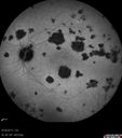
Acute Posterior Multifocal Placoid Pigment Epitheliopathy - APMPPE - Indocyanine Green Angiogram754 views 25-year-old man with one and half weeks ago he woke up with night chills. Also around that time his joints started hurting him. He also has headaches, although the headache at this point is not severe. His vision has been changing for about the last week and a half. He sees some spots in his vision and they are blurred spots. They are not in the central vision.
VISUAL ACUITY: OD 20/32, OS 20/32
|
|

Best Disease - Color Fundus Photo754 views
|
|

Retinal Arterial Macroaneurysm - Untreated Hypertension (250/135) - Serous Macular Detachment 754 views82 year old asian woman. VA 20/80 Headaches and vision loss for 1 month. Negative medical history but has not been to the doctor for years. (no insurance)
|
|

Retinal Detachment with PVR and large posterior macular hole - Vision HM754 viewsPatient had vitrectomy for 20/60 maular pucker and subsequent retinal detachment
|
|

CRVO with Paracentral Acute Middle Maculopath - PAMM754 views56 year old woman with sudden vision loss, hemorrhages in all four quadrants. FA shows good retinal circulation. The OCT shows PAMM lesions in the affected eye. Vision did improve on second visit from 20/160 to 20/60 in about a month
|
|

Chronic Central Serous Chorioretinopathy Both Eyes754 views60 year old man with 20 year history of chronic CSR. He has had laser and PDT in the past. The left eye has been 20/100 for decades. The right eye is now declining. Was 20/32 and is down to 20/50. There is chronic low grade subretinal fluid away from the center in the right eye. (focal laser in the right eye in 2005, left eye 2001 at BPEI). Refampin tried in 2012, Aldactone in 2013. PDT in 204 and 2017 Images are from 2017
|
|
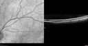
Stellate Non-heredtiary Idiopathic Foveomacular Retinoschisis (SNIFR)754 views77 year old man who is healthy and 6'6" tall with 20/40 vision and no complaints. OCT shows diffuse retinoschisis.
|
|

Acute Macular Neuroretinopathy - 40 year old female754 views
|
|

Albinism and Foveal Hypoplasia753 views65-year-old man was told about twenty years ago by a retina specialist that he spent so much time in the sun that he burned out his maculae. He does have two brothers who have dark brown hair and he has always had white hair. He had eye exercises when he was eight or nine because of a lazy eye and his brothers, when he was little, used to call him an albino. He has been diagnosed by dermatologists as having possible albinism but he has been told by other people his skin is just fair.
VA 20/70 OU
|
|
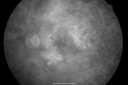
Diabetic and Pseudophakic Cystoid Macular Edema Late FA fellow Eye753 views56-year-old woman has diabetic retinopathy in both eyes and macular edema. She is recently pseudophakic. OD 20/80, OS 20/200.
|
|

Diabetic Retinopathy - Non-Perfusion - Possible Central Retinal Vein Occlusion753 views68-year-old woman OD 20/70, OS 20/200 CYSTOID MACULAR EDEMA, POSSIBLE EITHER OCULAR ISCHEMIC SYNDROME IN THE RIGHT EYE OR OCCULT CENTRAL RETINAL VEIN OCCLUSION IN THE RIGHT EYE,S EVERE NON-PerfUSION WITH DIABETIC RETINOPATHY IN THE LEFT EYE.
|
|

Bilateral Retinal Arteriol Occlusions - Possible Susac Syndrome 753 views80-year-old woman one month ago had vision loss and a vascular occlusion in the right eye. Vision loss occurred in the left eye about 9 years ago with cotton wool spots. Patients has a 30 year history of tinnitis.
|
|

69 Year Old Woman Normal Fluorescein Angiogram753 views69-year-old woman OD 20/50 - OS 20/30 ATYPICAL PHOTOPSIAS
|
|

Macular Hole - Asymptomatic - 20/400 Vision753 views75-year-old woman in a recent exam it was noticed her vision in the left eye had substantially declined. She is not clear as to when that exactly happened. She had not noticed a vision change in the left eye. VISUAL ACUITY: OD 20/30, OS 20/400.
|
|

Wet AMD - PED with exudate 20/400 87 Year old753 views87-year-old woman has wet age-related macular degeneration in the left eye. Vision OD is 20/25, OS is 20/400
|
|

Fluorescein Angiogram - Enhanced S Cone Syndrome - Goldmann Favre - 753 views55-year-old woman while in college her vision was poor even with glasses and she sought evaluation for that. She was told after she had an electroretinogram at USF 15 years ago, that she had something with her blue cones. She does have poor night vision, but her reading vision is pretty good.
VISUAL ACUITY: OD 20/40, OS 20/40
|
|

Ocular Histoplasmosis both Eyes - Laser Right Eye Only 753 views58-year-old woman who lost vision as a child in the left eye from ocular histoplasmosis. She used to play in her attic and she was told by her pulmonary doctor that is probably where she picked it up. She had laser in the right eye for a leaky lesion back in 1990. OD 20/25, OS 20/200.
|
|
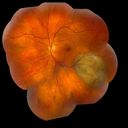
Choroidal Melanoma753 views
|
|

Acute Posterior Multifocal Placoid Pigment Epitheliopathy - APMPPE - Fluorescein Angiogram753 views 25-year-old man with one and half weeks ago he woke up with night chills. Also around that time his joints started hurting him. He also has headaches, although the headache at this point is not severe. His vision has been changing for about the last week and a half. He sees some spots in his vision and they are blurred spots. They are not in the central vision.
VISUAL ACUITY: OD 20/32, OS 20/32
|
|
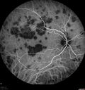
Acute Posterior Multifocal Placoid Pigment Epitheliopathy - APMPPE - Indocyanine Green Angiogram753 views 25-year-old man with one and half weeks ago he woke up with night chills. Also around that time his joints started hurting him. He also has headaches, although the headache at this point is not severe. His vision has been changing for about the last week and a half. He sees some spots in his vision and they are blurred spots. They are not in the central vision.
VISUAL ACUITY: OD 20/32, OS 20/32
|
|

Macular possible Cone Dystrophy - Fundus Autofluorescence753 views
|
|

Papilledema 10 year old child752 views10-year-old child has uveitis in both eyes. She probably has tubulointerstitial nephritis-uveitis syndrome, and she apparently also independently has idiopathic elevated intracranial pressure. VISUAL ACUITY: Vision OU is 20/20.
|
|
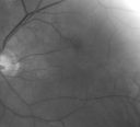
Albinism and Foveal Hypoplasia752 views65-year-old man was told about twenty years ago by a retina specialist that he spent so much time in the sun that he burned out his maculae. He does have two brothers who have dark brown hair and he has always had white hair. He had eye exercises when he was eight or nine because of a lazy eye and his brothers, when he was little, used to call him an albino. He has been diagnosed by dermatologists as having possible albinism but he has been told by other people his skin is just fair.
VA 20/70 OU
|
|

Cystoid Macular Edema - Xalatan - ECCE was done 20 years previously752 views71 Year old woman who noticed about a month ago the central vision in the right eye fogged up. OD 20/200 POSSIBLE XALATAN INDUCED CYSTOID MACULAR EDEMA
|
|

Toxocariasis752 views15-year-old one year ago had pink eye then her vision has been abnormal. She recently went for a drivers’ test and failed, OD: 20/80; OS: 20/20.
|
|

Retinal Torsion after Macula Off Retinal Detachment Repair with Inferior PVR752 views37 year old with torsion of the retina after macula off retinal detachment repair with inferior PVR and membranes dragging the fovea inferiorly.
|
|

Retinocytoma - Retinoma - Regressed Retinoblastoma 43 year old male incidental finding 752 views
|
|

Idiopathic Central Serous Retinopathy - Bilateral - Acute 752 views49-year-old man decreasing vision in the left eye for one week. He is not taking any steroid or steroid-related products. His blood pressure as far as he knows is under control.
VISUAL ACUITY: OD 20/30, OS 20/400
|
|

Cotton Wool Spots and New Diagnosis of Chronic Lymphocytic leukemia752 views73-year-old man with cotton wool spots diagnosed subsequently with with Chronic Lymphocytice Leukemia
|
|

Concentric Geograophic Atrophy - Age-related Macular Degeneration - Charles Bonnet Syndrome - Bulls Eye752 views88-year-old woman has age-related with vision gradually declining over the last year. She sees spots in her vision, like red spots and dark spots and she even notices during the daytime, for example when she was looking at her minister, it looked like there were little flowering pots coming out of his head.
VISUAL ACUITY: OD 20/200, OS 20/400.
|
|

Pigment Epithelial Detachment with Subretinal Fluid and Exudate 20/400 Vision752 views87-year-old woman has wet age-related macular degeneration in the left eye. She had treatment up north but there has been a gap in therapy and the vision in the left eye is poor. Vision OD is 20/25, OS is 20/400
|
|

Giant Retinal Diaylsis Retinal Detachment - Post-op 2 months vision reasonable under silicone oil - Retina Attached752 views2 months post-op with silicone oil in eye.
|
|
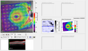
Dehydration Injury to Retina after Macula-On Retinal Detachment Repair - Also Subretinal Perfluoron752 views18 month ago he had a scleral buckle, vitrectomy, laser and gas for macula on retinal detachment. Since he notices blind spots in the center of the left eye/ He also notices a dense, inferior visual field defect. OD 20/20, OS 20/50
|
|
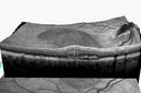
Punctate Inner Choroidopathy - PIC and Choroidal Neovascular Membrane - Right Eye - SD OCT VIDEO TAKES TIME TO LOAD752 views34-year-old woman with a change in vision in the right eye about eight days ago. Her vision was hazy and she saw waviness in the vision. She does have a history of aches and pains and she was diagnosed with Sjogren’s and 2 months ago she had some fevers and some sinus drainage and that went on for about two months. She is a moderate myope. OD 20/25, OS 20/20.
|
|
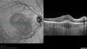
Punctate Inner Choroidopathy - PIC and Choroidal Neovascular Membrane - Right Eye - SD OCT752 views34-year-old woman with a change in vision in the right eye about eight days ago. Her vision was hazy and she saw waviness in the vision. She does have a history of aches and pains and she was diagnosed with Sjogren’s and 2 months ago she had some fevers and some sinus drainage and that went on for about two months. She is a moderate myope. OD 20/25, OS 20/20.
|
|

Intraocular Foreign Body752 viewsIntraocular Foreign Body
|
|
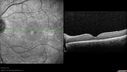
Retinitis Pigmentosa Sine Pigmento or Unknown Dystrophy - Peripheral Superficial White Spots and Recurrent CME752 viewsCME worse - patient received subtenon's kenalog injection right eye.
|
|
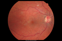
West Nile Virus Multifocal Choroiditis and diabetic retinopathy752 viewsMultifocal and clustered focal lesions are from west nile virus. The retinopathy is probably diabetic but could also be related to west nile virus
|
|

Multifocal Bilateral Central Serous Retinopathy in African American Male752 views67 year old african american male with : Blurred Vision OD. Duration of Problem: 4-6 weeks. VA 20/32 OD 20/20 OS
|
|

Toxoplasmosis - Inner Retinal - Recurrent - Treated with Bactrim DS 751 views43-year-old man has macular toxoplasmosis. He has responded nicely to Bactrim OS: 20/60
|
|

Hypertensive Retinopathy with Choroidal Ischemia Right Eye and Vision 8/200 - Blood Pressure 240/120 mmHg IgA Nephropathy751 views39-year-old man two days ago he noticed poor vision in his right eye OD: 8/200; OS: 20/20. BP is 240/120
|
|

69 Year Old Woman Normal FA751 views69-year-old woman OD 20/50 - OS 20/30 ATYPICAL PHOTOPSIAS
|
|
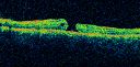
Macular Hole - Traumatic - 11 year old boy (also retinal whitening Berlins Edema)751 views11-year-old boy with a history of blunt trauma to the left eye yesterday. He reports he lost vision immediately but it has gotten somewhat better. He reports he sees “red streamers†and he has very blurry visual acuity., 20/200 OS
1 week post injury and 1 month later (not shown here) mature full thickness macular hole.
|
|

Eccentric Disciform Scar - Breakthrough Vitreous Hemorrhage - InferoTemporal Subretinal Fibrosis - Ectopic Choroidal Neovascular Membranes751 views88-year-old man has noticed for the last few months the vision in the right eye is foggy, like there is a film in the eye or a haze, His vision is 20/16 in each eye.
Right eye - inferotemporal periphery there is a hemorrhage at the equator at about 7:00 o’clock, which is about 4-disc diameters across and adjacent to that there is scarring.
Left eye - large disciform scar inferotemporally
|
|

Microaneurysms from Ocular Ischemic Syndrome (non-diabetic) Right Eye - Left eye CRAO 1 year ago751 views66-year-old woman lost vision in the left eye from a central retinal artery occlusion 1 year ago. Vision OD is fine. OD 20/25, OS 20/50
|
|

Choroidal Melanoma751 views
|
|

Choroidal Melanoma751 viewsChoroidal Melanoma with Retinal Detachment present inferior.
|
|

Retinitis Pigmentosa Sine Pigmento or Unknown Dystrophy - Peripheral Superficial White Spots and Recurrent CME - FA Early751 views46-year-old woman three of her 5 siblings have been diagnosed with retinitis pigmentosa. Her mother and father were seen and as far as everyone can tell, neither of them has it, although there was some thought they might be carriers. They were evaluated twenty five years ago and since then as far as she knows, there has not been a lot of testing done.
She has noticed a substantial decline in vision in both eyes over the last month or two. OD 20/40, OS 20/50. IOP: Both eyes have CME
|
|

Idiopathic Polypoidal Choroidal Vasculopathy - SD-OCT751 views
|
|

Proliferative Diabetic Retinopathy - Moderate - mild NVD with NVE 751 viewsVenous beading, vascular loops and NVE are visible on photos
|
|

Fundus Flavimaculatus - Asymptomatic - Spectral Domain OCT Line Scan751 views45 year old man was told to come in because of ‘white spots on their retina’.
|
|
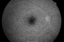
Basal Laminar Drusen - Cuticular Drusen - 19 Year Old with Lupus Nephritis750 views19-year-old. She has a history of developing severe systemic lupus erythematosus in 2001. She was in renal failure and had biopsy proven Class 2 lupus nephritis. She is currently on Plaquenil 400 mg for six years, as well as Prednisolone orally, every other day. OD 20/20, OS 20/25.
|
|
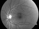
Occult Maculopathy - Thin Fovea on OCT and Normal Color VA, Photos, FA VA 20/80 OU750 views45-year-old man his mother and his maternal grandmother each had vision loss at a relatively young age. Red Free Photo - Normal
|
|

Retinoblastoma - Bilateral - Regressed - Chemotherapy, Thermal Therapy, Cryotherapy750 views10-year-old child was seen in the office on August 6, 2008. She was treated with a combination of chemotherapy at 10 months old as well as thermal therapy and cryotherapy for bilateral retinoblastoma tumors. She reportedly had nine tumors in the left eye and five tumors in the right eye
|
|

Hemi Central Retinal Vein Occlusion 35 year old non-compliant man750 views35-year-old man hazy vision in his left eye for about the last three days. OD 20/20, OS 20/30
|
|

69 Year Old Woman Normal Fluorescein Angiogram750 views69-year-old woman OD 20/50 - OS 20/30 ATYPICAL PHOTOPSIAS
|
|

Giant Retinal Diaylsis Retinal Detachment - Post-op 2 months vision reasonable under silicone oil - Retina Attached750 views2 months post-op with silicone oil in eye.
|
|

Hyperpigmentation of the Retinal Pigment Epithelium Right Eye (Treated wet AMD OS)750 views85-year-old man has wet age-related macular degeneration in the left eye treated for one year most recently one year ago. OD is 20/30, OS is 20/60
|
|
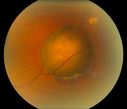
Choroidal Melanoma750 views
|
|

Retinitis Pigmentosa Sine Pigmento or Unknown Dystrophy - Peripheral Superficial White Spots and Recurrent CME750 viewsSubtenon's Kenlalog given right eye today.
|
|

Macular possible Cone Dystrophy - Fundus Autofluorescence750 views
|
|

Acute Branch Retinal Artery Occlusion Left Eye750 viewsThis 84 year old woman had an old BRVO in the right eye and saw a shadow for 2 days in the left eye. She has a superotemporal BRAO. The photos show retinal whitening, the OCT shows inner retinal edema and the FA shows poor circulation.
|
|

Unilateral acute idiopathic maculopathy750 views44 year old man One week ago the left eye was running non-stop and it burned and he had to pry the eye open. Then the vision started to decline three days ago. When he closes the right eye to look at something bright he cannot read. He had a chest infection started about 2-3 weeks ago. He saw a doctor on Monday 4 days ago. He was started on an antibiotic but has not started it yet. He did sore on the right side of his mouth with the infection and he did not have any rashes. He does not have exposure
|
|

Retinal Pigment Epithlial Rip and Good Vision (20/40)749 views82-year-old woman has age-related macular degeneration in the left eye. She developed a pigment epithelial rip in the macula and remarkably her vision is improved with 2 years of treatment. She has had no injections for one year. OS 20/40. (fellow eye has a disciform scar)
|
|

Acute Zonal Occult Outer Retinopathy749 views45-year-old man noticed a few years ago peripheral vision blurriness in the left eye and also some central vision loss. Previous to that, as far as he knows, the two eyes were okay.
VISUAL ACUITY: OD: 20/20; OS: 20/40.
|
|

Anomalous Branch Retinal Artery 749 views59-year-old man OD 20/25, OS 20/25. TORTUOUS RETINAL ARTERY – LEF EYE
(POSSIBLE GROUP 2 WYBURN-MASON SYNDROME
POSSIBLE ACEPHALIC MIGRAINES )
|
|

Retinal Arterial Macroaneurysm - Ruptured and self-sealed after hemorrhage749 views90-year-old woman has vision loss in the right eye for 6 weeks. OD 8/200, OS 20/50. Initial Visit
|
|

Coats' Disease in 14 Year Old Boy Treated with Cryotherapy and Avastin749 views14 Year Old Boy with Coats Disease
|
|

Fundus Flavimaculatus - Asymptomatic - InfraRed Reflectance749 views45 year old man was told to come in because of ‘white spots on their retina’.
|
|

Fundus Flavimaculatus - Asymptomatic - InfraRed Reflectance749 views45 year old man was told to come in because of ‘white spots on their retina’.
|
|

Choroideremia - Female Carrier - Patchy Atrophy of Choroid and RPE - Lyonization749 views
|
|

Acute Posterior Multifocal Placoid Pigment Epitheliopathy - 4 years after onset749 views4 years after presentation - vision 20/80 OD 20/16 OS and there is subretinal fibrosis
|
|

Retinoblastoma - Bilateral - Regressed - Chemotherapy, Thermal Therapy, Cryotherapy748 views10-year-old child was seen in the office on August 6, 2008. She was treated with a combination of chemotherapy at 10 months old as well as thermal therapy and cryotherapy for bilateral retinoblastoma tumors. She reportedly had nine tumors in the left eye and five tumors in the right eye
|
|

26-year-old man choroideremia OU is 20/60748 views
|
|

Purtchers Retinopathy with Bilateral Central Retinal Artery Occlusions 748 views50-year-old man traumatic chest compression and leg crushing injury. OD 1/200, OS 3/200
|
|

Chronic Central Serous Retinopathy treated with Photodynamic Laser and Visudyne748 views48-year-old man has had waxing and waning vision in the left eye for the last year from central serous retinopathy. OD is 20/16, OS is 20/30.
|
|

Retinocytoma - Retinoma - Regressed Retinoblastoma 43 year old male incidental finding 748 views
|
|

Optic Nerve Pit Retinal Detachment - Macular Detachment - 8 Year Old - 5/200 Vision OS748 views8-year-old child on two school exams that his vision was poor in the left eye and his last normal exam was in preschool.
VISUAL ACUITY: OD 20/30, OS 5/200. OS: Vertical C/D ratio is 0.2. There is a whitening on the temporal edge of the optic nerve consistent with an optic disc pit and there is a serous macular detachment.
|
|

Optic Nerve Pit Retinal Detachment - Macular Detachment - 8 Year Old - 5/200 Vision OS748 views8-year-old child on two school exams that his vision was poor in the left eye and his last normal exam was in preschool.
VISUAL ACUITY: OD 20/30, OS 5/200. OS: Vertical C/D ratio is 0.2. There is a whitening on the temporal edge of the optic nerve consistent with an optic disc pit and there is a serous macular detachment.
|
|

Giant Retinal Diaylsis Retinal Detachment - Previous Inflammatory Retinitis - Small Inferior Retinal Holes748 views25-year-old man vision loss for three days. He had a previous episode in this eye in 3 years ago with a strange sort of inflammatory lesion. He had laser in that eye and a pneumatic retinopexy at the time for a retinal detachment. That same area has a giant dialysis retinal detachment. OD 20/400, OS is 20/20
|
|
| 18117 files on 182 page(s) |
 |
|
|
|
|
25 | |
|
|
|
|


