|
|
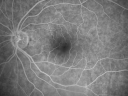
69 Year Old Woman Normal FA741 views69-year-old woman OD 20/50 - OS 20/30 ATYPICAL PHOTOPSIAS
|
|

Retinal Detachment with Subretinal Fibrosis - Chronic with new Foveal Involvement 25 Year Old POST OP741 views25-year-old man vision loss in the left eye for a month. He noticed a shadow up above, which went through his central vision. He has not had any injuries in the left eye.
VISUAL ACUITY: Vision OD is 20/20, OS is 20/400
Post Op photo vision still 20/400
|
|

Choroidal Melanoma - Exudative Retinal Detachment 82 Year Old Man 741 views82-year-old man who had 3 falls 2 weeks ago. After the falls he started checking his vision and noticed there was a veil over the left eye, which he had seen for about two weeks in the superior visual field. OD 20/25, OS 20/40
(patient was medically never well enough for brachytherapy and died 6 months later from heart disease)
|
|

Sectoral Optic Atrophy from an old Branch Retinal Artery Occlusion 741 views74-year-old woman had cataract surgery 3 months ago. Her vision in the right eye has been abnormal since. She notices a shadow in the inferior vision in the right eye. OD is 20/60, OS is 20/50
|
|

Cilioretinal artery occlusion and Ophthalmic Artery Occlusion with Relative Sparing of the Central Retinal Artery741 views80-year-old woman who says she was looking in the mirror, covered her right eye and noticed she could not see well out of the left eye. That just happened yesterday. Her last eye check was a few years ago. She also had a carotid endarterectomy on the right side in 2006 shortly after coronary bypass surgery. OD is 20/40, OS is 20/200.
|
|

Optic Nerve Drusen Both Eyes (Mild) 741 views74-year-old woman has macular edema in both eyes. She also has macular puckers. OD 20/30, OS 20/40.
|
|

Macular Laser Scars - Patient had Central Retinal Vein Occlusion in her only eye741 views80-year-old woman left eye had a vascular occlusion, probably about five years ago, subsequent to that she had macular laser. She said since then the vision has been poor. OS 20/160.
|
|

Branch Retinal Vein Occlusion with NVE (Neovascularization Elsewhere)741 viewsPre and post laser therapy - NV has partially regressed
|
|

Dry AMD - Confluent Drusen Sparing Center of Macula which has Atrophy741 views65 year old woman, VA 20/40 OD; 20/80 OS. The center of the macula has few or no drusen with predominantly non-geographic atrophy
|
|

Fundus Albipunctatus741 views12 year old female with normal vision. She has 4 siblings all of whom have either white spots or spots on IR. Genetic testing by parents was deferred.
|
|

Multifocal Choroiditis - Pneumocystis - aspergillis - cryptococcus741 views31 year old male with anaplastic astrocytoma on chemotherapy with mild vision loss in the right eye. LP did not reveal organism. He was placed on a trial of anti-fungal medications and lost to follow-up
|
|

Chronic Retinal Detachment and Macular hole Pre-Op Post-Op740 views51-year-old woman about 3 or 4 months ago she noticed some flashing lights. Her vision became hazy and dark, especially when she was covering her left eye. OD 2/200, OS 20/20. Post op vision 20/200.
|
|

Bilateral Retinal Arteriol Occlusions - Possible Susac Syndrome 740 views80-year-old woman one month ago had vision loss and a vascular occlusion in the right eye. Vision loss occurred in the left eye about 9 years ago with cotton wool spots. Patients has a 30 year history of tinnitis.
|
|

Stage 2 Macular Hole with 20/70 Vision740 views57-year-old woman blurred central vision in the right eye for about two to three weeks. OD 20/70
|
|

Acute Zonal Occult Outer Retinopathy with Severe Peripheral Vision Loss Left Eye Post Treatment for CME740 views 58-year-old man developed a peripheral vision problem about 25 years ago. OD 20/30, OS 20/40.
|
|

Proliferative Diabetic Retinopathy both Eyes Neovascularization of the Disc740 views63-year-old woman has been diabetic for twenty one years who sees new floaters in the right eye. OD 20/40, OS 20/40.
|
|

Acute Annular Outer Retinopathy for 3 Months - 25 YO Female Fluorescein Angiogram740 views25-year-old female who, about three months ago, put on her sunglasses and noticed a smudge in the vision and at that point she noticed the right eye was hazy. She had a cold a week or two before the vision loss, but she didn’t have any fever. She has no focal neurological problems in her past history. VA 20/20 OU
|
|
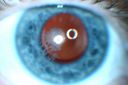
Post Lasik Epithelial Ingrowth740 views58-year-old man had a vitrectomy for a macular pucker in the right eye almost two years ago. His vision was good after his cornea surgeon removed the cells from his flap but then recently the cells have regrown and his vision has declined again.
OD is 20/100.
|
|

Acute Posterior Multifocal Placoid Pigment Epitheliopathy - APMPPE - Indocyanine Green Angiogram740 views 25-year-old man with one and half weeks ago he woke up with night chills. Also around that time his joints started hurting him. He also has headaches, although the headache at this point is not severe. His vision has been changing for about the last week and a half. He sees some spots in his vision and they are blurred spots. They are not in the central vision.
VISUAL ACUITY: OD 20/32, OS 20/32
|
|

Retinitis Pigmentosa Sine Pigmento or Unknown Dystrophy - Peripheral Superficial White Spots and Recurrent CME740 views46-year-old woman three of her 5 siblings have been diagnosed with retinitis pigmentosa. Her mother and father were seen and as far as everyone can tell, neither of them has it, although there was some thought they might be carriers. They were evaluated twenty five years ago and since then as far as she knows, there has not been a lot of testing done.
She has noticed a substantial decline in vision in both eyes over the last month or two. OD 20/40, OS 20/50. IOP: Both eyes have CME
|
|

Fundus Flavimaculatus - Asymptomatic - Fundus Autofluorescence740 views45 year old man was told to come in because of ‘white spots on their retina’. - FAF image shows that spots are strongly autofluorescent
|
|

Travatan Induced CME Left eye740 views63 year old woman 4 years post ECCE and 2 years post PPV for floaters. She developed CME in the left eye after 1 year of travatan. The travatan was stopped and the CME and serous retinal detachment resolved.
|
|

Anomalous Branch Retinal Artery 739 views59-year-old man OD 20/25, OS 20/25. TORTUOUS RETINAL ARTERY – LEF EYE
(POSSIBLE GROUP 2 WYBURN-MASON SYNDROME
POSSIBLE ACEPHALIC MIGRAINES )
|
|

A case of Disc Granuloma-1739 views
|
|

Macular Branch Retinal Vein Occlusions (2) Left Eye - No Macular Edema (yet) 739 views77-year-old woman with two macular branch retinal vein occlusions in the left eye OD 20/25
|
|

Pigmented Choroidal Mass Right Eye - Melanoma vs. Nevus vs. Unilateral Melanocytic Proliferation - 4 Month Follow-Up Images739 views71-year-old man his vision 4 month follow-up. Vision is 20/60 and patient sees flashing lights. Lesion has grown slightly in the superior half of the macula.
|
|

X-linked Juvenille Retinoschisis - Peripheral Retinal Vascular Anomalies - Vitreous Hemorrhage - 8 Year Old Boy739 views8-year-old child OD 20/40, OS 20/50
OD: Vertical C/D ratio is 0.2. There are foveal cysts. There is also a retinal elevation inferiorly and there are patchy peripheral retinal hemorrhages.
OS: Vertical C/D ratio is 0.2. There are foveal cysts. There is peripheral retinal issues with some hemorrhage in some areas of peripheral retinoschisis.
|
|

Primary Ocular Lymphoma - White Mass Left Eye - 112712 739 views
|
|

Primary Ocular Lymphoma - 020513 - FUndus Autofluorescence739 views
|
|

Reticular Macular Disease (Dry Macular Degeneration) - Color Photo739 views85 year old woman with 20/25 vision OU and reticular macular disease
|
|

Acute Macular Neuroretinopathy - (AMN) - Spot in Vision OS - 40 Year old Female739 views
|
|

Adult Vitelliform Macular Dystrophy - Pattern Dystrophy739 views
|
|

Persistent subfoveal fluid following retinal detachment repair739 views62 year old man following pneumatic retinopexy for macula-off retinal detachment. Subfoveal fluid persisted for more than a year.
|
|

Wet AMD Fresh Submacular Hemorrhage - Displaced with Vitrectomy and Subretinal TPA738 views85-year-old woman has wet age-related macular degeneration in her better eye and she was reading with it until two days ago when she had sudden severe vision loss in the eye. Post-surgical displacment vision was 20/120.
|
|

AIDS - HIV Retinopathy - Cotton Wool Spots738 views54-year-old woman HIV positive for 18 years. Her CD4 count is currently less than 50 and she was hospitalized for pneumocystis carinii pneumonia 5 months ago. She has noticed just for the last few weeks a few blind spots in the paracentral vision. Predominantly out of her right eye when she closes her left eye, she sees a few temporal blind spots. VA 20/25 in both eyes.
|
|

Punctate Inner Choroidopathy - PIC and Choroidal Neovascular Membrane - Right Eye - Color Fundus Photograph738 views34-year-old woman with a change in vision in the right eye about eight days ago. Her vision was hazy and she saw waviness in the vision. She does have a history of aches and pains and she was diagnosed with Sjogren’s and 2 months ago she had some fevers and some sinus drainage and that went on for about two months. She is a moderate myope. OD 20/25, OS 20/20.
|
|

Retinal Capillary Hemangioma due to Von Hipple-Lindau Syndrome738 viewsA 47-year old female comes in for a second opinion for retinal capillary Hemangioma in the left eye. VA was HM (hand motion) in the left eye. She has had multiple procedures done for the left eye including, Retinal Detachment surgery, YAG Laser PI, YAG Laser Capsulotomy, and Cataract surgery.
|
|

Lacy Neovascularization of the Disk - Color Photo738 views
|
|

Acute non arteritic anterior ischemic optic neuropathy left eye - Color Photo738 views66 y.o. woman spot in central vision in left eye started about 1 week ago since then it is not changing. VA OD: sc20/25 OS: sc20/200
|
|
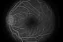
Central Serous Retinopathy737 viewsLater FA
|
|

Stargart's Macular Dystrophy Severe - New Vitreomacular Asymptomatic Vitreomacular Traction OD737 views32-year-old man carries a diagnosis of Stargardt’s disease with poor vision in both eyes since he was 13. He has not noticed any substantial change since he was here 4 years ago.
VISUAL ACUITY: OD 10/200, OS 8/200
|
|

Chronic Central Serous Retinopathy - Gutter in Both Eyes - Treated with PDT - POST PDT Line Scan737 views69-year-old man with blurred vision in the right eye for about three weeks. He had a similar episode about twenty years ago which lasted about six weeks. He is not taking any steroids. He takes Lupron injections for his prostate cancer every three months.
VISUAL ACUITY: Vision OD is 20/80
FU: Treated with PDT after 6 months of observation with success. Treatment was done to area between nerve and fovea at 1/2 fluence.
|
|
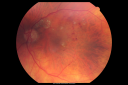
New BRVO Left Eye Old BRVO Right Eye - Left Eye Shows Twig Superior Vein Occlusion with Edema737 views90-year-old man decreased vision maybe one year since cataract surgery he is no longer able to read. OD 20/200, OS 20/200
|
|
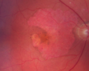
Geographic Atrophy and Age-Related Macular Degeneration - Choriocapillaris is Largely Still Intact737 views85-year-old woman has age-related macular degeneration in both eyes with geographic atrophy. She has noticed her vision getting slightly worse in the past 6 months. VISUAL ACUITY: OD 20/70, OS 20/400
|
|
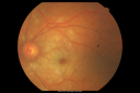
Central Retinal Artery Occlusion Left Eye 2 Weeks Old - Still Non-Perfusion737 views85-year-old woman who had sudden severevision loss in the left eye two weeks ago. she can see a little more centrally and much more in the periphery, since about the last four days.
VISUAL ACUITY: OD 20/30, OS: Hand motion
|
|

Central Retinal Artery Occlusion less than 24 hours - 69 year old man VA light perception737 views
|
|

Hypertensive Choroidopathy - Pheochromocytoma - Chronic Very High Blood Pressure 49 YO Male737 views49-year-old man chronic very high blood pressure sometimes the blood pressure will go as high as 260/160, sometimes it is 200/120 and sometimes it is 160/80. He has a pheochromocytoma on the adrenal gland and he has had vision loss in the right eye starting between three and four years ago.
VISUAL ACUITY: OD 20/60, OS 20/25.
|
|

Myopic Macular Degeneration - Staphyloma - Recent Vision Loss Right Eye - Possible CNVM737 views83-year-old woman is a high myope with myopic macular degeneration. She had a retinal detachment repair in the right eye in 1968. I saw her in June of 2003. She had posterior colobomas and areas of pigment epithelial atrophy. She has noticed over the last six weeks substantial declining vision in the right eye. Prior to that, the vision in the two eyes was about the same. She uses Xalatan in the left eye and Alphagan in both eyes.
VISUAL ACUITY: Vision OD is 20/160, OS is 20/40
|
|

Non-Arteritic Anterior Ischemic Optic Neuropathy - Peripapillary Choroidal Ischemia737 views68-year-old woman noticed central vision loss in the right eye about a week ago. It is getting a little bit better. OD 20/160, OS 20/25.
|
|

Myopic Traction Maculopathy - Vitreoretinal Traction - Possible Occult Neovascular Membrane737 views58-year-old woman was seen in the office on June 30, 2011. In 2002, her vision was 20/30. Subsequent to that she had her left eye made her distance eye, which was dominate. She had LASIK surgery on the right eye to make that her near eye and she was getting along well until about a year ago when she noticed substantial vision loss in the left eye only. VISUAL ACUITY: The near eye is 20/200, but she reads J2. OS 20/100- and J16.
|
|

Acute Annular Outer Retinopathy 737 viewsInitial visit (June 11, 2011): 2 days of vision loss right eye. Wedge suddenly of VA loss OD, too dardkto see through. No flashes or floaters. VA 20/20 OU.
|
|
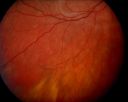
Acute Annular Outer Retinopathy - VA loss 2 days Right Eye Only - 25 Year Old Woman - AZOOR - White Line Around Fresh Lesions737 viewsInitial visit (June 11, 2011): 2 days of vision loss right eye. Wedge suddenly of VA loss OD, too dardkto see through. No flashes or floaters. VA 20/20 OU.
|
|

CRVO (retinal thrombophlebitis)737 views42 yr old female
20/25 VA
Labs normal
|
|

Cilioretinal artery occlusion and Ophthalmic Artery Occlusion with Relative Sparing of the Central Retinal Artery737 views80-year-old woman who says she was looking in the mirror, covered her right eye and noticed she could not see well out of the left eye. That just happened yesterday. Her last eye check was a few years ago. She also had a carotid endarterectomy on the right side in 2006 shortly after coronary bypass surgery. OD is 20/40, OS is 20/200.
|
|

Montage of Hemi-Central Retinal Artery Occlusion737 views
|
|
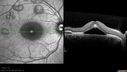
Acute Multifocal Vitelliform Exudative Maculopathy Right Eye At Presentation737 viewsOCT reveals submacular hyperreflective areas between RPE and photoreceptors with hyporeflective subretinal fluid
|
|

Iris Nevus737 viewsPatient came in for cataract evaluation. VA 20/200, right eye, 20/60, left eye. Slit lamp photography shows iris nevus 3-4 mm located nasally at 9-o'clock in the left eye.
|
|

Sclopteria Chorioretinitis - 16 Year Old Girl - Pellet Injury736 views
|
|

Shotgun wound to face with Perforating Pellet - Final Vision 20/400 736 views18-year-old had a gunshot wound to the left eye. He had surgery on December 31st (1 week post injury) and February 4th and his vision has remarkably done well.
VISUAL ACUITY: Vision OS is 3/200.
|
|

Cicatricial ROP with Macular Folds - 60 Year Old Woman Nystagmus736 views60-year-old was born at 6 pounds but then she was incubated and she had difficulty with her eye subsequent to that.
VISUAL ACUITY: OD 8/200, OS 1/200
|
|

DUSN - Diffuse Unilateral Subacute Neuroretinitis - Nematode736 views
|
|
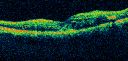
Diabetic Macular Edema and Hypertensive Retinopathy - Circinate Exudate (Ring Exudate) 736 views55-year-old woman diabetic for fifteen years and high blood pressure, especially over the last few months. She has had problems with nosebleeds, headaches, and there has been some difficulty bringing her blood pressure down. She said now the blood pressure is under control but it was running 220/120 mmHg for some time. Her vision in the right eye has been poor for four weeks with a spot in the central vision and both eyes have been blurred.
VISUAL ACUITY: Vision OD is 20/160, OS is 20/60
|
|

Superior Retinal Detachment - Bullous - Macula-off736 views70-year-old man noticed a shadow creeping over the vision in the left eye and he no longer sees well out of that eye. He had cataract surgery two years ago and a YAG capsulotomy six months ago.
VISUAL ACUITY: OD 20/20, OS 1/200
|
|

Dehydration Injury to Retina after Macula-On Retinal Detachment Repair - Also Subretinal Perfluoron736 views18 month ago he had a scleral buckle, vitrectomy, laser and gas for macula on retinal detachment. Since he notices blind spots in the center of the left eye/ He also notices a dense, inferior visual field defect. OD 20/20, OS 20/50
|
|

Uveitis and Retinal Whitening - 04-27-12 - Prior to PPV OD - Possible Primary Ocular Lymphoma 736 views
|
|

Fundus Flavimaculatus - Asymptomatic - Color Photo736 views45 year old man was told to come in because of ‘white spots on their retina’.
|
|

Acute Macular Neuroretinopathy - (AMN) - Spot in Vision OS - 40 Year old Female736 viewsPhotos shows gray area nasal to the fovea
|
|

Choroideremia - Female Carrier - Patchy Atrophy of Choroid and RPE - Lyonization736 views
|
|

Dislocated IOL - Astigmastism - Corrected with new prescription736 views82 year old woman with prior vitrectomy for macular hole - vision dropped from 20/32 to 20/200. Corrected with new rx: 11/2016 old RX OD +0.5 -0.75 x 63; new rx -0.50 -3.00 x 65
|
|

Confluent Soft Drusen with Subetinal Fluid - Right Eye736 viewsAsymptomatic 85 year old man. OCT shows pocket of subfoveal fluid in the center of the macula. This could be mechanical from the drusen preventing the retina from settling down. Testing did not reveal a CNVM and subsequent OCT 6 weeks later showed no change in structure.
|
|

Familial Exudative Vitreoretinopathy - FEVR - Stage 4b OD736 views10 year old child with poor vision OD from birth. The left eye had vascular remodelling in the temporal periphery with preretinal abnormalitlies seen on OCT. The patient never returned for a fluorescein angiogram. Left eye is either stage 1 or stage 2. no family history
|
|

Myopic Macular Degeneration - Dry 735 views55-year-old woman has myopic macular degeneration in both eyes with vision and an Amsler grid change starting a few days ago in the left eye.
VISUAL ACUITY: OD: 20/60. PH: 20/25. OS: 20/200. PH: 20/30.
|
|

Classic Subfoveal Choroidal Neovascular Membrane - 20/400 Vision - 6 - 12 months old lesion735 views78-year-old woman six months or a year ago decreasing vision in the right eye. OD 20/400. Fluorescein angiography shows a classic subfoveal choroidal neovascular membrane in the right eye 3 disc-diameters across, which has brisk leakage. The left eye shows staining drusen.
|
|

Retinal Pigment Epithlial Rip and Good Vision (20/40)735 views82-year-old woman has age-related macular degeneration in the left eye. She developed a pigment epithelial rip in the macula and remarkably her vision is improved with 2 years of treatment. She has had no injections for one year. OS 20/40. (fellow eye has a disciform scar)
|
|

Acute Zonal Occult Outer Retinopathy with Severe Peripheral Vision Loss Left Eye 735 views 58-year-old man developed a peripheral vision problem about 25 years ago. OD 20/30, OS 20/40.
|
|

Retinal Detachment Under Oil with Inferior Retinectomy735 views
|
|

Congenital Toxoplasmosis with non-macular Scars Both Eyes - Recent Reactivation (recurrence) Right Eye735 views34-year-old man has congenital toxoplasmosis with four week history of foggy vision and new floaters in the right eye. OD is 20/40, OS 20/30
|
|
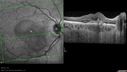
Punctate Inner Choroidopathy - PIC and Choroidal Neovascular Membrane - Right Eye - SD OCT735 views34-year-old woman with a change in vision in the right eye about eight days ago. Her vision was hazy and she saw waviness in the vision. She does have a history of aches and pains and she was diagnosed with Sjogren’s and 2 months ago she had some fevers and some sinus drainage and that went on for about two months. She is a moderate myope. OD 20/25, OS 20/20.
|
|

Sclerosed vein left eye735 views
|
|
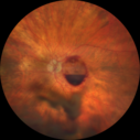
Retinal Arterial Macroaneurysm - Boat Shaped Hemorrhage735 views93 year old woman getting every 2 months Lucentis injection for wet AMD developed a ruptured MA. No treatment was done and the vision improved
|
|

Asteroid Hyalosis - Asymptomatic734 views80-year-old woman her vision is not bothering her, you noticed a problem with the retina. Her left eye tests worse than the right eye today, but she says that it has been like that for a long time and she is not bothered by any floaters or change in central vision.
VISUAL ACUITY: OD is 20/25, OS 20/40
|
|

734 views
|
|

Best Disease for 30 Years - 20/50 Vision both eyes - 61 Year Old Woman734 views61-year-old woman was diagnosed with Best’s disease about 30 years ago. She and her many family members were tested, both at the VA Hospital and then at USF. OD 20/50, OS 20/50
|
|

Retinitis Pigmentosa Newly Diagnosed 55 Year Old734 views55-year-old noticed his vision declining recently, over the last few weeks, particularly in the right eye. He said previous to that the vision was not normal but was pretty good. OD is 20/100, OS is 20/50
OCT SCAN: There is macular thickening in both eyes with intraretinal cysts.
|
|

Choroidal Melanoma734 views
|
|

Polypoidal Choroidal Vasculopathy with Pulsating ICG polyp left eye734 views
|
|

Basal Laminar Drusen - Cuticular Drusen - 19 Year Old with Lupus Nephritis733 views19-year-old. She has a history of developing severe systemic lupus erythematosus in 2001. She was in renal failure and had biopsy proven Class 2 lupus nephritis. She is currently on Plaquenil 400 mg for six years, as well as Prednisolone orally, every other day. OD 20/20, OS 20/25.
|
|

PDR - NVE733 views58-year-old woman has diabetic retinopathy in both eyes with neovascularization of the optic nerve, worse in the right eye than the left eye. She has had panretinal laser in both eyes and vitreous hemorrhage in the left eye. Her vision, since I saw her last in June, is about the same.
VISUAL ACUITY: Vision OD is 20/25, OS is 20/20.
|
|
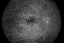
Diabetic and Pseudophakic Cystoid Macular Edema Pretreatment Early FA733 views56-year-old woman has diabetic retinopathy in both eyes and macular edema. She is recently pseudophakic. OD 20/80, OS 20/200.
|
|

Retinal Pigment Epithelial Dysgenesis733 views
|
|

Retinal Arterial Macroaneurysm - Increased Swelling after Laser 733 viewsVisit 2 - 2 months later:
March 21, 2011. This pleasant 66-year-old woman had laser to retinal arterial microaneurysm in the right eye January 12th. Her vision was getting better, but now it is worse the last few days. She is at home taking care of her demented mother and she and her sister lift her mother a fare amount of the time. I also checked her blood pressure, which was 120/65. VISUAL ACUITY: OD 20/200
|
|

Concentric Geograophic Atrophy - Age-related Macular Degeneration - Charles Bonnet Syndrome - Bulls Eye733 views88-year-old woman has age-related with vision gradually declining over the last year. She sees spots in her vision, like red spots and dark spots and she even notices during the daytime, for example when she was looking at her minister, it looked like there were little flowering pots coming out of his head.
VISUAL ACUITY: OD 20/200, OS 20/400.
|
|

Pigmented Choroidal Mass Right Eye - Melanoma vs. Nevus vs. Unilateral Melanocytic Proliferation - Initial Visit733 views71-year-old man is vision has been bothering him for about a month. He does not notice one eye particularly worse than the other. OD is 20/40, OS is 20/25 - Initial Visit
|
|

Calcified Macular Drusen - Age Related Macular Degeneration - Dry733 views74-year-old woman
OD 20/25, OS 20/32
|
|

Reticular Macular Disease (Dry Macular Degeneration) - Infrared reflectance733 views85 year old woman with 20/25 vision OU and reticular macular disease
|
|

Cartwheel CNVM - Classic - wet AMD - evidence of possible old CSR - FA shows cartwheel shaped CNVM733 views75 y.o Decreased Vision right eye for one year. VA OD: sc20/125+2 OS 20/25. Autofluorescence and SD OCT show evidence of old serous retinal detachment superior to the fovea suggesting a history of CSR. The macular lesion on FA shows a classic cartwheel appearance
|
|

Branch Retinal Vein Occlusion (BRVO) no macular edema - cotton wool spot733 views85 year old man with asymptomatic BRVO - his vision stayed great without needing any treatment.
|
|

Vision Loss, Blood, and Cartwheel CNVM OD with no fluid on OCT Scan732 views86-year-old man has had changing vision. He says his vision has been gradually declining since I saw him in June. You saw a hemorrhage in the eye and suggested he comes in here for an evaluation. He is on Combigan and Travatan in the right eye for his glaucoma.
VISUAL ACUITY: Vision OD is 6/200 OS is 8/200 The macula in the right eye is wet even though interestingly the OCT does not show subretinal fluid.
|
|

Gyrate Atrophy - 15 year old male - 20/25 vision both eyes - Positive Family History732 views15 year old male was diagnosed with “retinitis pigmentosa†at age seven. The patient reports that his night vision is poor. He reports he does not notice a visual field change. He reports that his maternal grandfather has lost vision and a cousin has lost vision as well. He reports he has an uncle with some visual changes.
The most significantly affected relative is a 26-year-old legally blind cousin.
20/25+ OD, 20/25 OS.
|
|

Non-Ischemic Central Retinal Vein Occlusion in Young Man (30 years) - Many Cotton Wool Spots732 views30-year-old man with a history of a central retinal vein occlusion for which he was first examined on 2 months ago and his vision then was 4/200. He responded nicely to two Avastin treatments. OD is 20/20, OS is 20/60
|
|
| 18117 files on 182 page(s) |
 |
|
|
|
|
27 | |
|
|
|
|


