|
|

Classic Subfoveal Choroidal Neovascular Membrane - 20/400 Vision - 6 - 12 months old lesion731 views78-year-old woman six months or a year ago decreasing vision in the right eye. OD 20/400. Fluorescein angiography shows a classic subfoveal choroidal neovascular membrane in the right eye 3 disc-diameters across, which has brisk leakage. The left eye shows staining drusen.
|
|

Retinal Pigment Epithelial Dysgenesis731 views
|
|
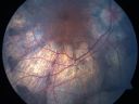
Gyrate Atrophy - 15 year old male - 20/25 vision both eyes - Positive Family History731 views15 year old male was diagnosed with “retinitis pigmentosa†at age seven. The patient reports that his night vision is poor. He reports he does not notice a visual field change. He reports that his maternal grandfather has lost vision and a cousin has lost vision as well. He reports he has an uncle with some visual changes.
The most significantly affected relative is a 26-year-old legally blind cousin.
20/25+ OD, 20/25 OS.
|
|

ECTOPIC DISCIFORM SCAR/PERIPHERAL CHOROIDAL NEOVASCULAR MEMBRANE – EXUDATE –INFLAMMATION IN REACTION TO THE PERIPHERAL VASCULAR LESION 731 views53-year-old woman has noticed some blurred vision in the right eye for a few years. In retrospect, she thinks it is possible since she was a teenager the right eye was always the weaker eye and that the left eye has always been the better eye. VISUAL ACUITY: Vision OD is 20/400
|
|

Acute Posterior Multifocal Placoid Pigment Epitheliopathy - APMPPE - Fluorescein Angiogram731 views 25-year-old man with one and half weeks ago he woke up with night chills. Also around that time his joints started hurting him. He also has headaches, although the headache at this point is not severe. His vision has been changing for about the last week and a half. He sees some spots in his vision and they are blurred spots. They are not in the central vision.
VISUAL ACUITY: OD 20/32, OS 20/32
|
|
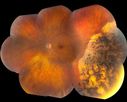
Pigmented Peripheral Retinal Degeneration731 views42-year old male comes in for routine eye exam and to follow up on peripheral retinal degeneration in both eyes. VA is 20/20, right eye and 20/25, left eye. Patient is asymptomatic with no visual complaints.
|
|

Macular Atrophy - Phototoxicity - Macular Hole Surgery - FAF image731 views25 years ago patient suffered immediate severe vision loss following macular hole surgery. You can almost see light pipe tracks in the atrophy in the macula. Vision is 5/200
|
|

Branch Retinal Vein Occlusion (BRVO) no macular edema - cotton wool spot731 views85 year old man with asymptomatic BRVO - his vision stayed great without needing any treatment.
|
|

Cystoid Macular Edema - Xalatan - ECCE was done 20 years previously730 views71 Year old woman who noticed about a month ago the central vision in the right eye fogged up. OD 20/200 POSSIBLE XALATAN INDUCED CYSTOID MACULAR EDEMA
|
|

Serous Macular Detachment Left Eye - Possible Hypertensive Choroidopathy v. Central Serous Retinopathy730 views59-year-old woman decreasing vision in the left eye for about two days. She sees a spot in front of her vision. It is a yellow spot which is just straight ahead and moves with her eye. She does have high blood pressure. She checks her blood pressure at home, and even at home her blood pressure runs very high, with the systolic running around 200 and the diastolic around 100, although occasionally it does return to the normal range.
VISUAL ACUITY: Vision OD is 20/60, OS is 20/30
|
|
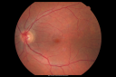
Hypertensive Choroidopathy - Pheochromocytoma - Chronic Very High Blood Pressure 49 YO Male730 views49-year-old man chronic very high blood pressure sometimes the blood pressure will go as high as 260/160, sometimes it is 200/120 and sometimes it is 160/80. He has a pheochromocytoma on the adrenal gland and he has had vision loss in the right eye starting between three and four years ago.
VISUAL ACUITY: OD 20/60, OS 20/25.
|
|

Retinitis Pigmentosa 32 Year Old Woman - 20/25 vision both eyes730 views32-year-old woman was seen in the office on 6/26/2009. She has had retinitis pigmentosa since 2003. She has difficulty with night vision but overall she is doing pretty well.
Vision OU is 20/25.
|
|

Vision Loss, Blood, and Cartwheel CNVM OD with no fluid on OCT Scan730 views86-year-old man has had changing vision. He says his vision has been gradually declining since I saw him in June. You saw a hemorrhage in the eye and suggested he comes in here for an evaluation. He is on Combigan and Travatan in the right eye for his glaucoma.
VISUAL ACUITY: Vision OD is 6/200 OS is 8/200 The macula in the right eye is wet even though interestingly the OCT does not show subretinal fluid.
|
|

Concentric Annular Macular Dystrophy 4/200 2/200 vision 61 Year Old Macular Crystals730 views61-year-old woman has concentric annular macular dystrophy. She has had vision loss for 10 years who is no longer able to function even with the magnifiers and vision aids that she has. Her brother who is ten years younger than her, is recently diagnosed with the same macular dystrophy and her father and grandfather may have had similar problems in the past.
VISUAL ACUITY: OD 4/200, OS 2/200.
|
|

Concentric Annular Macular Dystrophy 4/200 2/200 vision 61 Year Old Macular Crystals730 views61-year-old woman has concentric annular macular dystrophy. She has had vision loss for 10 years who is no longer able to function even with the magnifiers and vision aids that she has. Her brother who is ten years younger than her, is recently diagnosed with the same macular dystrophy and her father and grandfather may have had similar problems in the past.
VISUAL ACUITY: OD 4/200, OS 2/200.
|
|

Non-Ischemic Central Retinal Vein Occlusion in Young Man (30 years) - Many Cotton Wool Spots730 views30-year-old man with a history of a central retinal vein occlusion for which he was first examined on 2 months ago and his vision then was 4/200. He responded nicely to two Avastin treatments. OD is 20/20, OS is 20/60
|
|

Non-Ischemic Central Retinal Vein Occlusion in Young Man (30 years) - Many Cotton Wool Spots730 views30-year-old man with a history of a central retinal vein occlusion for which he was first examined on 2 months ago and his vision then was 4/200. He responded nicely to two Avastin treatments. OD is 20/20, OS is 20/60
|
|

Swollen Nerve with Hemorraghe730 views
|
|
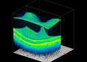
3-D OCT Image of Tractional Maculopathy730 views
|
|
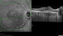
Punctate Inner Choroidopathy - PIC and Choroidal Neovascular Membrane - Right Eye - SD OCT730 views34-year-old woman with a change in vision in the right eye about eight days ago. Her vision was hazy and she saw waviness in the vision. She does have a history of aches and pains and she was diagnosed with Sjogren’s and 2 months ago she had some fevers and some sinus drainage and that went on for about two months. She is a moderate myope. OD 20/25, OS 20/20.
|
|

Panretinal (Scatter) Photocoagulation (Fresh)730 views
|
|

Diabetic Retinopathy730 viewsAnnual diabetic exam. VA 20/30, right eye and 20/50, left eye. Fundus photos shows hemorrhages with cotton wool spots. Exudates are visible around the macula in the right eye. Scattered hemorrhages in the left eye.
|
|

Large Retinal Detachment with PVR730 viewsPatient with poor vision in the left eye. Fundus photography shows rhegamatogenous retinal detachment with proliferative vitreoretinopathy in the left eye. Silicone oil is also present in the left eye.
|
|

Acute Macular Neuroretinopathy - (AMN) - Spot in Vision OS - 40 Year old Female730 views
|
|

Dislocated IOL - Astigmastism - Corrected with new prescription730 views82 year old woman with prior vitrectomy for macular hole - vision dropped from 20/32 to 20/200. Corrected with new rx: 11/2016 old RX OD +0.5 -0.75 x 63; new rx -0.50 -3.00 x 65
|
|

Idiopathic Central Serous Retinopathy - Bilateral - Acute 729 views49-year-old man decreasing vision in the left eye for one week. He is not taking any steroid or steroid-related products. His blood pressure as far as he knows is under control.
VISUAL ACUITY: OD 20/30, OS 20/400
|
|

Macular Hole - Traumatic - 11 year old boy (also retinal whitening Berlins Edema)729 views11-year-old boy with a history of blunt trauma to the left eye yesterday. He reports he lost vision immediately but it has gotten somewhat better. He reports he sees “red streamers†and he has very blurry visual acuity., 20/200 OS
|
|

Gyrate Atrophy - 15 year old male - 20/25 vision both eyes - Positive Family History729 views15 year old male was diagnosed with “retinitis pigmentosa†at age seven. The patient reports that his night vision is poor. He reports he does not notice a visual field change. He reports that his maternal grandfather has lost vision and a cousin has lost vision as well. He reports he has an uncle with some visual changes.
The most significantly affected relative is a 26-year-old legally blind cousin.
20/25+ OD, 20/25 OS.
|
|

Retinal Arterial Macroaneurysm - Increased Swelling after Laser 729 views65-year-old woman was seen in the office on January 12, 2011. She has noticed decreased vision in the right eye for the last two or three weeks. She has had some discomfort in both eyes as well. She has a history of low blood pressure, but she does have high cholesterol.
VISUAL ACUITY: OD 20/60
|
|

Hyperpigmentation of the Retinal Pigment Epithelium in Dry AMD729 views87-year-old woman has wet age-related macular degeneration in the left eye. Vision OD is 20/25, OS is 20/400
|
|
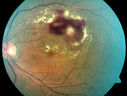
Retinal Arterial Macroaneurysm - Untreated Hypertension (250/135) - Serous Macular Detachment729 views82 year old asian woman. VA 20/80 Headaches and vision loss for 1 month. Negative medical history but has not been to the doctor for years. (no insurance)
|
|

Macular possible Cone Dystrophy - Fundus Autofluorescence729 views
|
|
729 views
|
|

Moderate PDR729 views43 year old man 20/32 vision in both eyes. Being started on Anti-VEGF injections which will be followed by PRP.
|
|

Diabetic and Pseudophakic Cystoid Macular Edema Pretreatment early FA728 views56-year-old woman has diabetic retinopathy in both eyes and macular edema. She is recently pseudophakic. OD 20/80, OS 20/200.
|
|

Fundus Autofluorescence - Enhanced S Cone Syndrome - Goldmann Favre - 728 views55-year-old woman while in college her vision was poor even with glasses and she sought evaluation for that. She was told after she had an electroretinogram at USF 15 years ago, that she had something with her blue cones. She does have poor night vision, but her reading vision is pretty good.
VISUAL ACUITY: OD 20/40, OS 20/40
|
|

Toxoplasmosis Macular Scar (acquired not congenital) 31 Year Old Man- Recent Vision Change - Creeping Scar728 views31-year-old man had an episode of toxoplasmosis in the left eye, where unfortunately it adversely affected his central vision that was back in 2004 (7 years ago)
He did notice recently a change in his visual function. He plays softball, he was getting hit by the ball and having trouble seeing and he said that is how he noticed the toxoplasmosis when it first occurred and he was concerned it might have recurred.
OD 20/16, OS 20/80.
|
|

Punctate Inner Choroidopathy - Recurrent CNVM 4 years after initial treatment Left Eye 728 viewsJune 7, 2012. This pleasant 35-year-old woman noticed decreasing vision in the left eye recently. She returns now noticing the vision in the left eye is declining. OD 20/50, OS 20/80. OS: Vertical C/D ratio is 0.3. There is a scar just inferonasal to the fovea, which has grown some towards the fovea with a little blood just under the fovea.
|
|

Elevated Nevus728 views
|
|
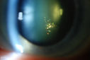
Polychromatic "Christmas Tree" Cataract728 viewsPatient comes in for annual eye exam. Eval on cataracts. VA is 20/20, right eye, 20/40, left eye. Slit Lamp exam shows polychromatic christmas tree cataract in the left eye.
|
|

PDR - Mild Vitreous Hemorrhage and Good Vision727 views58-year-old woman has diabetic retinopathy in both eyes with neovascularization of the optic nerve, worse in the right eye than the left eye. She has had panretinal laser in both eyes and vitreous hemorrhage in the left eye. Her vision, since I saw her last in June, is about the same.
VISUAL ACUITY: Vision OD is 20/25, OS is 20/20.
|
|

Central Serous Retinopathy - Acute727 views49-year-old man sees a spot in the central vision with a red halo for 2 weeks OD is 20/50, OS is 20/20
|
|
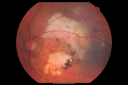
Disciform Macular Scar Both Eyes from Wet Macular Degeneration727 views86-year-old woman has disciform scars OD: 20/400; OS: 20/100
|
|

Retrobulbar Needle Stick Macular Left Eye with Scar - Anesthetic Complication727 views79-year-old woman has a macular pucker in the right eye. She also has a macular scar in the left eye from a retrobulbar needle stick. OD 20/80
|
|

Retinal Detachment and Macular Pucker 727 views76-year-old man has had a retinal detachment repair in the right eye. His vision was improving until just on the way over today and then his vision declined.
VISUAL ACUITY: OD 1/200
|
|

Pigmented Choroidal Mass Right Eye - Melanoma vs. Nevus vs. Unilateral Melanocytic Proliferation - 4 Month Follow-Up Images727 views71-year-old man his vision 4 month follow-up. Vision is 20/60 and patient sees flashing lights. Lesion has grown slightly in the superior half of the macula.
|
|

Giant Retinal Diaylsis Retinal Detachment - Post-op 2 months vision reasonable under silicone oil - Retina Attached727 views2 months post-op with silicone oil in eye.
|
|

Traumatic Vitreous Hemorrhage - Firecracker in Face - Teenager727 viewsSixteen year-old with traumatic vitreous hemorrhage following injury from firecracker exploding in his face.
|
|

Punctate Inner Choroidopathy - Recurrent CNVM 4 years after initial treatment Left Eye 727 viewsJune 7, 2012. This pleasant 35-year-old woman noticed decreasing vision in the left eye recently. She returns now noticing the vision in the left eye is declining. OD 20/50, OS 20/80. OS: Vertical C/D ratio is 0.3. There is a scar just inferonasal to the fovea, which has grown some towards the fovea with a little blood just under the fovea.
|
|

Idiopathic Polypoidal Choroidal Vasculopathy - IR727 views
|
|

Fundus Flavimaculatus - Asymptomatic - Color Photo727 views45 year old man was told to come in because of ‘white spots on their retina’.
|
|

Self Sealed Retinal Arteriol Macroaneurysm 727 viewsFA on presentation showed minimal to no active leakage. Also, there is a small cuff of blood around the MA which is favorable for closure
|
|

Multifocal Choroiditis - Linear Scars - Schlaegel lines727 views66 year old woman with macular pucker right eye. Images show typical linear peripheral scars for MFC or POHS - this is unilateral.
|
|
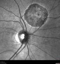
Congenital Hypetrophy of the Retinal Pigment Epithelium (CHRPE)727 views63 year old female with normal vision and CHRPE lesion in the right eye.
|
|

Cystoid Macular Edema - Xalatan - ECCE was done 20 years previously726 views71 Year old woman who noticed about a month ago the central vision in the right eye fogged up. OD 20/200 POSSIBLE XALATAN INDUCED CYSTOID MACULAR EDEMA
|
|

Acute Zonal Occult Outer Retinopathy with Severe Peripheral Vision Loss Left Eye 726 views 58-year-old man developed a peripheral vision problem about 25 years ago. OD 20/30, OS 20/40.
|
|

Retrobulbar Needle Stick Macular Left Eye with Scar - Anesthetic Complication726 views79-year-old woman has a macular pucker in the right eye. She also has a macular scar in the left eye from a retrobulbar needle stick. OD 20/80
|
|

Hypertensive Retinopathy with Choroidal Ischemia Right Eye and Vision 8/200 - Blood Pressure 240/120 mmHg IgA Nephropathy726 views39-year-old man two days ago he noticed poor vision in his right eye OD: 8/200; OS: 20/20. BP is 240/120
|
|

Vitreomacular Traction - Macular Pucker - 20/50 Vision726 views75-year-old woman decreasing vision in the right eye a few months ago. She had her cataract removed and unfortunately her vision is still hazy in the right eye. Her left eye seems a little hazy from the cataract.
VISUAL ACUITY: OD 20/50, OS 20/20. IOP: OD 17, OS 18.
SLIT-LAMP EXAMINATION
|
|

Plaquenil (Hydroxycholorquine) Toxicity - Bull's Eye Maculopathy - 73 year old womand 25 Years therapy - 64 inches tall - 110 lbs726 views73-year-old woman has been on Plaquenil for twenty five years. She noticed loss of paracentral vision a few months ago. VISUAL ACUITY: OD 20/70, OS 20/30. Her color vision was 0 out 10 correct in both eyes and she also missed the controls.
|
|

Retinal Arterial Macroaneurysm - Ruptured and self-sealed after hemorrhage726 views90-year-old woman has vision loss in the right eye for 6 weeks. OD 8/200, OS 20/50. Initial Visit
|
|

Swollen Nerve with Hemorraghe726 views
|
|
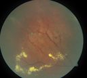
periphery 2. od726 viewsleaking miliary aneurysms
|
|

Central Serous Retinopathy - Acute725 views49-year-old man sees a spot in the central vision with a red halo for 2 weeks OD is 20/50, OS is 20/20
|
|

725 views
|
|
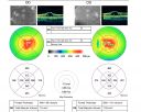
X-Linked Juvenille Retinoschisis 10 Year Old Child treated with Trusopt725 views10-year-old child has x-linked juvenile retinoschisis VA 20/60, OS is 20/100. Macula and Vision improved with Trusopt drops.
|
|

Multiple Evanescent White Dot Syndrome - Acute Idiopathic Blind Spot Enlargement725 views33-year-old woman one week ago she awoke and saw a red spots flashing out of her right eye. She notices them to the right side, which is the temporal visual field out of the right eye, then they changed to bursts of white lights without colors and then she notices straight ahead a couple spots. She has headaches in the back of her head. OD 20/20, OS 20/20
|
|

Proliferative Diabetic Retinopathy both Eyes Neovascularization of the Disc725 views63-year-old woman has been diabetic for twenty one years who sees new floaters in the right eye. OD 20/40, OS 20/40.
|
|

Pigment Epithelial Detachment with Subretinal Fluid and Exudate 20/400 Vision725 views87-year-old woman has wet age-related macular degeneration in the left eye. She had treatment up north but there has been a gap in therapy and the vision in the left eye is poor. Vision OD is 20/25, OS is 20/400
|
|

Concentric Annular Macular Dystrophy 4/200 2/200 vision 61 Year Old Macular Crystals725 views61-year-old woman has concentric annular macular dystrophy. She has had vision loss for 10 years who is no longer able to function even with the magnifiers and vision aids that she has. Her brother who is ten years younger than her, is recently diagnosed with the same macular dystrophy and her father and grandfather may have had similar problems in the past.
VISUAL ACUITY: OD 4/200, OS 2/200.
|
|
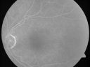
Microaneurysms from Ocular Ischemic Syndrome (non-diabetic) Right Eye - Left eye CRAO 1 year ago725 views66-year-old woman lost vision in the left eye from a central retinal artery occlusion 1 year ago. Vision OD is fine. OD 20/25, OS 20/50
|
|

Central Retinal Vein Occlusion and Tractional Retinal Detachment for 3 Years (Patient is Diabetic)725 views67-year-old woman has severe vision loss in the left eye for 3 years OD 20/32, OS 3/200
|
|

Angioid Streaks - Peau D'Orange - Pseudoxanthoma Elasticum - Asymptomatic Extrafoveal Choroidal Neovascular Membrane725 views60-year-old woman has pseudoxanthoma elasticum since 2008. Her sister has the same problem and lost vision in both eyes. She had a problem with blood vessel growth in the left eye for which she had injections of Avastin in August, September, and October of 2011 (5 months ago). Subsequent to that, the macula has been dry and you have just been watching her carefully. She is in Florida for a short time but will ultimately be spending nine months out of the year here. Both of her eyes have good vision at
|
|

Optic Nerve Drusen725 views
|
|

Acute Non-arteritic Anterior Ischemic Optic Neuropathy Right Eye - Previous NA-AION left eye 725 views69-year-old man previously lost vision in the left eye from nonarteritic anterior ischemic optic neuropathy in 2006. About a week ago and noticed blurred vision in the inferonasal part of the vision in the right eye.
VISUAL ACUITY: Vision OD is 20/16, OS is 20/40
|
|

Toxoplasmosis - Vitreoretinal Traction725 viewsA 28-year old male who noticed an upper partial visual loss to his right eye for about 2-weeks. Diagnosed with retinitis and was put on acyclovir. Patient received second opinion due to culture of aqueous humor which was then diagnosed as Toxoplasmosis. VA was 20/25 in the right eye. Pt has been followed after for the past 7-months.
|
|

Primary Ocular Lymphoma - White Mass Left Eye - 112712 725 views
|
|

Stasis Retinopathy, Central Retinal Vein Occlusion725 viewsYoung male who complains of decreased vision in the left eye. VA is 20/20 in the left eye. Fundus photo shows mild central retinal vein occlusion in the left eye.
|
|

Basal Laminar Drusen - Cuticular Drusen - 19 Year Old with Lupus Nephritis724 views19-year-old. She has a history of developing severe systemic lupus erythematosus in 2001. She was in renal failure and had biopsy proven Class 2 lupus nephritis. She is currently on Plaquenil 400 mg for six years, as well as Prednisolone orally, every other day. OD 20/20, OS 20/25.
|
|

Retinal Vasculopathy associated with Facioscapulohumeral Muscular Dystrophy line os724 views59-year-old woman had a blood pressure spike to 170/90 and was on medications for that. She does have facioscapulohumeral muscular dystrophy, which runs in her family. She has a cousin with the same disorder.
VISUAL ACUITY: Her vision is 20/25 OU. IOP: OD 21, OS 18.
There is no posterior vitreous separation, telangiectatic vessels near the macula. There are patchy retinal hemorrhages. There is no edema or proliferation.
|
|

Multiple Evanescent White Dot Syndrome - Atypical Yellow Foveal Spot Right Eye724 views12-year-old had decreased vision in the left eye from reported amblyopia since around the year 2000. His right eye was doing fine until two days ago, when he had sudden severe vision loss. He sees a blurred spot in the middle of the vision in the right eye wherever he looks.
OD 20/60 with eccentric movement, OS 20/200. Pinhole 20/70. IOP: OD 14, OS 10.
|
|
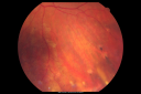
Macular Scars - Laser (1980s) Ocular Histoplasmosis724 views70 year old man who had laser at Wills, Wilmer and the Mayo clinic before better therapies were available for ocular histoplasmosis related CNVM
|
|

Central Retinal Vein Occlusion and Laser Induced Chorioretinal Anastomosis724 views84-year-old woman has age-related macular degeneration in both eyes with foveal atrophy. She also has a central retinal vein occlusion
in the left eye for which she had intravitreal Kenalog and finally chorioretinal anastomosis laser a year
ago. VISUAL ACUITY: Vision OD is 20/160, OS is 7/200
|
|

Pigmented Choroidal Mass Right Eye - Melanoma vs. Nevus vs. Unilateral Melanocytic Proliferation - 4 Month Follow-Up Images724 views71-year-old man his vision 4 month follow-up. Vision is 20/60 and patient sees flashing lights. Lesion has grown slightly in the superior half of the macula.
|
|

Ocular Histoplasmosis both Eyes - Laser Right Eye Only InfraRed724 views58-year-old woman who lost vision as a child in the left eye from ocular histoplasmosis. She used to play in her attic and she was told by her pulmonary doctor that is probably where she picked it up. She had laser in the right eye for a leaky lesion back in 1990. OD 20/25, OS 20/200.
|
|
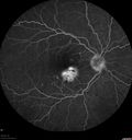
Punctate Inner Choroidopathy - PIC and Choroidal Neovascular Membrane - Right Eye - Fluorescein Angiogram724 views34-year-old woman with a change in vision in the right eye about eight days ago. Her vision was hazy and she saw waviness in the vision. She does have a history of aches and pains and she was diagnosed with Sjogren’s and 2 months ago she had some fevers and some sinus drainage and that went on for about two months. She is a moderate myope. OD 20/25, OS 20/20.
|
|

Reticular Macular Disease (Pseudo-drusen) Both Eyes - Occult CNVM and Macular Edema Right Eye 724 viewsColor Fundus Photo
|
|

Acute Macular Neuroretinopathy - (AMN) - Spot in Vision OS - 40 Year old Female724 viewsICG angiogram is normal
|
|

Occult Macular Dystrophy - 9 Years later 724 views53 year old man. Vision is better at 20/50 OU
|
|
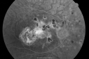
Multifocal Choroiditis and Subretinal Fibrosis - 32 yo Female Old Scar OD723 views32-year-old woman vision loss in the right eye associated with macular scarring and multifocal choroiditis in 1999 with new vision loss in left eye: OD 20/400, OS 20/50.
|
|

Diabetic Vitreous Hemorrhage Left Eye723 views35-year-old man diabetic for the last 25 years. His hemoglobin A1C has been running between 10 and 11. He was doing fine and then when he woke up from sleeping last night he noticed sudden vision loss in his left eye OD 20/30, OS 20/200.
|
|

Acute Zonal Occult Outer Retinopathy with Severe Peripheral Vision Loss Left Eye Post Treatment for CME723 views 58-year-old man developed a peripheral vision problem about 25 years ago. OD 20/30, OS 20/40.
|
|

Chronic Central Serous Retinopathy treated with Photodynamic Laser and Visudyne723 views48-year-old man has had waxing and waning vision in the left eye for the last year from central serous retinopathy. OD is 20/16, OS is 20/30.
|
|

69 Year Old Woman Normal FA723 views69-year-old woman OD 20/50 - OS 20/30 ATYPICAL PHOTOPSIAS
|
|

Progressive Outer Retinal Necrosis 77 Year Old Woman with CLL (Acute Retinal Necrosis) (PORN - ARN)723 views77-year-old woman with CLL who had shingles on the left side of her face about 6 weeks ago then she developed a dendrite in the cornea which was treating about four weeks ago. She noticed severe vision loss in the left eye just a few days ago and you saw retinitis and she comes in because of that. Vision OD is 20/25, OS is hand motion
|
|

Optic Atrophy Left Eye and Benign Pituitary Tumor - VA 20/20 Right Eye 20/160 Left Eye723 views58-year-old man has had gradually declining vision in the left eye over about the last 18 months. Vision OD is 20/20, OS is 20/160
OCT SCAN: The OCT scan of the nerve fiber layer in the right eye shows a thickness of 88 microns, the left eye 59 microns.
MRI did show a suprasellar mass which is affecting the optic chiasm and is responsible for the vision loss. The tumor was resected and was a benign pituitary tumor.
|
|

Proliferative Sickle Retinopathy - Hemoglobin SC723 views53-year-old woman with a new spider-like floater in the right eye for about six days. She sees one big floater and another little one. She does have hemoglobin SC hemoglobinopathy. VISUAL ACUITY: Vision OD is 20/20; OS is 20/50, PH is 20/20.
|
|

Best Vettiliform Dystrophy with CNVM-1723 views
|
|

Retinal Detachment723 views80 yr old Male
Wet AMD c/o sudden loss of VA
|
|

Sectoral Optic Atrophy from an old Branch Retinal Artery Occlusion 723 views74-year-old woman had cataract surgery 3 months ago. Her vision in the right eye has been abnormal since. She notices a shadow in the inferior vision in the right eye. OD is 20/60, OS is 20/50
|
|
| 18117 files on 182 page(s) |
 |
|
|
|
|
28 | |
|
|
|
|


