|
|

New Subfoveal Classic CNVM OS - Wet AMD - Macular Scar Congenital Toxoplasmosis Right Eye 707 views77-year-old woman as had poor vision in the right eye since birth from congenital toxoplasmosis of her macula in the right eye. Two to three weeks ago when she noticed vision loss in the left eye. VISUAL ACUITY: OD 2/200, OS 20/200.
|
|

AIDS - HIV Retinopathy - Cotton Wool Spots707 views54-year-old woman HIV positive for 18 years. Her CD4 count is currently less than 50 and she was hospitalized for pneumocystis carinii pneumonia 5 months ago. She has noticed just for the last few weeks a few blind spots in the paracentral vision. Predominantly out of her right eye when she closes her left eye, she sees a few temporal blind spots. VA 20/25 in both eyes.
|
|
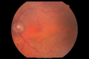
Post-operative photography - vision has improved to 20/50 1 year later.707 views1 year after scleral buckle vision is 20/50 and patient has a cataract.
|
|

Dehydration Injury to Retina after Macula-On Retinal Detachment Repair - Also Subretinal Perfluoron707 views18 month ago he had a scleral buckle, vitrectomy, laser and gas for macula on retinal detachment. Since he notices blind spots in the center of the left eye/ He also notices a dense, inferior visual field defect. OD 20/20, OS 20/50
|
|
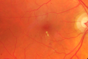
Diabetic Macular Edema - Mild707 views70 year old man with mild edema did well without treatment
|
|

Retinitis Pigmentosa - Autosomal Dominant - Vision Loss at Young Age - Fundus Photo707 views 54-year-old woman has autosomal dominant retinitis pigmentosa. Her mother and grandmother both had it and she has one son who has it and one daughter who doesn’t. She also has a sister with it. She lost vision since she was younger. She had cataract surgery done in January and February of this year. She has been seeing unusual flashing lights throughout the periphery and central vision for about the last year or two.
VISUAL ACUITY: OD 20/160, OS 20/160
|
|

oct fovea. od707 views
|
|
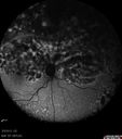
Primary Ocular Lymphoma - 020513 - FUndus Autofluorescence707 views
|
|

Basal Laminar Drusen - Cuticular Drusen - 19 Year Old with Lupus Nephritis706 views19-year-old. She has a history of developing severe systemic lupus erythematosus in 2001. She was in renal failure and had biopsy proven Class 2 lupus nephritis. She is currently on Plaquenil 400 mg for six years, as well as Prednisolone orally, every other day. OD 20/20, OS 20/25.
|
|
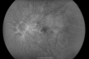
Myopic Macular Degeneration - Dry 706 views55-year-old woman has myopic macular degeneration in both eyes with vision and an Amsler grid change starting a few days ago in the left eye.
VISUAL ACUITY: OD: 20/60. PH: 20/25. OS: 20/200. PH: 20/30.
|
|

Central Retinal Artery Occlusion Left Eye 2 Weeks Old - Still Non-Perfusion706 views85-year-old woman who had sudden severevision loss in the left eye two weeks ago. she can see a little more centrally and much more in the periphery, since about the last four days.
VISUAL ACUITY: OD 20/30, OS: Hand motion
|
|

Concentric Geograophic Atrophy - Age-related Macular Degeneration - Charles Bonnet Syndrome - Bulls Eye706 views88-year-old woman has age-related with vision gradually declining over the last year. She sees spots in her vision, like red spots and dark spots and she even notices during the daytime, for example when she was looking at her minister, it looked like there were little flowering pots coming out of his head.
VISUAL ACUITY: OD 20/200, OS 20/400.
|
|
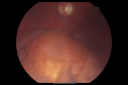
Choroidal Melanoma - Exudative Retinal Detachment 82 Year Old Man 706 views82-year-old man who had 3 falls 2 weeks ago. After the falls he started checking his vision and noticed there was a veil over the left eye, which he had seen for about two weeks in the superior visual field. OD 20/25, OS 20/40
(patient was medically never well enough for brachytherapy and died 6 months later from heart disease)
|
|

Proliferative Diabetic Retinopathy and Mild Vitreous Hemorrhage706 views58-year-old woman has diabetic retinopathy in both eyes with neovascularization of the optic nerve, worse in the right eye than the left eye. Vision OD is 20/25, OS is 20/20
|
|

Basal Laminar Drusen - Cuticular Drusen - Equatorial Drusen - 70 Year Old Woman - Fluorescein Angiogram Montage Left Eye706 views70 year old woman has had macular drusen since her thirties and she said her sister has a similar problem. Recently the vision in the left eye has substantially declined. OD 20/16, OS 10/200.
|
|

Fluorescein Angiogram - Enhanced S Cone Syndrome - Goldmann Favre - 706 views55-year-old woman while in college her vision was poor even with glasses and she sought evaluation for that. She was told after she had an electroretinogram at USF 15 years ago, that she had something with her blue cones. She does have poor night vision, but her reading vision is pretty good.
VISUAL ACUITY: OD 20/40, OS 20/40
|
|

Non-Ischemic Central Retinal Vein Occlusion in Young Man (30 years) - Many Cotton Wool Spots706 views30-year-old man with a history of a central retinal vein occlusion for which he was first examined on 2 months ago and his vision then was 4/200. He responded nicely to two Avastin treatments. OD is 20/20, OS is 20/60
|
|

Congenital Achromatopsia 706 views26-year-old man was diagnosed with congenital achromatopsia by ERG as a child. He has family members who also have this disorder. OD 20/200, OS 20/200.
|
|

Cilioretinal artery occlusion and Ophthalmic Artery Occlusion with Relative Sparing of the Central Retinal Artery706 views80-year-old woman who says she was looking in the mirror, covered her right eye and noticed she could not see well out of the left eye. That just happened yesterday. Her last eye check was a few years ago. She also had a carotid endarterectomy on the right side in 2006 shortly after coronary bypass surgery. OD is 20/40, OS is 20/200.
|
|
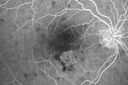
Punctate Inner Choroidopathy - PIC and Choroidal Neovascular Membrane - Right Eye - Fluorescein Angiogram706 views34-year-old woman with a change in vision in the right eye about eight days ago. Her vision was hazy and she saw waviness in the vision. She does have a history of aches and pains and she was diagnosed with Sjogren’s and 2 months ago she had some fevers and some sinus drainage and that went on for about two months. She is a moderate myope. OD 20/25, OS 20/20.
|
|

Punctate Inner Choroidopathy - Recurrent CNVM 4 years after initial treatment Left Eye - FAF706 viewsJune 7, 2012. This pleasant 35-year-old woman noticed decreasing vision in the left eye recently. She returns now noticing the vision in the left eye is declining. OD 20/50, OS 20/80. OS: Vertical C/D ratio is 0.3. There is a scar just inferonasal to the fovea, which has grown some towards the fovea with a little blood just under the fovea.
|
|

Cilioretinal artery occlusion706 viewsCilioretinal artery occlusion with early venous stasis retinopathy
|
|
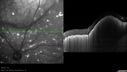
Primary Ocular Lymphoma - 020513 - SD OCT706 views
|
|

Best Disease - Color Fundus Photo706 views
|
|
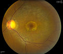
Bullseye Maculopathy without Plaquinel706 viewsFundus photograph reveals Bull's-eye maculopathy without the use of plaquinel. Patients VA is 20/20, Right eye and 20/25, Left eye. There are reported cases of Scleroderma patients with retinal pigment epithelial atrophy. Will return for follow up in 6-months.
|
|

Central Areolar Choroidal Dystrophy706 viewsVision loss from early 60's. This 78 year old woman has choroidal atrophy centrally.
|
|

Upside Down Lens Syndrome - Anterior Chamber IOL Upside Down706 views77 year old female who had vitrectomy for retained lens fragments and has had difficulty with recurrent uveitis since her cataract surgery. The anterior chamber intraocular lens is upside down. A lens exchange is planned.
|
|

Focal Laser for diabetic macular edema705 views66 year old female with diabetes for 45 years. VA 20/20 OU. Extrafoveal edema in the left eye treated with light focal laser.
|
|

Multifocal Choroiditis and Subretinal Fibrosis - 32 yo Female New Lesion OS705 views32-year-old woman vision loss in the right eye associated with macular scarring and multifocal choroiditis in 1999 with new vision loss in left eye: OD 20/400, OS 20/50.
|
|

Serpiginous choroiditis705 viewsInactive scars in both eyes
|
|

Small Retinal Pigment Epithelial Rip and Poor Vision (20/400)705 views80-year-old woman vision in the right eye has substantially declined.
OD: 20/400; OS: 20/50
|
|

Central Retinal Artery Occlusion Left Eye 2 Weeks Old - Still Non-Perfusion705 views85-year-old woman who had sudden severevision loss in the left eye two weeks ago. she can see a little more centrally and much more in the periphery, since about the last four days.
VISUAL ACUITY: OD 20/30, OS: Hand motion
|
|

Pigmented Maculae - Mother and Daughter - Normal Visual Acuity705 views12-year-old had a dilated macular exam and the macula look abnormal. Interestingly, her mother, who is with her at this visit, was told about a year ago that her maculae are abnormal too. Her mother’s vision is fine. In the family tree, there are no known retinal dystrophies. They are both healthy.
VISUAL ACUITY: Her vision is 20/20 in both eyes.
|
|

Hyperpigmentation of Retinal Pigment Epithlium in Dry AMD (hyperplasia)705 views87-year-old woman has wet age-related macular degeneration in the left eye. She had treatment up north but there has been a gap in therapy and the vision in the left eye is poor. Vision OD is 20/25, OS is 20/400
|
|

Diabetic Retinopathy - Foveal Laser Scar Right Eye705 views74-year-old woman has diabetic retinopathy in both eyes. She has had focal laser in her right eye done elsewhere in the mid 90’s and unfortunately the laser was fairly close to the center and as time progressed she developed foveal atrophy around the scars. She has noticed her vision declining since she was here last a year ago. She does take Travatan in both eyes at night for glaucoma.
VISUAL ACUITY: OD 20/200, OS 20/40.
|
|

Capillary Non-Perfusion - Severe Non-Proliferative (Background) Diabetic Retinopathy - Type I - 20 Year Old Man Diabetic 16 years705 views20 year old diabetic with poor vision for the past three or four years. He has been diabetic for sixteen years and hemoglobin A1C of 10.0. 20/25 OD, 20/30 OS.
|
|
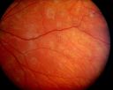
Acute Annular Outer Retinopathy - VA loss 2 days Right Eye Only - 25 Year Old Woman- AZOOR - White Line Around Fresh Lesions705 viewsInitial visit (June 11, 2011): 2 days of vision loss right eye. Wedge suddenly of VA loss OD, too dardkto see through. No flashes or floaters. VA 20/20 OU.
|
|

OCT Line Scan - Enhanced S Cone Syndrome - Goldmann Favre - 705 views55-year-old woman while in college her vision was poor even with glasses and she sought evaluation for that. She was told after she had an electroretinogram at USF 15 years ago, that she had something with her blue cones. She does have poor night vision, but her reading vision is pretty good.
VISUAL ACUITY: OD 20/40, OS 20/40
|
|
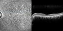
OCT Line Scan - Enhanced S Cone Syndrome - Goldmann Favre - 705 views55-year-old woman while in college her vision was poor even with glasses and she sought evaluation for that. She was told after she had an electroretinogram at USF 15 years ago, that she had something with her blue cones. She does have poor night vision, but her reading vision is pretty good.
VISUAL ACUITY: OD 20/40, OS 20/40
|
|
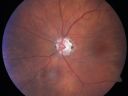
Eccentric Disciform Scar - Breakthrough Vitreous Hemorrhage - InferoTemporal Subretinal Fibrosis - Ectopic Choroidal Neovascular Membranes705 views88-year-old man has noticed for the last few months the vision in the right eye is foggy, like there is a film in the eye or a haze, His vision is 20/16 in each eye.
Right eye - inferotemporal periphery there is a hemorrhage at the equator at about 7:00 o’clock, which is about 4-disc diameters across and adjacent to that there is scarring.
Left eye - large disciform scar inferotemporally
|
|

A case of Valsalva Retinopathy -2705 views
|
|

ECTOPIC DISCIFORM SCAR/PERIPHERAL CHOROIDAL NEOVASCULAR MEMBRANE – EXUDATE –INFLAMMATION IN REACTION TO THE PERIPHERAL VASCULAR LESION 705 views53-year-old woman has noticed some blurred vision in the right eye for a few years. In retrospect, she thinks it is possible since she was a teenager the right eye was always the weaker eye and that the left eye has always been the better eye. VISUAL ACUITY: Vision OD is 20/400
|
|

Macular Branch Retinal Vein Occlusions (2) Left Eye - No Macular Edema (yet) 705 views77-year-old woman with two macular branch retinal vein occlusions in the left eye OD 20/25
|
|

FAF choroidal rupture705 views
|
|

Punctate Inner Choroidopathy - Recurrent CNVM 4 years after initial treatment Left Eye 705 viewsJune 7, 2012. This pleasant 35-year-old woman noticed decreasing vision in the left eye recently. She returns now noticing the vision in the left eye is declining. OD 20/50, OS 20/80. OS: Vertical C/D ratio is 0.3. There is a scar just inferonasal to the fovea, which has grown some towards the fovea with a little blood just under the fovea.
|
|

Posterior Polar Annular Choroidal Dystrophy705 views68 year old woman with know macular irregularities for over 20 years. She has since been followed another 6 years and her vision remains 20/25.
|
|

Proliferative Diabetic Retinopathy - Fresh and Old Scatter (Panretinal) Photocoagulation (Laser) - Neovascularization is regressing705 views1 month after initial laser
|
|

Band Keratopathy/Neurotrophic Ulcer705 viewsPatient comes in with blind painful left eye. Slit lamp photos shows corneal diffuse scarring, descemets folds, corneal striae, band keratopathy, left eye. Proceed with Jupiter contact lens fitting on the left eye.
|
|

Plaquenil Toxicity - Bulls Eye Maculopathy704 views70-year-old woman with systemic Lupus erythematosus and clotting problems. She was on the Plaquenil for about eight years and then off the Plaquenil for the last eight years because she developed macular toxicity. Although her vision was hazy, it was stable. Recent deceased vision left eye: OD 20/60, OS 20/100. IOP: OD 18, OS 19.
|
|

Psuedo-retinitis Pigmentosa - Bone Spicules One Eye - Probably Acute Zonal Occult Outer Retinopathy (AZOOR)704 views65-year-old woman has pseudoretinitis pigmentosa in the right eye only, most likely from acute zonal occult outer retinopathy. OD 20/25, OS 20/25
|
|

Concentric Annular Macular Dystrophy 4/200 2/200 vision 61 Year Old Macular Crystals704 views61-year-old woman has concentric annular macular dystrophy. She has had vision loss for 10 years who is no longer able to function even with the magnifiers and vision aids that she has. Her brother who is ten years younger than her, is recently diagnosed with the same macular dystrophy and her father and grandfather may have had similar problems in the past.
VISUAL ACUITY: OD 4/200, OS 2/200.
|
|

Congenital Toxoplasmosis with non-macular Scars Both Eyes - Recent Reactivation (recurrence) Right Eye704 views34-year-old man has congenital toxoplasmosis with four week history of foggy vision and new floaters in the right eye. OD is 20/40, OS 20/30
|
|

Acute Central Serous Retinopathy - CSR - Steroid Induced (probably) - Good Vision 704 views42-year-old man was seen in the office on October 5, 2011. He had noticed starting in August after a course of antibiotic and steroids, that he developed new spots in his vision in the right eye. He may have had an episode like this sometime in the past. He did take steroids a few years ago and his vision did change at that time, but then returned.
VISUAL ACUITY: OD 20/32, OS 20/32. The OCT scan of the right eye confirms subretinal fluid.
|
|

Macular Laser Scars - Patient had Central Retinal Vein Occlusion in her only eye704 views80-year-old woman left eye had a vascular occlusion, probably about five years ago, subsequent to that she had macular laser. She said since then the vision has been poor. OS 20/160.
|
|

Punctate Inner Choroidopathy - Recurrent CNVM 4 years after initial treatment Left Eye - Fluorescein Angiogram704 viewsJune 7, 2012. This pleasant 35-year-old woman noticed decreasing vision in the left eye recently. She returns now noticing the vision in the left eye is declining. OD 20/50, OS 20/80. OS: Vertical C/D ratio is 0.3. There is a scar just inferonasal to the fovea, which has grown some towards the fovea with a little blood just under the fovea.
|
|

Posterior Polar Annular Choroidal Dystrophy704 views68 year old woman with know macular irregularities for over 20 years. She has since been followed another 6 years and her vision remains 20/25.
|
|

Choroideremia - Female Carrier - Patchy Atrophy of Choroid and RPE - Lyonization704 views
|
|
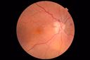
Anemic Retinopathy704 viewsMyelodysplasia - HgB 8 and very low platelet count
|
|

Plaquenil Toxicity - Bulls Eye Maculopathy703 views70-year-old woman with systemic Lupus erythematosus and clotting problems. She was on the Plaquenil for about eight years and then off the Plaquenil for the last eight years because she developed macular toxicity. Although her vision was hazy, it was stable. Recent deceased vision left eye: OD 20/60, OS 20/100. IOP: OD 18, OS 19.
|
|

Proliferative Diabetic Retinopathy both eyes Type I Diabetic for 25 years703 views47-year-old woman with diabetes for 25 years and decreased vision for 3 months. OD 20/40, OS 20/40
|
|
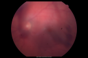
Steel Dart Perforating Injury - 15 year old - 3 weeks post-injury Vision 20/400703 views15-year-old injured in his left eye with a steel dart. The dart entered superiorly at the pars plana and his vision is clearing as the blood absorbs.
VISUAL ACUITY: OD 20/20, OS 20/400.
|
|

Retinal Arterial Macroaneurysm - Increased Swelling after Laser 703 views65-year-old woman was seen in the office on January 12, 2011. She has noticed decreased vision in the right eye for the last two or three weeks. She has had some discomfort in both eyes as well. She has a history of low blood pressure, but she does have high cholesterol.
VISUAL ACUITY: OD 20/60
|
|

Retinal Arterial Macroaneurysm - Increased Swelling after Laser 703 viewsVisit 2 - 2 months later:
March 21, 2011. This pleasant 66-year-old woman had laser to retinal arterial microaneurysm in the right eye January 12th. Her vision was getting better, but now it is worse the last few days. She is at home taking care of her demented mother and she and her sister lift her mother a fare amount of the time. I also checked her blood pressure, which was 120/65. VISUAL ACUITY: OD 20/200
|
|

Wet AMD - PED with exudate 20/400 87 Year old703 views87-year-old woman has wet age-related macular degeneration in the left eye. Vision OD is 20/25, OS is 20/400
|
|

Macular Scars - Laser (1980s) Ocular Histoplasmosis703 views70 year old man who had laser at Wills, Wilmer and the Mayo clinic before better therapies were available for ocular histoplasmosis related CNVM
|
|

Choroidal Melanoma - Exudative Retinal Detachment 82 Year Old Man 703 views82-year-old man who had 3 falls 2 weeks ago. After the falls he started checking his vision and noticed there was a veil over the left eye, which he had seen for about two weeks in the superior visual field. OD 20/25, OS 20/40
(patient was medically never well enough for brachytherapy and died 6 months later from heart disease)
|
|

Geographic Atrophy 20/160 Vision703 views84-year-old woman has age-related macular degeneration in both eyes with foveal atrophy. She also has a central retinal vein occlusion
in the left eye for which she had intravitreal Kenalog and finally chorioretinal anastomosis laser a year
ago. VISUAL ACUITY: Vision OD is 20/160, OS is 7/200.
|
|
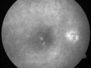
ECTOPIC DISCIFORM SCAR/PERIPHERAL CHOROIDAL NEOVASCULAR MEMBRANE – EXUDATE –INFLAMMATION IN REACTION TO THE PERIPHERAL VASCULAR LESION 703 views53-year-old woman has noticed some blurred vision in the right eye for a few years. In retrospect, she thinks it is possible since she was a teenager the right eye was always the weaker eye and that the left eye has always been the better eye. VISUAL ACUITY: Vision OD is 20/400
|
|

Chronic Pseudophakic Cystoid Macular Edema - Haptic Migrated into Anterior Chamber Through Peripheral Iridotomy703 views57-year-old man with congenital nystagmus. He had cataract surgery done 30 years ago and has decreased vision left eye for 6 months. OD is 20/50, OS is 20/100
|
|

Montage of Hemi-Central Retinal Artery Occlusion703 views
|
|

Retinitis Pigmentosa - Autosomal Dominant - Vision Loss at Young Age- SD OCT703 views 54-year-old woman has autosomal dominant retinitis pigmentosa. Her mother and grandmother both had it and she has one son who has it and one daughter who doesn’t. She also has a sister with it. She lost vision since she was younger. She had cataract surgery done in January and February of this year. She has been seeing unusual flashing lights throughout the periphery and central vision for about the last year or two.
VISUAL ACUITY: OD 20/160, OS 20/160
|
|

wide angle fa late. od703 viewsleaking vessels
|
|

Sickle Retinpoathy SC Disease - Vitreous Hemorrhage Left Eye - 29 Year old703 views
|
|

West Nile Virus Multifocal Choroiditis and diabetic retinopathy703 viewsMultifocal and clustered focal lesions are from west nile virus. The retinopathy is probably diabetic but could also be related to west nile virus
|
|

Myelinated Nerve Fiber Layer - Optic Nerve703 viewsRight eye subtle and inferior - left eye obvious
|
|
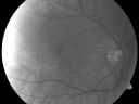
Albinism and Foveal Hypoplasia702 views65-year-old man was told about twenty years ago by a retina specialist that he spent so much time in the sun that he burned out his maculae. He does have two brothers who have dark brown hair and he has always had white hair. He had eye exercises when he was eight or nine because of a lazy eye and his brothers, when he was little, used to call him an albino. He has been diagnosed by dermatologists as having possible albinism but he has been told by other people his skin is just fair.
VA 20/70 OU
|
|
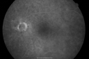
Bilateral Retinal Arteriol Occlusions - Possible Susac Syndrome 702 views80-year-old woman one month ago had vision loss and a vascular occlusion in the right eye. Vision loss occurred in the left eye about 9 years ago with cotton wool spots. Patients has a 30 year history of tinnitis.
|
|

Proliferative Diabetic Retinopathy both eyes Type I Diabetic for 25 years702 views47-year-old woman with diabetes for 25 years and decreased vision for 3 months. OD 20/40, OS 20/40
|
|

Acute Zonal Occult Outer Retinopathy with Severe Peripheral Vision Loss Left Eye 702 views 58-year-old man developed a peripheral vision problem about 25 years ago. OD 20/30, OS 20/40.
|
|

Normal Fellow Eye702 views65-year-old woman has pseudoretinitis pigmentosa in the right eye only, most likely from acute zonal occult outer retinopathy. OD 20/25, OS 20/25
|
|
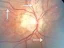
Angioid Streaks - Pseudoxanthoma Elasticum - Supressed CNVM702 views54-year-old woman had angioid streaks and macular degeneration in both eyes. She has had intravitreal Avastin in the right eye about every three months and her vision in that eye is good.
VISUAL ACUITY: Vision OD is 20/30, OS is 2/200
|
|

Drusen Maculopathy - Macular Degeneration (Dry) - 50 year old African-American Woman702 views50-year-old woman has drusen and maculopathy. VISUAL ACUITY: OD 20/30, OS 20/25.
|
|

Macular Branch Retinal Vein Occlusions (2) Left Eye - No Macular Edema (yet) 702 views77-year-old woman with two macular branch retinal vein occlusions in the left eye OD 20/25
|
|

Primary Ocular Lymphoma - White Mass Left Eye - 112712 702 views
|
|

Best Disease - Color Fundus Photo702 views
|
|
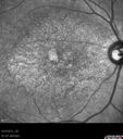
Macular possible Cone Dystrophy - Infra-red702 views
|
|

Fundus Flavimaculatus - Asymptomatic - Color Photo702 views45 year old man was told to come in because of ‘white spots on their retina’.
|
|

Purtchers Retinopathy with Bilateral Central Retinal Artery Occlusions 701 views50-year-old man traumatic chest compression and leg crushing injury. OD 1/200, OS 3/200
|
|

Stargardt's Disease - Macular Dystrophy - 69 Year Old African American Male - 20/200 both eyes701 views69-year-old man has had vision loss in both eyes starting 20 years ago. He was diagnosed at one point with Stargardt’s macular dystrophy. When I saw him, he had retinal atrophy in each eye.
Vision is 20/200 in each eye
|
|

Pigment Epithelial Detachment with Subretinal Fluid and Exudate 20/400 Vision701 views87-year-old woman has wet age-related macular degeneration in the left eye. She had treatment up north but there has been a gap in therapy and the vision in the left eye is poor. Vision OD is 20/25, OS is 20/400
|
|

Juxtafoveal Telangiectasis - MacTel - Group 2a - Stage IV - Pigment Plaque 89 Year Old Man (Also Nevus)701 views89-year-old man has stage IV juxtafoveal retinal telangiectasis in both eyes with background diabetic retinopathy. His vision is stable since he was here last which is about 6 months ago. OD 20/50, OS 20/70.
|
|

Twig Branch Retinal Vein Occlusion - Recurrent Macular Edema with extended interval Lucentis Therapy after first 6 monthly Injections701 views39-year-old man has a macular branch retinal vein occlusion with macular edema. He had an excellent response to Lucentis for his first 6 treatments. He was last here 6 weeks ago and his vision seems OK to him. OD 20/40.
|
|
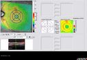
701 views63-year-old man diabetic for twenty two years. His central vision is fine.
VISUAL ACUITY: OD 20/25, OS 20/25
|
|

Choroidal Metastases - Both Eyes - Exudative Retinal Detachment701 views58-year-old man smoke a pack of cigarettes a day. He has had some ribcage pain for about the last two weeks. He also had a sinus infection for about two weeks, and he has also had hazy vision on the superior visual field in the left eye for about the last two weeks. vision is 20/25 in both eyes
|
|

Serous Retinal Detachment - Hypertensive Retinopathy - 165/120 701 views48-year-old woman noticed when she was blinking there was a white flash in the left eye starting about a month ago. She sees it temporally. OD 20/16, OS 20/20. There is a serous retinal detachment superotemporal to the optic nerve falling short of the fovea. Her blood pressure 165/120.
|
|

Primary Ocular Lymphoma - 020513 - SD OCT701 viewsVIDEO TAKES TIME TO LOAD
|
|

Streptococcus mitis Endophthalmitis following Intravitreal Avastin for wet AMD701 views88 Year Old Man treated with Avastin 02/28/13. Next day developed pain and severe vision loss. Treated with Intravitreal Vancomycin 1mg/0.1 cc; Ceftazidime 2mg/0.1cc; Decadron 0.4mg/0.1cc. Heavy growth Strep 2 days later from culture. Reinjected with Vancomycin 2 days after first Vancomyin. Now seeing Light Perception.
|
|

Asteroid Hyalosis - Benign Calcified Deposits in the Vitreous701 views
|
|

branch retinal vein occlusion - fluorescein angiogram700 views70 year old woman with 20/80 vision from a branch retinal vein occlusion and macular edema. She has had laser treatment and intravitreal steroids. Her vision with further laser improved to 20/50.
|
|

Toxocariasis700 views15-year-old one year ago had pink eye then her vision has been abnormal. She recently went for a drivers’ test and failed, OD: 20/80; OS: 20/20.
|
|
| 18117 files on 182 page(s) |
 |
|
|
|
|
31 | |
|
|
|
|


