|
|

638 views49-year-old woman has central retinal vein occlusion right eye with fluctuating vision for a few month now with OD 20/100. IOP: OD 20
|
|

Bilateral Retinal Arteriol Occlusions - Possible Susac Syndrome 638 views80-year-old woman one month ago had vision loss and a vascular occlusion in the right eye. Vision loss occurred in the left eye about 9 years ago with cotton wool spots. Patients has a 30 year history of tinnitis.
|
|

Hypertensive Retinopathy One Year later with good Vision IgA Nephropathy638 viewsOD 20/30, OS 20/25 One year following initial episode.
|
|

Pistol to head in attempted suicide - Retinal Atrophy - Shockwave Retinopathy 638 views66-year-old woman had a gunshot wound to the head on August 12, 2010, with the entry wound just on the right side, just about below her temple, toward the lower part of the temple and the exit wound on the left side just above her cheek, a little behind the eye. Since then she recovered and did not need any surgery. When that all cleared she realized her vision was poor in both eyes. She has seen a neuro-ophthalmologist who thought she had traumatic optic neuropathy. OD light perception, OS 20/400.
|
|
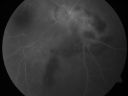
Serpiginouse Choroiditis (Chorioretinitis) - Acute Right Eye - Old Left Eye VA 20/25 OD , 20/50 OS638 views63-year-old woman has serpiginous choroiditis (date - March 2011). The right eye has not been previously involved, and then she noticed new onset floaters in the right eye for the last two weeks. Her vision in the right eye is hazy because of that.
VISUAL ACUITY: Vision OD is 20/25, OS is 20/50
|
|

Birdshot Chorioretinitis - Retinal Vasculitis - HLA A29 Positive 638 views64-year-old woman three or four year’s fluctuations in her vision. She mostly notices them in bright light her vision glares out and she has difficulty seeing. PMHx: malaria in 1948 that was treated. She was told at one point she could possibly have had Familial Mediterranean fever.
She does intermittently get rashes around her arms. She has arthritis in her knees and ankles.
VISUAL ACUITY: OD 20/40, OS 20/50 - HLA - A29 Positive
|
|

Hyperpigmentation of Retinal Pigment Epithlium in Dry AMD (hyperplasia)638 views87-year-old woman has wet age-related macular degeneration in the left eye. She had treatment up north but there has been a gap in therapy and the vision in the left eye is poor. Vision OD is 20/25, OS is 20/400
|
|

Non-Ischemic Central Retinal Vein Occlusion in Young Man (30 years) - Many Cotton Wool Spots638 views30-year-old man with a history of a central retinal vein occlusion for which he was first examined on 2 months ago and his vision then was 4/200. He responded nicely to two Avastin treatments. OD is 20/20, OS is 20/60
|
|

Vitreomacular traction with traction over retinal vessels in non-diabetic - Fluorescein leakage in areas of traction - Progressing vision loss left eye638 views80-year-old woman had vitrectomy surgery done in the right eye in 2007. Vision has been poor since despite a good anatomical result. Recently her vision in the left eye has been declining. The vision has declined in the left eye. Vision OD is 20/160, OS is 20/80
|
|

Pigmented Peripheral Retinal Degeneration638 views42-year old male comes in for routine eye exam and to follow up on peripheral retinal degeneration in both eyes. VA is 20/20, right eye and 20/25, left eye. Patient is asymptomatic with no visual complaints.
|
|

Reticular Macular Disease (Pseudo-drusen) Both Eyes - Occult CNVM and Macular Edema Right Eye 638 viewsFluorescein Angiogram
|
|

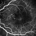
Polypoidal Choroidal Vasculopathy with Pulsating ICG polyp left eye638 views
|
|

Choroidal Osteoma - 13 year old - Good Vision 20-40 - B-scan Ultrasound638 views
|
|

Choroidal Nevus with Drusen638 viewsLow risk choroidal nevus with many overlying drusen
|
|

Choroidal Nevus with Drusen638 viewsLow risk choroidal nevus with many overlying drusen
|
|
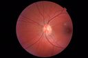
Cat Scratch - Neuroretinitis OD AND retinitis OS - Bartonella henselae positive638 views27 year old female with exposure to cats and prior fever. Reports with recent vision loss right eye. She was treated with doxycycline and within 2 weeks the right eye looks better. Then she skipped all follow-up visits. The right eye presented 20/200 and the left 20/16
|
|
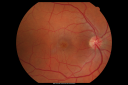
Macular Hole Closed with Vitrectomy - Preop637 views
|
|

Juxtapapillary CNVM and Serous Macular Detachment Wet AMD Rx Laser637 views85-year-old man OD is 20/25, OS is 20/100. IOP: OD 21, OS 22.
OS: Vertical C/D ratio is 0.1. There is a posterior vitreous separation. There is a hemorrhagic pigment epithelial detachment on the superior pole of the optic nerve, extending a disc-and-a-half diameter off the nerve, with adjacent exudates. There is a serous macular detachment involving the center of the fovea.
VA improved from 20/100 to 20/50 in four months from laser.
|
|

X-Linked Juvenille Retinoschisis 10 Year Old Child treated with Trusopt637 views10-year-old child has x-linked juvenile retinoschisis VA 20/60, OS is 20/100. Macula and Vision improved with Trusopt drops.
|
|

637 views66-year-old woman had a gunshot wound to the head on August 12, 2010, with the entry wound just on the right side, just about below her temple, toward the lower part of the temple and the exit wound on the left side just above her cheek, a little behind the eye. Since then she recovered and did not need any surgery. When that all cleared she realized her vision was poor in both eyes. She has seen a neuro-ophthalmologist who thought she had traumatic optic neuropathy. OD light perception, OS 20/400.
|
|

Steel Dart Perforating Injury - 15 year old - 3 weeks post-injury Vision 20/400637 views16-year-old had a dart injury to the left eye August 3rd of 2009. He has had a few surgeries since then, although he never did require a vitrectomy. His vision is improving remarkably.
VISUAL ACUITY: OS: Without correction 20/40
|
|

DUSN - Diffuse Unilateral Subacute Neuroretinitis - Nematode637 views
|
|

Myopic Macular Degeneration - Staphyloma - Recent Vision Loss Right Eye - Possible CNVM637 views83-year-old woman is a high myope with myopic macular degeneration. She had a retinal detachment repair in the right eye in 1968. I saw her in June of 2003. She had posterior colobomas and areas of pigment epithelial atrophy. She has noticed over the last six weeks substantial declining vision in the right eye. Prior to that, the vision in the two eyes was about the same. She uses Xalatan in the left eye and Alphagan in both eyes.
VISUAL ACUITY: Vision OD is 20/160, OS is 20/40
|
|

Twig Branch Retinal Vein Occlusion - Recurrent Macular Edema with extended interval Lucentis Therapy after first 6 monthly Injections637 views39-year-old man has a macular branch retinal vein occlusion with macular edema. He had an excellent response to Lucentis for his first 6 treatments. He was last here 6 weeks ago and his vision seems OK to him. OD 20/40.
|
|

Cuticular Drusen (Basal Laminar Drusen) and Bilateral Chronic Subfoveal Fluid (Serous Retinal Detachment) unresponsive to monthly Avastin Late FA OD637 views66-year-old man with bilateral subfoveal fluid unresponsive to one year of monthly avastin therapy.
OD 20/160, Pinhole 20/80. OS 20/63, Pinhole 20/50. OCT scan shows subretinal fluid with normal choroidal thickness. Fluorescein angiogram shows cuticular/basal laminar drusen. ICG shows no foci of choriocapillaris leakage.
|
|
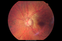
Posterior Polar Annular Choroidal Dystrophy637 views68 year old woman with know macular irregularities for over 20 years. She has since been followed another 6 years and her vision remains 20/25.
|
|

Asymptomatic Juxtapapillary Choroidal Neovascular Membrane - Reticular Macular Disease (Wet AMD) - FAF637 views
|
|

Gonioscopy, Blood in the Anterior Chamber from Hyphema637 viewsPatient comes in with blunt trauma to the right eye due to a BB gun incident. Patient was present with a hyphema at 8-o'clock about 1mm thick. Gonioscopy photos were then taken to show blood from the hyphema entered into the anterior chamber. Patient had no angle recession in the right eye.
|
|

BDR OD with Macular Edema637 views
|
|

Choroidal Neovascular Membrane - Multifocal Scars - PIC vs MFC vs OHS 637 views
|
|
Polypoidal Choroidal Vasculopathy with Pulsating ICG polyp left eye637 views
|
|

Outer Retinal Holes - Probably Solar Retinopathy (history is negative)637 views
|
|

Posterior Uveal Effusion637 views
|
|

IPCV - idiopathic polypoidal choroidal vasculopathy - juxtapapillary CNVM with hemorrhage both eyes (also choroidal nevus right eye) - Subretinal hemorrhage both eyes637 views86 year old woman with shadow in vision OD and new hemmorhage OU.FA shows juxtapapillary CNVM
|
|
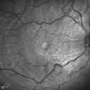
Choroidal Folds Hyperope (+5.00)637 views
|
|

Travatan Induced CME Left eye - 1 month post stopping travatan CME is gone637 views63 year old woman 4 years post ECCE and 2 years post PPV for floaters. She developed CME in the left eye after 1 year of travatan. The travatan was stopped and the CME and serous retinal detachment resolved.
|
|

Juxtapapillary CNVM and Serous Macular Detachment Wet AMD Rx Laser636 views85-year-old man OD is 20/25, OS is 20/100. IOP: OD 21, OS 22.
OS: Vertical C/D ratio is 0.1. There is a posterior vitreous separation. There is a hemorrhagic pigment epithelial detachment on the superior pole of the optic nerve, extending a disc-and-a-half diameter off the nerve, with adjacent exudates. There is a serous macular detachment involving the center of the fovea.
VA improved from 20/100 to 20/50 in four months from laser.
|
|

Diabetic Retinopathy - Non-Perfusion - Possible Central Retinal Vein Occlusion636 views68-year-old woman OD 20/70, OS 20/200 CYSTOID MACULAR EDEMA, POSSIBLE EITHER OCULAR ISCHEMIC SYNDROME IN THE RIGHT EYE OR OCCULT CENTRAL RETINAL VEIN OCCLUSION IN THE RIGHT EYE,S EVERE NON-PerfUSION WITH DIABETIC RETINOPATHY IN THE LEFT EYE.
|
|

Plaquenil Toxicity - Bulls Eye Maculopathy636 views70-year-old woman with systemic Lupus erythematosus and clotting problems. She was on the Plaquenil for about eight years and then off the Plaquenil for the last eight years because she developed macular toxicity. Although her vision was hazy, it was stable. Recent deceased vision left eye: OD 20/60, OS 20/100. IOP: OD 18, OS 19.
|
|

DUSN - Diffuse Unilateral Subacute Neuroretinitis - Nematode636 views
|
|
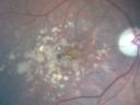
Drusen Maculopathy 85 year old Male636 views85-year-old man has age-related macular degeneration in both eyes with drusen maculopathy. OD is 20/40, OS is 20/400
|
|
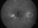
Hyperpigmentation of the Retinal Pigment Epithelium in Dry AMD636 views87-year-old woman has wet age-related macular degeneration in the left eye. Vision OD is 20/25, OS is 20/400
|
|

Pigment Epithelial Detachment with Subretinal Fluid and Exudate 20/400 Vision636 views87-year-old woman has wet age-related macular degeneration in the left eye. She had treatment up north but there has been a gap in therapy and the vision in the left eye is poor. Vision OD is 20/25, OS is 20/400
|
|

Pigment Epithelial Detachment with Subretinal Fluid and Exudate 20/400 Vision636 views87-year-old woman has wet age-related macular degeneration in the left eye. She had treatment up north but there has been a gap in therapy and the vision in the left eye is poor. Vision OD is 20/25, OS is 20/400
|
|

Retinitis Pigmentosa Newly Diagnosed 55 Year Old636 views55-year-old noticed his vision declining recently, over the last few weeks, particularly in the right eye. He said previous to that the vision was not normal but was pretty good. OD is 20/100, OS is 20/50
OCT SCAN: There is macular thickening in both eyes with intraretinal cysts.
|
|

Cuticular Drusen (Basal Laminar Drusen) and Bilateral Chronic Subfoveal Fluid (Serous Retinal Detachment) unresponsive to monthly Avastin OCT OD VIDEO TAKES A LONG LONG TIME TO LOAD636 views66-year-old man with bilateral subfoveal fluid unresponsive to one year of monthly avastin therapy.
OD 20/160, Pinhole 20/80. OS 20/63, Pinhole 20/50. OCT scan shows subretinal fluid with normal choroidal thickness. Fluorescein angiogram shows cuticular/basal laminar drusen. ICG shows no foci of choriocapillaris leakage.
|
|

X-linked Juvenille Retinoschisis - Peripheral Retinal Vascular Anomalies - Vitreous Hemorrhage - 8 Year Old Boy636 views8-year-old child OD 20/40, OS 20/50
OD: Vertical C/D ratio is 0.2. There are foveal cysts. There is also a retinal elevation inferiorly and there are patchy peripheral retinal hemorrhages.
OS: Vertical C/D ratio is 0.2. There are foveal cysts. There is peripheral retinal issues with some hemorrhage in some areas of peripheral retinoschisis.
|
|

Trans-Illumination Defects636 views74-year old patient who VA 20/70 OD, 20/50 OS. Complaints of blurred vision. Iris atrophy, both eyes. ERM, right eye, Patient to have cataract surgery to improve distance vision.
|
|

Retinitis Pigmentosa Sine Pigmento or Unknown Dystrophy - Peripheral Superficial White Spots and Recurrent CME - FA LATE636 views46-year-old woman three of her 5 siblings have been diagnosed with retinitis pigmentosa. Her mother and father were seen and as far as everyone can tell, neither of them has it, although there was some thought they might be carriers. They were evaluated twenty five years ago and since then as far as she knows, there has not been a lot of testing done.
She has noticed a substantial decline in vision in both eyes over the last month or two. OD 20/40, OS 20/50. IOP: Both eyes have CME
|
|

Primary Ocular Lymphoma - White Mass Left Eye - 112712 636 views
|
|

Pigmented Peripheral Retinal Degeneration636 views42-year old male comes in for routine eye exam and to follow up on peripheral retinal degeneration in both eyes. VA is 20/20, right eye and 20/25, left eye. Patient is asymptomatic with no visual complaints.
|
|

Macular Pucker 20/40 vision - minimal symptoms for 1 year Right eye636 viewsMulticolor
|
|

BRAO and PAMM636 views
|
|

Acute Macular Neuroretinopathy - (AMN) - Spot in Vision OS - 40 Year old Female636 viewsIR shows gray spot nasal to the fovea
|
|
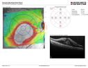
Self Sealed Retinal Arteriol Macroaneurysm636 viewsMAP shows edema and some subretinal fluid
|
|

Macular Edema and dry AMD - excellent example of why FA and ICG are necessary in management of wet AMD636 viewsThis 72 year old woman has an OCT that looks like wet AMD. Only her FA and ICG show that she has inflammatory macular edema. She had no clinical signs or symptoms of uveitis. Nevertheless, the macular edema responded to subtenon's kenalog.
|
|

Macular Atrophy - Phototoxicity - Macular Hole Surgery636 views25 years ago patient suffered immediate severe vision loss following macular hole surgery. You can almost see light pipe tracks in the atrophy in the macula. Vision is 5/200
|
|

Pattern Dystrophy - Foveal Atrophy636 views73 year old female with vision loss - looks like macular hole
|
|
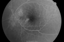
Multifocal Choroiditis and Subretinal Fibrosis - 32 yo Female New Lesion OS635 views32-year-old woman vision loss in the right eye associated with macular scarring and multifocal choroiditis in 1999 with new vision loss in left eye: OD 20/400, OS 20/50.
|
|

Peripapillary Lesion Hemangioma or CNVM Line Scan Shows Serous RD Right Eye635 views67-year-old man with a history of a peripapillary lesion in the right eye.
|
|

Atypical Neuroretinitis - ?Ischemic - ?Paraneoplastic - Line Scan OS 1 year After Initial Episode635 views74-year-old woman has stage 4 non-small cell lung carcinoma decreasing vision in the left eye for at least the last two weeks, VA 10/200. Trace APD OS. No pain
|
|
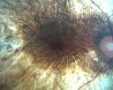
26-year-old man choroideremia OU is 20/60635 views
|
|

New BRVO Left Eye Old BRVO Right Eye - Right Eye Showing Bulls Eye Atrophy from Chronic CME and Non-Perfusion Infero-temporally635 views90-year-old man decreased vision maybe one year since cataract surgery he is no longer able to read. OD 20/200, OS 20/200
|
|

Old Subhyaloid - Preretinal Hemorrhage (Yellow) 635 views53-year-old man 3 monts previous was hit in a motor vehicle/pedestrian accident and was mostly bedridden and in and out of consciousness for two months. It is now three months since the injury. His right eye has permanent vision loss from optic nerve injury. His left eye has reasonable vision, but he does notice a floater often obstructing his vision in that eye. OD is 1/200, OS is 20/20.
|
|

Serpiginouse Choroiditis (Chorioretinitis) - Acute Right Eye - Old Left Eye VA 20/25 OD , 20/50 OS635 views63-year-old woman has serpiginous choroiditis (date - March 2011). The right eye has not been previously involved, and then she noticed new onset floaters in the right eye for the last two weeks. Her vision in the right eye is hazy because of that.
VISUAL ACUITY: Vision OD is 20/25, OS is 20/50
|
|

Drusen Maculopathy - Macular Degeneration (Dry) - 50 year old African-American Woman635 views50-year-old woman has drusen and maculopathy. VISUAL ACUITY: OD 20/30, OS 20/25.
|
|

Pigmented Choroidal Mass Right Eye - Melanoma vs. Nevus vs. Unilateral Melanocytic Proliferation - 4 Month Follow-Up Images635 views71-year-old man his vision 4 month follow-up. Vision is 20/60 and patient sees flashing lights. Lesion has grown slightly in the superior half of the macula.
|
|

Traumatic Macular Hole and Pre-retinal Hemorrhage - Assault635 viewsThis 18 year old male was assaulted on 3 weeks ago and seen in the emergency room for that. He returns today reporting his left eye is getting better and is not quite as blurry. VA 20/60 (uncorrected)
|
|

Retinal Tear635 views
|
|

Choroidal Metastases - Both Eyes - Exudative Retinal Detachment635 views58-year-old man smoke a pack of cigarettes a day. He has had some ribcage pain for about the last two weeks. He also had a sinus infection for about two weeks, and he has also had hazy vision on the superior visual field in the left eye for about the last two weeks. vision is 20/25 in both eyes
|
|

Retinitis Pigmentosa Sine Pigmento or Unknown Dystrophy - Peripheral Superficial White Spots and Recurrent CME635 views1 month of treatment with diamox 500 mg BID- cme is a little better
|
|

PDR635 views
|
|

Fundus Flavimaculatus - Asymptomatic - Multi-Color (false color) Photo635 views45 year old man was told to come in because of ‘white spots on their retina’.
|
|

Posterior Uveal Effusion635 views
|
|

Choroidal Folds Hyperope (+5.00)635 views
|
|
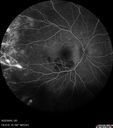
Coats' Disease - 14 year old - Asymptomatic635 viewsPresented with macular exudates. Temporal macroaneurysms responded to laser. Ultra-wide field FA shows nonperfusion
|
|

AMN 3 years later635 viewsShe still sees the blind spot one she looks for it.
|
|

Fundus Albipunctatus635 views12 year old female with normal vision. She has 4 siblings all of whom have either white spots or spots on IR. Genetic testing by parents was deferred.
|
|

Macular Laser Scars635 views86 year old man who had Laser done from 2010 - 2018 for diabetic retinopathy. The right eye lost vision immediately after laser was done a few years ago. VA 1/200 OD; 20/25 OS
|
|
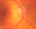
Retinal Arterial Atheromata634 views68-year-old woman with records going back 7 years, which clearly document retinal arterial plaques in both eyes.
|
|
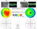
Macular Pucker Pre-op 20/200 vision (history of retinal tears)634 views66-year-old man has had laser to retinal tears in both eyes in 1999 and in 2001. His right eye has had rapidly declining vision over the last month. VISUAL ACUITY: OD: 20/200
|
|

Plaquenil Toxicity with New Symptomatic Scotoma Left Eye634 views64-year-old female decreased vision OS over the past 2 months. She describes a blurred central scotoma. She has rheumatoid arthritis and is on Plaquenil 200 mg BID for 15 years. She does have yearly visual field tests done with last visual field test performed 3 years ago which was within normal limits OD 20/40-2 OS 20/40-1
|
|

Ocular Ishemic Syndrome - Bilateral Carotid Occlusions - OD 20/60 OS 20/200 Enlarged Foveal Avascular Zone and Temporal Non-perfusion634 views75-year-old man had laryngeal cancer treated in 15 year ago with radiation. He recently was told that the carotid circulation is closed off on both sides. He does have a patent vertebral artery, which is supplying him pretty well and in addition to that his cardiac output is compromised by poor mitral valves. His consideration is to what can be done about that in the future.
VISUAL ACUITY: OD 20/60, OS 20/200
|
|
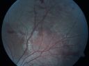
Macular Hole - Traumatic - 11 year old boy (also retinal whitening Berlins Edema)634 views11-year-old boy with a history of blunt trauma to the left eye yesterday. He reports he lost vision immediately but it has gotten somewhat better. He reports he sees “red streamers†and he has very blurry visual acuity., 20/200 OS
|
|

Plaquenil (Hydroxycholorquine) Toxicity - Bull's Eye Maculopathy - 73 year old womand 25 Years therapy - 64 inches tall - 110 lbs634 views73-year-old woman has been on Plaquenil for twenty five years. She noticed loss of paracentral vision a few months ago. VISUAL ACUITY: OD 20/70, OS 20/30. Her color vision was 0 out 10 correct in both eyes and she also missed the controls.
|
|

Wet AMD - PED with exudate 20/400 87 Year old634 views87-year-old woman has wet age-related macular degeneration in the left eye. Vision OD is 20/25, OS is 20/400
|
|

Indocyanine Green Angiogram - Enhanced S Cone Syndrome - Goldmann Favre - 634 views55-year-old woman while in college her vision was poor even with glasses and she sought evaluation for that. She was told after she had an electroretinogram at USF 15 years ago, that she had something with her blue cones. She does have poor night vision, but her reading vision is pretty good.
VISUAL ACUITY: OD 20/40, OS 20/40
|
|
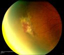
Sickle Cell634 views
|
|
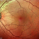
Macular Pucker 20/40 vision - minimal symptoms for 1 year Right eye634 viewsMulticolor
|
|

Acute non arteritic anterior ischemic optic neuropathy left eye - SD Optical Coherence Tomography Line Scan634 views66 y.o. woman spot in central vision in left eye started about 1 week ago since then it is not changing. VA OD: sc20/25 OS: sc20/200
|
|

Macular Hole Left Eye - progression from VMT - Stage II macular hole - Spontaneously Closed Macular Hole634 views
|
|

Posterior Uveal Effusion634 views
|
|

Wet AMD - Subfoveal CNVM - Excellent Response to Avastin633 views76-year-old woman two to three months of vision loss OD
Vision started at 20/200 and with Avastin improved to 20/25. She has required monthly injections and as of October 2010 has been treated about 16 times. Her left eye remains good at 20/20.
The OCT from post-treatment as well as pretreatment images are in this album.
|
|
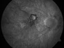
Myopic Choroidal Neovascular Membrane 54 year old woman 20/20633 views54-year-old woman has had problems in her vision in the right eye for about the last three months. Inferotemporally she sees a gray area, which is distorted. OD is 20/20, OS is 20/20
|
|

Branch Retinal Vein Occlusion with Macular Edema633 viewsCentral foveal thickness came to normal after laser and intravitreal Kenalog
|
|

Central Retinal Vein Occlusion - New- and old Macular Scar from AMD633 views87-year-old woman with longstaning macular scars and new vision loss left eye for 2 months. OD: 20/200, OS 20/400.
|
|

Proliferative Diabetic Retinopathy in patient with Moyamoya Disease633 views28-year-old womandiabetic since age nine, and she also has had multiple strokes. OD is 20/30, OS is 20/30.
|
|
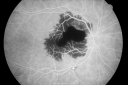
Ruptured Retinal Arterial Macroaneurysm - Submacular Hemorrhage - Vision 20/400 never recovered - Hypertension 205/94633 views
|
|

633 views66-year-old woman had a gunshot wound to the head on August 12, 2010, with the entry wound just on the right side, just about below her temple, toward the lower part of the temple and the exit wound on the left side just above her cheek, a little behind the eye. Since then she recovered and did not need any surgery. When that all cleared she realized her vision was poor in both eyes. She has seen a neuro-ophthalmologist who thought she had traumatic optic neuropathy. OD light perception, OS 20/400.
|
|

Macular Pucker Pre-OP 20/200 633 views
|
|
| 18117 files on 182 page(s) |
 |
|
|
|
|
42 | |
|
|
|
|


