|
|

Plaquenil (Hydroxycholorquine) Toxicity - Bull's Eye Maculopathy - 73 year old womand 25 Years therapy - 64 inches tall - 110 lbs634 views73-year-old woman has been on Plaquenil for twenty five years. She noticed loss of paracentral vision a few months ago. VISUAL ACUITY: OD 20/70, OS 20/30. Her color vision was 0 out 10 correct in both eyes and she also missed the controls.
|
|

Wet AMD - PED with exudate 20/400 87 Year old634 views87-year-old woman has wet age-related macular degeneration in the left eye. Vision OD is 20/25, OS is 20/400
|
|

Non-Arteritic Anterior Ischemic Optic Neuropathy - Peripapillary Choroidal Ischemia634 views68-year-old woman noticed central vision loss in the right eye about a week ago. It is getting a little bit better. OD 20/160, OS 20/25.
|
|

Myopic Traction Maculopathy - Vitreoretinal Traction - Possible Occult Neovascular Membrane634 views58-year-old woman was seen in the office on June 30, 2011. In 2002, her vision was 20/30. Subsequent to that she had her left eye made her distance eye, which was dominate. She had LASIK surgery on the right eye to make that her near eye and she was getting along well until about a year ago when she noticed substantial vision loss in the left eye only. VISUAL ACUITY: The near eye is 20/200, but she reads J2. OS 20/100- and J16.
|
|

New Subfoveal Classic CNVM OS - Wet AMD - Macular Scar Congenital Toxoplasmosis Right Eye 634 views77-year-old woman as had poor vision in the right eye since birth from congenital toxoplasmosis of her macula in the right eye. Two to three weeks ago when she noticed vision loss in the left eye. VISUAL ACUITY: OD 2/200, OS 20/200.
|
|
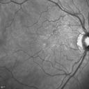
Psuedomacular Hole - Epiretinal Membrane - Vision unchanged 20/40 for 6 months634 views
|
|

Gonioscopy, Blood in the Anterior Chamber from Hyphema634 viewsPatient comes in with blunt trauma to the right eye due to a BB gun incident. Patient was present with a hyphema at 8-o'clock about 1mm thick. Gonioscopy photos were then taken to show blood from the hyphema entered into the anterior chamber. Patient had no angle recession in the right eye.
|
|

Acute non arteritic anterior ischemic optic neuropathy left eye - Color Photo634 views66 y.o. woman spot in central vision in left eye started about 1 week ago since then it is not changing. VA OD: sc20/25 OS: sc20/200
|
|

Toxoplasmosis Retinitis - Vitritis and Anerior Uveitis634 views2 weeks after starting treatment - lesion is involuting
|
|
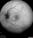
Unilateral Acute Idiopathic Maculopathy (UAIM)634 views41 year old with serous retinal detachment OS and sudden vision loss. He had a viral prodrome. Coxscackie IgG was high but no acute infection was documented. Vision improved in about 2 months without therapy.
|
|

Choroideremia - Female Carrier - Patchy Atrophy of Choroid and RPE - Lyonization634 views
|
|

Syphilitic Chorioretinitis634 viewsSevere outer retinal atrophy and RPE non-geographic atrophy from syphilis. Vision improved from 20/63 to 20/32 with therapy. Images look a lot like DUSN. Patient had several surgeries (vitrectomy for floaters, vitrectomy for macular pucker, Avastin injections and macular laser for edema) prior by other retina specialists prior to being correctly diagnosed and treated.
|
|
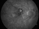
Myopic Choroidal Neovascular Membrane 54 year old woman 20/20633 views54-year-old woman has had problems in her vision in the right eye for about the last three months. Inferotemporally she sees a gray area, which is distorted. OD is 20/20, OS is 20/20
|
|

Myopic Choroidal Neovascular Membrane 54 year old woman 20/20633 views54-year-old woman has had problems in her vision in the right eye for about the last three months. Inferotemporally she sees a gray area, which is distorted. OD is 20/20, OS is 20/20
|
|

Bilateral Retinal Arteriol Occlusions - Possible Susac Syndrome 633 views80-year-old woman one month ago had vision loss and a vascular occlusion in the right eye. Vision loss occurred in the left eye about 9 years ago with cotton wool spots. Patients has a 30 year history of tinnitis.
|
|

Branch Retinal Vein Occlusion with Macular Edema633 viewsCentral foveal thickness came to normal after laser and intravitreal Kenalog
|
|

Central Retinal Vein Occlusion - New- and old Macular Scar from AMD633 views87-year-old woman with longstaning macular scars and new vision loss left eye for 2 months. OD: 20/200, OS 20/400.
|
|
633 views
|
|
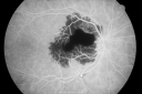
Ruptured Retinal Arterial Macroaneurysm - Submacular Hemorrhage - Vision 20/400 never recovered - Hypertension 205/94633 views
|
|

Birdshot Chorioretinitis - Retinal Vasculitis - HLA A29 Positive 633 views64-year-old woman three or four year’s fluctuations in her vision. She mostly notices them in bright light her vision glares out and she has difficulty seeing. PMHx: malaria in 1948 that was treated. She was told at one point she could possibly have had Familial Mediterranean fever.
She does intermittently get rashes around her arms. She has arthritis in her knees and ankles.
VISUAL ACUITY: OD 20/40, OS 20/50 - HLA - A29 Positive
|
|

633 views
|
|

Choriodal Neovascular Membrane - Subfoveal - Pseudo-Retinitis Pigmentosa - 50 years post Retinal Detachment Repair633 views86-year-old with a history of decreased vision in the right eye. The patient reports she has lost vision in the right eye over the past two months or so. She reports the vision was always compromised in that eye since retinal detachment surgery 50 years ago with a scleral buckler. Vision dropped from 20/50 to 20/200 right eye in 2 months.
|
|

Macular Hole - Traumatic - 11 year old boy (also retinal whitening Berlins Edema)633 views11-year-old boy with a history of blunt trauma to the left eye yesterday. He reports he lost vision immediately but it has gotten somewhat better. He reports he sees “red streamers†and he has very blurry visual acuity., 20/200 OS
|
|

Macular Pucker - 1 year after Vitrectomy - 20/25 Vision - Postoperative Images633 views60-year-old man had vitrectomy for a macular pucker in the right eye a year ago. His vision is good.
VISUAL ACUITY: Vision OD is 20/25 Eye is now pseudophakic
|
|

Fluorescein Angiogram - Enhanced S Cone Syndrome - Goldmann Favre - 633 views55-year-old woman while in college her vision was poor even with glasses and she sought evaluation for that. She was told after she had an electroretinogram at USF 15 years ago, that she had something with her blue cones. She does have poor night vision, but her reading vision is pretty good.
VISUAL ACUITY: OD 20/40, OS 20/40
|
|

Central Retinal Vein Occlusion and Tractional Retinal Detachment for 3 Years (Patient is Diabetic)633 views67-year-old woman has severe vision loss in the left eye for 3 years OD 20/32, OS 3/200
|
|
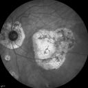
Ocular Histoplasmosis both Eyes - Laser Right Eye Only InfraRed633 views58-year-old woman who lost vision as a child in the left eye from ocular histoplasmosis. She used to play in her attic and she was told by her pulmonary doctor that is probably where she picked it up. She had laser in the right eye for a leaky lesion back in 1990. OD 20/25, OS 20/200.
|
|

633 views
|
|
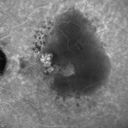
Polypoidal Choroidal Vasculopathy with Pulsating ICG polyp left eye633 views
|
|

Acute Macular Neuroretinopathy - (AMN) - Spot in Vision OS - 40 Year old Female633 viewsFluorescein Angiogram is normal
|
|

Coats' Disease - 14 year old - Asymptomatic633 viewsPresented with macular exudates. Temporal macroaneurysms responded to laser. Ultra-wide field FA shows nonperfusion
|
|

Punctate Inner Choroidopathy - New CNVM right eye633 views42 year old female with recent vision loss in the right eye - Right eye is 20/80 - Vision improved to 20/16 with Ranabizumab injections over 3 months
|
|
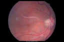
Retinal fold following removal of severe macular pucker633 views75 year old man had macula on inferior retinal detachment repair. He then developed a 5/200 dense macular pucker. After the pucker was removed the vision returned to 20/40 but there was a persistent superior retinal fold that looks a little like a snake.
|
|

Normal Left eye633 viewsLeft eye of patient with valsalva retinopathy in the right
|
|

Macular Hole Closed with Vitrectomy - Preop632 views
|
|

PDR - Mild Vitreous Hemorrhage and Good Vision632 views58-year-old woman has diabetic retinopathy in both eyes with neovascularization of the optic nerve, worse in the right eye than the left eye. She has had panretinal laser in both eyes and vitreous hemorrhage in the left eye. Her vision, since I saw her last in June, is about the same.
VISUAL ACUITY: Vision OD is 20/25, OS is 20/20.
|
|
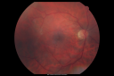
Macular Pucker in eye following giant retinal tear RD repair post-op Fundus Photo632 viewsnow that the bubble is gone, but he is starting to notice distortion of central vision. He had a giant retinal tear retinal detachment repair emergently done on November 8, 2007, and subsequent laser on November 15th.
VISUAL ACUITY: Vision OD 20/400.
6 weeks post-op VA 20/40
|
|

Multiple Evanescent White Dot Syndrome - Atypical Yellow Foveal Spot Right Eye632 views12-year-old had decreased vision in the left eye from reported amblyopia since around the year 2000. His right eye was doing fine until two days ago, when he had sudden severe vision loss. He sees a blurred spot in the middle of the vision in the right eye wherever he looks.
OD 20/60 with eccentric movement, OS 20/200. Pinhole 20/70. IOP: OD 14, OS 10.
|
|

Diabetic and Pseudophakic Cystoid Macular Edema Late FA - Leakage Macular and Nerve632 views56-year-old woman has diabetic retinopathy in both eyes and macular edema. She is recently pseudophakic. OD 20/80, OS 20/200.
|
|

Hypertensive Retinopathy with Choroidal Ischemia Right Eye and Vision 8/200 - Blood Pressure 240/120 mmHg IgA Nephropathy632 views39-year-old man two days ago he noticed poor vision in his right eye OD: 8/200; OS: 20/20. BP is 240/120
|
|

Cotton Wool Spots now Gone in Both Eyes632 views60-year-old man was sick in the hospital in 3 month ago with streptococcal pneumonia. He was in the cardiac intensive care unit and at one point he was in critical condition. He has no visual complaints. OD 20/20, OS 20/25. Cotton Wool Spots now GONE
|
|
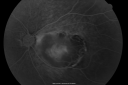
Retinocytoma - Retinoma - Regressed Retinoblastoma 43 year old male incidental finding 632 views
|
|

Patterned Pigment Dystrophy of the Fovea 79 YO Woman VA 20/20 Right 20/30 Left632 views
|
|
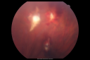
Steel Dart Perforating Injury - 15 year old - 3 weeks post-injury Vision 20/400632 views15-year-old injured in his left eye with a steel dart. The dart entered superiorly at the pars plana and his vision is clearing as the blood absorbs.
VISUAL ACUITY: OD 20/20, OS 20/400.
|
|

Plaquenil Toxicity - Bull's Eye Maculopathy - 25 Years Therapy OCT Posterior Pole MAP632 views73-year-old woman has been on Plaquenil for twenty five years. Plaquenil has now been stopped for 6 months. VA continues to decline as does visual function - 20/80 OD , 20/40 OS
|
|
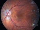
Non-arteritic Anterior Ischemic Optic Neuropathy following Intravitreal Injection 632 views81-year-old woman has had injections in both eyes for wet age-related macular degeneration. 2 weeks ago the left eye was treated. Less than a week ago she noticed a change in vision in the left eye. She had a dull left frontal headache and noticed her vision was hazy as if there was a veil over the vision. OD is 20/25, OS is 20/25. IOP: OD 18, OS 26. The left eye has a 3+ APD
|
|

Coats' Disease in 14 Year Old Boy Treated with Cryotherapy and Avastin632 views14 Year Old Boy with Coats Disease
|
|

Retinitis Pigmentosa - Autosomal Dominant - Vision Loss at Young Age - Infrared632 views 54-year-old woman has autosomal dominant retinitis pigmentosa. Her mother and grandmother both had it and she has one son who has it and one daughter who doesn’t. She also has a sister with it. She lost vision since she was younger. She had cataract surgery done in January and February of this year. She has been seeing unusual flashing lights throughout the periphery and central vision for about the last year or two.
VISUAL ACUITY: OD 20/160, OS 20/160
|
|

Punctate Inner Choroidopathy - Recurrent CNVM 4 years after initial treatment Left Eye - Fluorescein Angiogram632 viewsJune 7, 2012. This pleasant 35-year-old woman noticed decreasing vision in the left eye recently. She returns now noticing the vision in the left eye is declining. OD 20/50, OS 20/80. OS: Vertical C/D ratio is 0.3. There is a scar just inferonasal to the fovea, which has grown some towards the fovea with a little blood just under the fovea.
|
|

Reticular Macular Disease (Pseudo-drusen) Both Eyes - Occult CNVM and Macular Edema Right Eye 632 viewsIndocyanine Green Angiogram
|
|
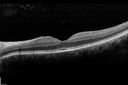
Macular Pucker 20/40 vision - minimal symptoms for 1 year Right eye632 views SD OCT line Scan Fellow Eye 20/16
|
|

Papilledema - Sagittal Cerebral Vein Thrombosis - Lung Transplant Cystic Fibrosis 36 Year Old Man632 viewsLine scan through optic nerve
|
|
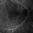
Cartwheel CNVM - Classic - wet AMD - evidence of possible old CSR - ICG is normal - shows nothing632 views75 y.o Decreased Vision right eye for one year. VA OD: sc20/125+2 OS 20/25. Autofluorescence and SD OCT show evidence of old serous retinal detachment superior to the fovea suggesting a history of CSR. The macular lesion on FA shows a classic cartwheel appearance
|
|

632 views
|
|

Polypoidal Choroidal Vasculopathy with Pulsating ICG polyp left eye632 views
|
|

Unilateral Acute Idiopathic Maculopathy (UAIM)632 views
|
|

ERM632 viewsERM: Epiretinal Membrane
Five years prior to presentation this patient was struck in the eye with a basketball, suffering a vitreal hemorrhage. He underwent vitrectomy and cataract surgery. He later developed a faint ERM. On recent presentation he complained of increased blurring and a floater in his O.D. Examination determined that the ERM had become more prominent.
|
|

Pre-retinal hemorrhage following Posterior Vitreous Detachment (separation - PVD)632 views68 year old woman with acute PVD OD and new preretinal hemorrhage - also there is a macular nevus
|
|

West Nile Virus Multifocal Choroiditis and diabetic retinopathy632 viewsMultifocal and clustered focal lesions are from west nile virus. The retinopathy is probably diabetic but could also be related to west nile virus
|
|

PDR NVD left eye and NVE right eye632 views41 year old diabetic woman with PDR in both eyes. High risk in the left eye. She had PRP, Avastin, and vitrectomy x 2 in the left eye with final vision of 20/25. The right eye had eventually PRP laser
|
|

Chronic CSR with mild vision loss - Macular Atrophy Right Eye632 views55 year old man has not been to an eye doctor in years. VA 20/40 OD and 20/25 OS. OCT shows outer retinal atrophy and choroidal thickening. FAF shows guttering. FA shows no active lesions. ICG shows patchy mild leakage visible at about 5 minutes.
|
|

Subfoveal CNVM wet AMD early fa631 viewsShe has noticed decreased vision in the left eye. It is hard to say for how long, maybe a few months. She had her last eye exam about a year ago. She thoughts perhaps it was cataracts, but you saw a problem with the macula and suggested she come here for evaluation.
VISUAL ACUITY: Vision OD is 20/25, OS is 2/200
|
|

Wet AMD 62 year old male smoker 631 views62-year-old man has wet age-related macular degeneration in the right eye. He has been undergoing treatment with Lucentis for that. Then his left eye developed hazy vision a few weeks ago while he was overseas and now his vision is hazy in both eyes. OD 20/40, OS 20/30. FA shows a classic subfoveal choroidal neovascular membrane in the left eye, which is leaking and in the right eye, which is dry. He is going to stop smoking today. FU: VA 20/30 OU with 2 years of every 6 weeks bilateral avastin!
|
|

Group 2a Juxtafoveal Retinal Telangiectasis (type 2)631 views58-year-old man decreased vision right eye and not better since cataract surgery one year ago. OD 20/40, OS 20/40.
|
|

Retinal Arterial Atheromata631 views68-year-old woman with records going back 7 years, which clearly document retinal arterial plaques in both eyes.
|
|

Branch Retinal Arterial Occlusion 20/20 Vision - Symptoms for 1 Day631 views75-year-old man was seen in the office on 4/6/2010. He noticed a sudden vision change in the right eye yesterday morning. 20/20 Vision both Eyes
|
|

Macular Pucker 20/40 Vision - Pigment Bottom of Photo from Old Retinal Detachment 631 views59-year-old woman blurry vision in the right eye for about the last one or two years. She had cataract surgery in 1981, a retinal detachment repair with scleral buckle in 1988 in the right eye, and then she had lens implants in both eyes, anterior chamber lenses, in 1994. She did start taking Latisse for eyelashes about a year and a half ago.
VISUAL ACUITY: Vision OD is 20/40, OS is 20/20
(4 months later with a short course of antiinflammatory drops and new glasses her vision improved to 20/25)
|
|

Acute Annular Outer Retinopathy for 3 Months - 25 YO Female Fellow Normal Eye631 views25-year-old female who, about three months ago, put on her sunglasses and noticed a smudge in the vision and at that point she noticed the right eye was hazy. She had a cold a week or two before the vision loss, but she didn’t have any fever. She has no focal neurological problems in her past history. VA 20/20 OU
|
|

ECTOPIC DISCIFORM SCAR/PERIPHERAL CHOROIDAL NEOVASCULAR MEMBRANE – EXUDATE –INFLAMMATION IN REACTION TO THE PERIPHERAL VASCULAR LESION 631 views53-year-old woman has noticed some blurred vision in the right eye for a few years. In retrospect, she thinks it is possible since she was a teenager the right eye was always the weaker eye and that the left eye has always been the better eye. VISUAL ACUITY: Vision OD is 20/400
|
|

Retinitis Pigmentosa Newly Diagnosed 55 Year Old631 views55-year-old noticed his vision declining recently, over the last few weeks, particularly in the right eye. He said previous to that the vision was not normal but was pretty good. OD is 20/100, OS is 20/50
OCT SCAN: There is macular thickening in both eyes with intraretinal cysts.
|
|

Wet Age-related Macular Degeneration (AMD) Radiating Chorioretinal Folds from Contracting Macular Scar631 views93-year-old man has wet age-related macular degeneration in his left eye with a subfoveal pigment epithelial detachment.
Vision OS is 20/40. There are striking radiating chorioretinal folds emanating off of the scar, which suggests it is contracting.
|
|

Choroidal Metastases - Both Eyes - Exudative Retinal Detachment631 views58-year-old man smoke a pack of cigarettes a day. He has had some ribcage pain for about the last two weeks. He also had a sinus infection for about two weeks, and he has also had hazy vision on the superior visual field in the left eye for about the last two weeks. vision is 20/25 in both eyes
|
|

Primary Ocular Lymphoma - 020513 - SD OCT631 viewsVIDEO TAKES TIME TO LOAD
|
|

Pigmented Peripheral Retinal Degeneration631 views42-year old male comes in for routine eye exam and to follow up on peripheral retinal degeneration in both eyes. VA is 20/20, right eye and 20/25, left eye. Patient is asymptomatic with no visual complaints.
|
|

631 views38-year old male presents with decreased vision in the left eye. VA was 20/20-right eye and 20/70-left eye with pinhole no improvement. Fundus exam shows exudates near the macula and also exudates with small hemorrhages, superior nasally in the left eye.
|
|
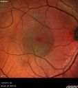
Chronic Central Serous Retinopathy - False Color631 views
|
|
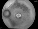
Bullseye Maculopathy without Plaquinel631 viewsFundus photograph reveals Bull's-eye maculopathy without the use of plaquinel. Patients VA is 20/20, Right eye and 20/25, Left eye. There are reported cases of Scleroderma patients with retinal pigment epithelial atrophy. Will return for follow up in 6-months.
|
|

Optic Disk Drusen Advanced Nerve Fiber Loss631 views
|
|
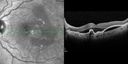
Polypoidal Choroidal Vasculopathy with Pulsating ICG polyp left eye631 views
|
|

Unilateral Acute Idiopathic Maculopathy (UAIM)631 views41 year old with serous retinal detachment OS and sudden vision loss. He had a viral prodrome. Coxscackie IgG was high but no acute infection was documented. Vision improved in about 2 months without therapy. - Right eye has spot nasal to the optic nerve
|
|

Orbital Cellulitis - Optic nerve compression with ocular ischemia631 views81 year old woman with constricted eye movement, orbital cellulitis and severe vision loss in the right eye (LP). She has whitening of the retina and the OCT shows ischemic changes as well as subretinal fluid and blood. The vision in this eye declined to NLP and the eye remained ischemic despite control of her infection.
|
|

Macular Atrophy - Phototoxicity - Macular Hole Surgery - Color631 views25 years ago patient suffered immediate severe vision loss following macular hole surgery. You can almost see light pipe tracks in the atrophy in the macula. Vision is 5/200
|
|
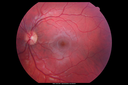
Choroidal Osteoma 13 Year Old631 views13 year old with 6 months of vision loss right eye and vision of 20/40
|
|
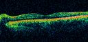
Multiple Evanescent White Dot Syndrome - Atypical Yellow Foveal Spot Right Eye630 views12-year-old had decreased vision in the left eye from reported amblyopia since around the year 2000. His right eye was doing fine until two days ago, when he had sudden severe vision loss. He sees a blurred spot in the middle of the vision in the right eye wherever he looks.
OD 20/60 with eccentric movement, OS 20/200. Pinhole 20/70. IOP: OD 14, OS 10.
|
|

630 views
|
|

Diabetic Macular Edema Left Eye - Post Laser630 views82-year-old woman diabetic for many years, last eye exam 5 years ago with gradual vision loss. OD 20/60, OS 20/70. IOP: OD 16, OS 17.
|
|

Macular Degeneration - Choroidal Laser Left eye - New Subfoveal CNVM left eye - Did well with Avastin630 views69-year-old woman new vision loss right eye and old macular scar left eye. OD 20/40, OS 20/200
|
|

DUSN - Diffuse Unilateral Subacute Neuroretinitis - Nematode630 views
|
|

Proliferative Sickle Retinopathy - Hemoglobin SC630 views53-year-old woman with a new spider-like floater in the right eye for about six days. She sees one big floater and another little one. She does have hemoglobin SC hemoglobinopathy. VISUAL ACUITY: Vision OD is 20/20; OS is 20/50, PH is 20/20.
|
|

Retinal Arterial Macroaneurysm - Increased Swelling after Laser 630 views65-year-old woman was seen in the office on January 12, 2011. She has noticed decreased vision in the right eye for the last two or three weeks. She has had some discomfort in both eyes as well. She has a history of low blood pressure, but she does have high cholesterol.
VISUAL ACUITY: OD 20/60
|
|
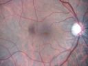
Macular Pucker - 1 year after Vitrectomy - 20/25 Vision - Postoperative Images630 views60-year-old man had vitrectomy for a macular pucker in the right eye a year ago. His vision is good.
VISUAL ACUITY: Vision OD is 20/25 Eye is now pseudophakic
|
|

Cornea Ulcer/Abrasion630 views
|
|
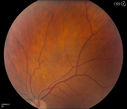
Pigmented Peripheral Retinal Degeneration630 views42-year old male comes in for routine eye exam and to follow up on peripheral retinal degeneration in both eyes. VA is 20/20, right eye and 20/25, left eye. Patient is asymptomatic with no visual complaints.
|
|

Retinal Capillary Hemangioma due to Von Hipple-Lindau Syndrome630 viewsA 47-year old female comes in for a second opinion for retinal capillary Hemangioma in the left eye. VA was HM (hand motion) in the left eye. She has had multiple procedures done for the left eye including, Retinal Detachment surgery, YAG Laser PI, YAG Laser Capsulotomy, and Cataract surgery.
|
|

Wet AMD - New Small Hemorrhage Both Eyes - Mature CNVM visible on Early Indocyanine Green Angiogram630 views
|
|

BRAO and PAMM630 views
|
|

Adult Vitelliform Macular Dystrophy - Pattern Dystrophy630 viewsSD OCT shows material to be subretinal not sub-RPE as you would see in typical AMD
|
|

Syphilitic Chorioretinitis630 viewsSevere outer retinal atrophy and RPE non-geographic atrophy from syphilis. Vision improved from 20/63 to 20/32 with therapy. Images look a lot like DUSN. Patient had several surgeries (vitrectomy for floaters, vitrectomy for macular pucker, Avastin injections and macular laser for edema) prior by other retina specialists prior to being correctly diagnosed and treated.
|
|

Asteroid Hyalosis - Clarus Color Image630 views
|
|

Peripheral exudative hemorrhagic chorioretinopathy - PECHR - Subfoveal fluid630 views85 year old female with
Fluctuating vision in the right eye.
VA 20/200 OD, 20/32 OS
|
|
| 18117 files on 182 page(s) |
 |
|
|
|
|
43 | |
|
|
|
|


