|
|

Branch Retinal Vein Occlusion with Macular Edema601 x angesehenCentral foveal thickness came to normal after laser and intravitreal Kenalog
|
|
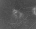
Juxtafoveal Telangiectasis - (MacTel) - Stage 4 both Eyes601 x angesehen57-year-old woman with a history of decreased vision. The patient reports that her vision has been gradually decreasing in the left more so than the right eye. 20/50 OD and 20/60
|
|

Non-Arteritic Anterior Ischemic Optic Neuropathy - Peripapillary Choroidal Ischemia601 x angesehen68-year-old woman noticed central vision loss in the right eye about a week ago. It is getting a little bit better. OD 20/160, OS 20/25.
|
|
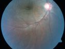
Congenital Toxoplasmosis with non-macular Scars Both Eyes - Recent Reactivation (recurrence) Right Eye601 x angesehen34-year-old man has congenital toxoplasmosis with four week history of foggy vision and new floaters in the right eye. OD is 20/40, OS 20/30
|
|
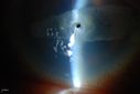
Corneal Ulcer. 601 x angesehenA hole in the cornea. Aqueous Humor leak
|
|

Wet Age-related Macular Degeneration (AMD) Radiating Chorioretinal Folds from Contracting Macular Scar601 x angesehen93-year-old man has wet age-related macular degeneration in his left eye with a subfoveal pigment epithelial detachment.
Vision OS is 20/40. There are striking radiating chorioretinal folds emanating off of the scar, which suggests it is contracting.
|
|

Posterior Polar Annular Choroidal Dystrophy601 x angesehen68 year old woman with know macular irregularities for over 20 years. She has since been followed another 6 years and her vision remains 20/25.
|
|

Neovascularization elsewhere (NVE) Left Eye with Vitreous Hemorrhage601 x angesehen
|
|

Asymptomatic Occult Subfoveal CNVM in Good Eye Visible on ICG601 x angesehen
|
|
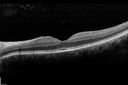
Macular Pucker 20/40 vision - minimal symptoms for 1 year Right eye601 x angesehen SD OCT line Scan Fellow Eye 20/16
|
|

Uveitis and Cystoid Macular Edema - Probably Sarcoidosis601 x angesehenVA 20/63 OD 20/50 OS - 3 months vision loss - second episode by history of uveitis
|
|

Acute Macular Neuroretinopathy - (AMN) - Spot in Vision OS - 40 Year old Female601 x angesehenVision improved 1 month later
|
|

Hairy Cell Leukemia - Retinal Hemorrhage and twig Branch Vein Occlusion601 x angesehen79 year old man He has had hairy cell leukemia since 2002. He is in remission. His last blood tests were 9/2018. He just moved down here and needs a new leukemia doctor. His vision is fine.
VA OD: Dcc20/25
VA OS: Dcc20/25
IOP: TP: OD:12 OS:12
|
|
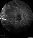
Coats' Disease - 14 year old - Asymptomatic601 x angesehenPresented with macular exudates. Temporal macroaneurysms responded to laser. Ultra-wide field FA shows nonperfusion
|
|

Coats' Disease -51 year old asymptomatic male601 x angesehen20/20 vision - had laser to non-perfusion because of proliferation.
|
|

Autoimmune Retinopathy601 x angesehen91 year old female with severe vision loss over the past 3 years. She has shimmery scotomas. The main findings are outer retinal atrophy evident on OCT and FAF. VA 20/80 OD and 20/125 OS
|
|
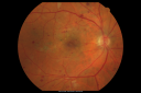
BDR and Diabetic Macular Edema with Fresh Laser Right Eye600 x angesehen60-year old man OD 20/60, OS 20/50 worsening vision both eyes
|
|

Serpiginous choroiditis600 x angesehenInactive scars in both eyes
|
|
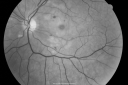
Purtchers Retinopathy with Bilateral Central Retinal Artery Occlusions 600 x angesehen50-year-old man traumatic chest compression and leg crushing injury. OD 1/200, OS 3/200
|
|

DUSN - Diffuse Unilateral Subacute Neuroretinitis - Nematode600 x angesehen
|
|

Chronic Pseudophakic Cystoid Macular Edema - Haptic Migrated into Anterior Chamber Through Peripheral Iridotomy600 x angesehen57-year-old man with congenital nystagmus. He had cataract surgery done 30 years ago and has decreased vision left eye for 6 months. OD is 20/50, OS is 20/100
|
|

Traumatic Macular Hole and Pre-retinal Hemorrhage - Assault600 x angesehenThis 18 year old male was assaulted on 3 weeks ago and seen in the emergency room for that. He returns today reporting his left eye is getting better and is not quite as blurry. VA 20/60 (uncorrected)
|
|

Cilioretinal artery occlusion and Ophthalmic Artery Occlusion with Relative Sparing of the Central Retinal Artery600 x angesehen80-year-old woman who says she was looking in the mirror, covered her right eye and noticed she could not see well out of the left eye. That just happened yesterday. Her last eye check was a few years ago. She also had a carotid endarterectomy on the right side in 2006 shortly after coronary bypass surgery. OD is 20/40, OS is 20/200.
|
|

wide angle fa mid. od600 x angesehenleaking, abnormal vessels
|
|

Haramotoma with ERM600 x angesehenHaramotoma with an ERM in the OS of a middle aged man
|
|

Pigmented Peripheral Retinal Degeneration600 x angesehen42-year old male comes in for routine eye exam and to follow up on peripheral retinal degeneration in both eyes. VA is 20/20, right eye and 20/25, left eye. Patient is asymptomatic with no visual complaints.
|
|

Wet AMD - New Small Hemorrhage Both Eyes - Mature CNVM visible on Early Indocyanine Green Angiogram600 x angesehen
|
|

Reticular Macular Disease (Pseudo-drusen) Both Eyes - Occult CNVM and Macular Edema Right Eye 600 x angesehenColor Fundus Photo
|
|

Diabetic Retinopathy with CME600 x angesehenSecond opinion for diabetic retinopathy. VA is 20/30, right eye, 20/50, left eye. Patient has mild swelling in the macula of the right eye. No evidence of CME but does have a moderate cataract in the left eye.
|
|

ERM600 x angesehenERM: Epiretinal Membrane
Five years prior to presentation this patient was struck in the eye with a basketball, suffering a vitreal hemorrhage. He underwent vitrectomy and cataract surgery. He later developed a faint ERM. On recent presentation he complained of increased blurring and a floater in his O.D. Examination determined that the ERM had become more prominent.
|
|

Choroidal Folds Hyperope (+5.00)600 x angesehen
|
|

Trousseau Sign - CRVO and Pancreatic Cancer600 x angesehenLeft eye shows CRVO with severe edema. The right eye has multiple small hemorrhages. Some of this is probably anemic retinopathy and some from hypercoaguability of cancer. The patient has pancreatic cancer.
|
|

Syphillitic Optic Neuropathy and Maculopathy600 x angesehen46 year old woman with 2 weeks of vision loss OD. VA 20/200 OD; 20/20 OS. there are posterior vitreous cells on OCT in both eyes. ICG, FAF, and FA show an abnormal macula OD. VA improved to 20/30 within a month with treatment with PCN.
|
|
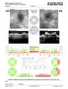
Grade 4 hypertensive retinopathy600 x angesehen55 year old man Hhe did have headaches a few weeks ago and also some nausea and vomiting. He noticed for a few weeks the vision in the left eye was blurred and that the vision in the left eye is dark.
VA OD: Dcc20/20
VA OS: Dcc20/100-1
He was admitted directly to ICU for blood pressure control (290 / 170 mmHg)
3 weeks later his VA increased to 20/20 OD, 20/32 OS
|
|

Macular Pucker in eye following giant retinal tear RD repair photo of tear599 x angesehennow that the bubble is gone, but he is starting to notice distortion of central vision. He had a giant retinal tear retinal detachment repair emergently done on November 8, 2007, and subsequent laser on November 15th.
VISUAL ACUITY: Vision OD 20/400.
6 weeks post-op VA 20/40
|
|
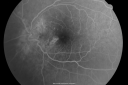
Multifocal Choroiditis and Subretinal Fibrosis - 32 yo Female New Lesion OS599 x angesehen32-year-old woman vision loss in the right eye associated with macular scarring and multifocal choroiditis in 1999 with new vision loss in left eye: OD 20/400, OS 20/50.
|
|

Serpiginous choroiditis599 x angesehenInactive scars in both eyes
|
|
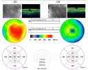
Macular Pucker Pre-op 20/200 vision (history of retinal tears)599 x angesehen66-year-old man has had laser to retinal tears in both eyes in 1999 and in 2001. His right eye has had rapidly declining vision over the last month. VISUAL ACUITY: OD: 20/200
|
|

Patterned Pigment Dystrophy of the Fovea 79 YO Woman VA 20/20 Right 20/30 Left599 x angesehen
|
|

Macular Fold - 40 years post retinal detachment repair - Glaucoma 599 x angesehen79-year-old woman has had a long-standing macular fold in the right eye associated with retinal detachment repair in 1970. (40 years ago) She has a macular scar in that eye. She has optic atrophy in both eyes from glaucoma. Vision OD is hand motion, OS is 20/50
|
|
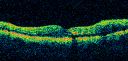
Diabetic Macular Edema and Hypertensive Retinopathy - Circinate Exudate (Ring Exudate) 599 x angesehen55-year-old woman diabetic for fifteen years and high blood pressure, especially over the last few months. She has had problems with nosebleeds, headaches, and there has been some difficulty bringing her blood pressure down. She said now the blood pressure is under control but it was running 220/120 mmHg for some time. Her vision in the right eye has been poor for four weeks with a spot in the central vision and both eyes have been blurred.
VISUAL ACUITY: Vision OD is 20/160, OS is 20/60
|
|
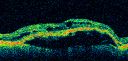
Wet AMD - PED with exudate 20/400 87 Year old599 x angesehen87-year-old woman has wet age-related macular degeneration in the left eye. Vision OD is 20/25, OS is 20/400
|
|

Central Retinal Vein Occlusion Recurrent Edema 6 weeks after Lucentis - Nonperfusion Temporally599 x angesehen66-year-old woman has a central retinal vein occlusion in the right eye with macular edema. She had intravitreal Lucentis treatment six weeks ago and she has noticed over the last week or two her vision is declining.
VISUAL ACUITY: OD 20/70, OS 20/30.
|
|

Choroidal Neovascular Membrane - Optic Disc Drusen - Hemorrhage (Coumadin) 599 x angesehen82-year-old man with decreased vision in the left eye over about the last six to 12 months. He is also on Coumadin. OD 20/20, OS 20/80
|
|
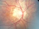
Angioid Streaks - Pseudoxanthoma Elasticum - Supressed CNVM599 x angesehen54-year-old woman had angioid streaks and macular degeneration in both eyes. She has had intravitreal Avastin in the right eye about every three months and her vision in that eye is good.
VISUAL ACUITY: Vision OD is 20/30, OS is 2/200
|
|

Myopic Traction Maculopathy - Vitreoretinal Traction - Possible Occult Neovascular Membrane599 x angesehen58-year-old woman was seen in the office on June 30, 2011. In 2002, her vision was 20/30. Subsequent to that she had her left eye made her distance eye, which was dominate. She had LASIK surgery on the right eye to make that her near eye and she was getting along well until about a year ago when she noticed substantial vision loss in the left eye only. VISUAL ACUITY: The near eye is 20/200, but she reads J2. OS 20/100- and J16.
|
|

Branch Retinal Vein Occlusion with Cotton Wool Spot 599 x angesehen70-year-old man with blurred vision in the right eye for about one or two months. OD is 20/80, OS is 20/25.
|
|

Acute Central Serous Retinopathy - CSR - Steroid Induced (probably) - Good Vision - ICG Fellow Eye599 x angesehen42-year-old man was seen in the office on October 5, 2011. He had noticed starting in August after a course of antibiotic and steroids, that he developed new spots in his vision in the right eye. He may have had an episode like this sometime in the past. He did take steroids a few years ago and his vision did change at that time, but then returned.
VISUAL ACUITY: OD 20/32, OS 20/32. The OCT scan of the right eye confirms subretinal fluid.
|
|

Choroidal Malignant Melanoma - mid phase FA599 x angesehenmid-phase of FA at 5 minutes 10 seconds (Choroidal malignant melanoma status post plaque radiotherapy OD in 1986, patient also has a submacular hemorrhage OD, and radiation retinopathy OD.)
|
|

Serous Retinal Detachment - Hypertensive Retinopathy - 165/120 - Late - ICG shows leakage599 x angesehen48-year-old woman noticed when she was blinking there was a white flash in the left eye starting about a month ago. She sees it temporally. OD 20/16, OS 20/20. There is a serous retinal detachment superotemporal to the optic nerve falling short of the fovea. Her blood pressure 165/120.
|
|

Retinitis Pigmentosa Sine Pigmento or Unknown Dystrophy - Peripheral Superficial White Spots and Recurrent CME - FA LATE599 x angesehen46-year-old woman three of her 5 siblings have been diagnosed with retinitis pigmentosa. Her mother and father were seen and as far as everyone can tell, neither of them has it, although there was some thought they might be carriers. They were evaluated twenty five years ago and since then as far as she knows, there has not been a lot of testing done.
She has noticed a substantial decline in vision in both eyes over the last month or two. OD 20/40, OS 20/50. IOP: Both eyes have CME
|
|
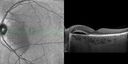
Serous Macular Detachment Both Eyes - Wet AMD vs. CSR - 62 Year Old Man - SD - OCT599 x angesehen
|
|

Unilateral Acute Idiopathic Maculopathy (UAIM)599 x angesehen1 week later eye looks better (after initial scan) 41 year old with serous retinal detachment OS and sudden vision loss. He had a viral prodrome. Coxscackie IgG was high but no acute infection was documented. Vision improved in about 2 months without therapy. - Right eye has spot nasal to the optic nerve
|
|

Posterior Uveal Effusion599 x angesehen
|
|

Syphillitic Optic Neuropathy and Maculopathy599 x angesehen46 year old woman with 2 weeks of vision loss OD. VA 20/200 OD; 20/20 OS. there are posterior vitreous cells on OCT in both eyes. ICG, FAF, and FA show an abnormal macula OD. VA improved to 20/30 within a month with treatment with PCN.
|
|

Late Onset Retinal Degeneration (L-ORD)599 x angesehen55 year old with acute vision loss from a CNVM in the right eye. He responded to Lucentis therapy. His mother and her family has been confirmed genetically to have L-ORD and were part of the early reports.
|
|

Fundus Albipunctatus599 x angesehen12 year old female with normal vision. She has 4 siblings all of whom have either white spots or spots on IR. Genetic testing by parents was deferred.
|
|

Familial Exudative Vitreoretinopathy - FEVR - Stage 4b OD599 x angesehen10 year old child with poor vision OD from birth. The left eye had vascular remodelling in the temporal periphery with preretinal abnormalitlies seen on OCT. The patient never returned for a fluorescein angiogram. Left eye is either stage 1 or stage 2. no family history
|
|

Wet AMD - Subfoveal CNVM - Excellent Response to Avastin598 x angesehen76-year-old woman two to three months of vision loss OD
Vision started at 20/200 and with Avastin improved to 20/25. She has required monthly injections and as of October 2010 has been treated about 16 times. Her left eye remains good at 20/20.
The OCT from post-treatment as well as pretreatment images are in this album.
|
|
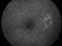
Subfoveal CNVM wet AMD fellow eye598 x angesehenShe has noticed decreased vision in the left eye. It is hard to say for how long, maybe a few months. She had her last eye exam about a year ago. She thoughts perhaps it was cataracts, but you saw a problem with the macula and suggested she come here for evaluation.
VISUAL ACUITY: Vision OD is 20/25, OS is 2/200
|
|

Macular Pucker in eye following giant retinal tear RD repair pre-op OCT Map598 x angesehennow that the bubble is gone, but he is starting to notice distortion of central vision. He had a giant retinal tear retinal detachment repair emergently done on November 8, 2007, and subsequent laser on November 15th.
VISUAL ACUITY: Vision OD 20/400.
6 weeks post-op VA 20/40
|
|

598 x angesehen
|
|

Purtchers Retinopathy with Bilateral Central Retinal Artery Occlusions 598 x angesehen50-year-old man traumatic chest compression and leg crushing injury. OD 1/200, OS 3/200
|
|

Basal Laminar (cuticular) Drusen both eyes 60 Year Old Woman598 x angesehen60-year-old woman with minimal vision loss. Vision OD is 20/30 OS 20/40
|
|

Nanopthalmous (Microphthalmous) Chorioretinal Folds and Choroidal Thickening Both Eyes598 x angesehen67-year-old woman with microphthalmos/nanophthalmos Her vision has gradually been getting a little bit worse, especially in the right eye. Also the anterior chamber is getting crowded and there is concern about the need for cataract surgery. She has one sister and one cousin who also have nanophthalmos.
VISUAL ACUITY: OD 20/50, OS 20/80.
|
|
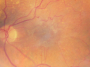
Asymptomatic 20/200 Macular Pucker 79 year old man - Left Eye598 x angesehen79-year-old man who had not noticed a change in vision, but was diagnosed with a macular pucker in the left eye. Even now he doesn’t notice much difference between the eyes, although he doesn’t usually compare them. He has a prescription for new glasses.
VISUAL ACUITY: OD 20/80, Pinhole 20/50. OS 20/200.
|
|

Congenital Achromatopsia 598 x angesehen26-year-old man was diagnosed with congenital achromatopsia by ERG as a child. He has family members who also have this disorder. OD 20/200, OS 20/200.
|
|

Reticular Macular Disease (Pseudo-drusen) Both Eyes - Occult CNVM and Macular Edema Right Eye 598 x angesehenColor Fundus Photo
|
|

Embolic CRAO Early598 x angesehen
|
|

Choroideremia - Female Carrier - Patchy Atrophy of Choroid and RPE - Lyonization598 x angesehen
|
|

Syphilitic Chorioretinitis598 x angesehenSevere outer retinal atrophy and RPE non-geographic atrophy from syphilis. Vision improved from 20/63 to 20/32 with therapy. Images look a lot like DUSN. Patient had several surgeries (vitrectomy for floaters, vitrectomy for macular pucker, Avastin injections and macular laser for edema) prior by other retina specialists prior to being correctly diagnosed and treated.
|
|

Melanosis Oculi and Choroidal Melanoma598 x angesehen88 year old female with 2.5 mm elevated choroidal melanoma and melanosis oculi. She also has geographic atrophy from AMD. The right eye is the better eye with 20/40 vision. After observation with growth, brachytherapy was done.
|
|

Peripheral exudative hemorrhagic chorioretinopathy - PECHR - Subfoveal fluid598 x angesehen85 year old female with
Fluctuating vision in the right eye.
VA 20/200 OD, 20/32 OS
|
|

Stargart's Macular Dystrophy Severe597 x angesehen32-year-old man carries a diagnosis of Stargardt’s disease with poor vision in both eyes since he was 13. He has not noticed any substantial change since he was here 4 years ago.
VISUAL ACUITY: OD 10/200, OS 8/200
|
|

Group 2a Juxtafoveal Retinal Telangiectasis (type 2)597 x angesehen58-year-old man decreased vision right eye and not better since cataract surgery one year ago. OD 20/40, OS 20/40.
|
|

Proliferative Diabetic Retinopathy Both Eyes - Vitreous Hemorrahge Left Eye - Vessel Leaking Fluorescein597 x angesehen67-year-old man has proliferative diabetic retinopathy in both eyes with vitreous hemorrhage in the left eye. He has had pan retinal laser in the left eye. The vitreous hemorrhage has been there for greater than two years and his vision is still hazy. He takes Lumigan in both eyes for his glaucoma.
VISUAL ACUITY: OD 20/40, OS 20/40
|
|

New Subfoveal Classic CNVM OS - Wet AMD - Macular Scar Congenital Toxoplasmosis Right Eye 597 x angesehen77-year-old woman as had poor vision in the right eye since birth from congenital toxoplasmosis of her macula in the right eye. Two to three weeks ago when she noticed vision loss in the left eye. VISUAL ACUITY: OD 2/200, OS 20/200.
|
|

Serous Retinal Detachment - Hypertensive Retinopathy - 165/120 597 x angesehen48-year-old woman noticed when she was blinking there was a white flash in the left eye starting about a month ago. She sees it temporally. OD 20/16, OS 20/20. There is a serous retinal detachment superotemporal to the optic nerve falling short of the fovea. Her blood pressure 165/120.
|
|

Macular Hole Left Eye - progression from VMT - Stage II macular hole - Spontaneously Closed Macular Hole597 x angesehen
|
|

Macular Hole Left Eye - progression from VMT - Stage II macular hole - Spontaneously Closed Macular Hole597 x angesehenVision has dropped to 20/50 and there is a full thickness macular hole
|
|

BRAO and PAMM597 x angesehen
|
|
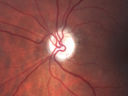
Acute AION OS - Mild disc pallor OD597 x angesehenSudden vision loss OS x 1 week with optic nerve swelling. Evidence of optic atrophy in right eye.
|
|
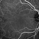
Acute Macular Neuroretinopathy - (AMN) - Spot in Vision OS - 40 Year old Female597 x angesehen
|
|

Toxocariasis - Stalk from optic nerve to peripheral scar597 x angesehen
|
|

Confluent Soft Drusen with Subetinal Fluid - Right Eye597 x angesehenAsymptomatic 85 year old man. OCT shows pocket of subfoveal fluid in the center of the macula. This could be mechanical from the drusen preventing the retina from settling down. Testing did not reveal a CNVM and subsequent OCT 6 weeks later showed no change in structure.
|
|

Chronic CSR with mild vision loss - Macular Atrophy Right Eye597 x angesehen55 year old man has not been to an eye doctor in years. VA 20/40 OD and 20/25 OS. OCT shows outer retinal atrophy and choroidal thickening. FAF shows guttering. FA shows no active lesions. ICG shows patchy mild leakage visible at about 5 minutes.
|
|

Toxoplasmosis - Recurrent - Moderate Vitritis597 x angesehen32 year old woman with 20/80 vision and moderately painful red eye in the left eye only. The left eye had some redness about 10 years ago. She is a native of Sierra Leone
|
|

Chronic Central Serous Chorioretinopathy Both Eyes597 x angesehen60 year old man with 20 year history of chronic CSR. He has had laser and PDT in the past. The left eye has been 20/100 for decades. The right eye is now declining. Was 20/32 and is down to 20/50. There is chronic low grade subretinal fluid away from the center in the right eye. (focal laser in the right eye in 2005, left eye 2001 at BPEI). Refampin tried in 2012, Aldactone in 2013. PDT in 204 and 2017 Images are from 2017
|
|

Subtle Branch Retinal Artery Occlusion597 x angesehen68 year old man with shadow in the left eye yesterday. IR and OCT show subtle retinal swelling. FA is normal
|
|

Sclerochoroidal Calcification and Peripheral Exudative Hemorrhagic Chorioretinopathy597 x angesehen67 year old man with asymptomatic peripheral subretinal hemorrhage associated with sclerochoroidal calcification.
VA 20/32 OU
|
|
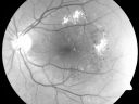
Clinically Significant Diabetic Macular Edema both Eyes with Exudates596 x angesehen56-year-old woman with diabetes for twelve years. She has noticed blurring of the vision in the right eye for about six months. OD is 20/70, OS is 20/25.
|
|

Pigmented Maculae - Mother and Daughter - Normal Visual Acuity596 x angesehen12-year-old had a dilated macular exam and the macula look abnormal. Interestingly, her mother, who is with her at this visit, was told about a year ago that her maculae are abnormal too. Her mother’s vision is fine. In the family tree, there are no known retinal dystrophies. They are both healthy.
VISUAL ACUITY: Her vision is 20/20 in both eyes.
|
|

Retinitis Pigmentosa Newly Diagnosed 55 Year Old596 x angesehen55-year-old noticed his vision declining recently, over the last few weeks, particularly in the right eye. He said previous to that the vision was not normal but was pretty good. OD is 20/100, OS is 20/50
OCT SCAN: There is macular thickening in both eyes with intraretinal cysts.
|
|

Choroidal Melanoma (Medium) Exudative Retinal Detachment 596 x angesehen87-year-old man noticed a new onset floater a few months ago. OD is 20/40, OS is 20/30
|
|

X-linked Juvenille Retinoschisis - Peripheral Retinal Vascular Anomalies - Vitreous Hemorrhage - 8 Year Old Boy596 x angesehen8-year-old child OD 20/40, OS 20/50
OD: Vertical C/D ratio is 0.2. There are foveal cysts. There is also a retinal elevation inferiorly and there are patchy peripheral retinal hemorrhages.
OS: Vertical C/D ratio is 0.2. There are foveal cysts. There is peripheral retinal issues with some hemorrhage in some areas of peripheral retinoschisis.
|
|

3-D OCT of Macular Hole with Operculum596 x angesehen
|
|
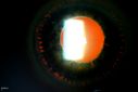
Trans-Illumination Defects596 x angesehen74-year old patient who VA 20/70 OD, 20/50 OS. Complaints of blurred vision. Iris atrophy, both eyes. ERM, right eye, Patient to have cataract surgery to improve distance vision.
|
|

Posterior Polar Annular Choroidal Dystrophy596 x angesehen68 year old woman with know macular irregularities for over 20 years. She has since been followed another 6 years and her vision remains 20/25.
|
|

Primary Ocular Lymphoma - 020513 596 x angesehen
|
|

Reticular Macular Disease (Pseudo-drusen) Both Eyes - Occult CNVM and Macular Edema Right Eye 596 x angesehenFundus Autofluorescence
|
|
| 17965 Dateien auf 180 Seite(n) |
 |
|
|
|
|
45 | |
|
|
|
|


